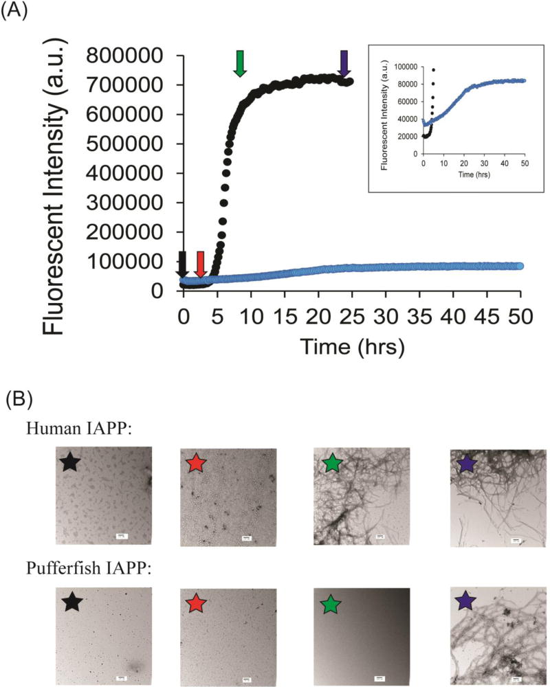
Figure-6.
Analysis of the ability of pufferfish IAPP to form amyloid in phosphate buffered saline solution. (A) Fluorescence monitored thioflavin-T assays of human IAPP (black) and pufferfish IAPP (blue) amyloid formation in 20 mM sodium phosphate, 140 mM potassium chloride at pH 7.4. Arrows indicate times at which aliquots where collected for TEM analysis. Aliquots were collected at t = 0 (black), 3 hrs (red), 7 hrs (green), and 24 hrs (blue) (B) TEM images of samples human IAPP (top) and pufferfish IAPP (bottom) collected at the different time points. Experiments were conducted with 16 µM IAPP, 32 µM thioflavin-T at 25 °C in 20 mM sodium phosphate, 140 mM potassium chloride at pH 7.4. Scale bars in TEM images represent 100 nm.

