Abstract
X-ray crystallography is one of the main methods to determine atomic-resolution 3D images of the whole spectrum of molecules ranging from small inorganic clusters to large protein complexes consisting of hundred-thousands of atoms that constitute the macromolecular machinery of life. Life is not static, and unravelling the structure and dynamics of the most important reactions in chemistry and biology is essential to uncover their mechanism. Many of these reactions, including photosynthesis which drives our biosphere, are light induced and occur on ultrafast timescales. These have been studied with high time resolution primarily by optical spectroscopy, enabled by ultrafast laser technology, but they reduce the vast complexity of the process to a few reaction coordinates. In the AXSIS project at CFEL in Hamburg, funded by the European Research Council, we develop the new method of attosecond serial X-ray crystallography and spectroscopy, to give a full description of ultrafast processes atomically resolved in real space and on the electronic energy landscape, from co-measurement of X-ray and optical spectra, and X-ray diffraction. This technique will revolutionize our understanding of structure and function at the atomic and molecular level and thereby unravel fundamental processes in chemistry and biology like energy conversion processes. For that purpose, we develop a compact, fully coherent, THz-driven atto-second X-ray source based on coherent inverse Compton scattering off a free-electron crystal, to outrun radiation damage effects due to the necessary high X-ray irradiance required to acquire diffraction signals. This highly synergistic project starts from a completely clean slate rather than conforming to the specifications of a large free-electron laser (FEL) user facility, to optimize the entire instrumentation towards fundamental measurements of the mechanism of light absorption and excitation energy transfer. A multidisciplinary team formed by laser-, accelerator,- X-ray scientists as well as spectroscopists and biochemists optimizes X-ray pulse parameters, in tandem with sample delivery, crystal size, and advanced X-ray detectors. Ultimately, the new capability, attosecond serial X-ray crystallography and spectroscopy, will be applied to one of the most important problems in structural biology, which is to elucidate the dynamics of light reactions, electron transfer and protein structure in photosynthesis.
Keywords: Terahertz accelerator, Optical undulator, Attosecond X-ray source, X-ray imaging, X-ray spectroscopy
1. Introduction
The vast majority of the >100,000 macromolecular structures that have been solved and are deposited in the protein data bank were determined by the method of X-ray crystallography. The intense and tunable X-ray beams produced by synchrotron radiation have made this amazing advance in structural biology possible. However, this success belies large challenges with the technique, which stem from radiation damage that is inherent to exposing soft matter to ionizing radiation. With the advent of X-ray FELs the new method of serial femtosecond crystallography (SFX), developed by some of us [8], initiated a new era in X-ray crystallography. It uses femtosecond pulses from X-ray FELs to obtain structures based on diffraction from a stream of nano-crystals in their mother liquor at room temperature (Fig. 1), thereby outrunning radiation damage induced atomic motion (“diffraction before destruction” principle). Diffraction from crystals as small as 100 nm in width has been successfully observed, which contain less than 1000 molecules and are smaller than the micro-domains of most macroscopic protein crystals. The biggest advantages of nanocrystals are that they are much better ordered and are much easier to grow. The growth of large crystals has been the major challenge for the determination of structures of difficult to crystallize proteins like membrane proteins for which less than 600 structures have been determined so far. An example is Photosystem (PS) I, which consist of 36 proteins and 381 cofactors, where it required 13 years of effort to obtain macroscopic crystals (500 micrometer in width) that diffracted to near atomic resolution [18] in a synchrotron beam.
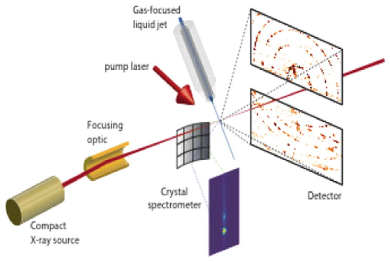
Fig. 1.

Scheme of the attosecond serial crystallography and spectroscopy set-up. X-ray emission is measured with a multi-crystal spectrometer in the von Halmos geometry [3]. Diffraction is measured with a custom 1-kHz CMOS pixel detector.
The SFX technique has the potential to determine the dynamics of proteins, [4,,23] by time-resolved SFX, where reactions are induced by light in the crystals “on the fly” before they are hit by the X-ray pulse, therefore molecular movies may be determined showing biomolecules at work. However, a current limitation of the technique is the limited access due to the worldwide only two current hard X-ray FEL facilities and their high cost of construction and operation. Moreover, while few-fs X-ray pulses can outrun the secondary X-ray radiation damage processes, as the fs pulses are faster than the motion of the atoms induced by ionization and breaking of chemical bonds, the electronic structure of the atoms is strongly disturbed in less than 5 fs by the loss of inner-shell electrons (formation of “hollow atoms”) and valence electrons [13,33]. Furthermore, the Self-Amplified Spontaneous Emission (SASE) mode at current FELs produces X-ray pulses, where each pulse varies in its exact field waveform, i.e., it misses longitudinal coherence. That is, the spectral photon distribution varies in each pulse and does not follow a Gaussian distribution but shows a complex spectral distribution with hundreds of maxima. This maps into the diffraction pattern and has very severe consequences for the accuracy of structure factor determination. Currently, more than 50000 indexed patterns and redundancies of 300–1000 for each reflection are required for the determination of accurate structure factors using the Monte Carlo integration method [22,28,7]. Reconstruction of the Bragg peak profiles could reduce the number of patterns required, but due to the random SASE spectra, this is extremely challenging. An attosecond X-ray source producing pulses with a “clean” and reproducible spectrum, i.e., full longitudinal coherence, will increase accuracy and require much fewer patterns. The attosecond pulses will outrun any damage, including the electronic states. The necessary high peak fluence can be achieved by using the naturally broad bandwidth attosecond pulses, which vastly improve the peak integration as compared with monochromatic X-rays.
In the AXSIS-project, we develop attosecond serial X-ray crystallography and spectroscopy to push X-ray crystallography to a new frontier and overcome limitations of current fs-FELs to fuse atomic-resolution structure determination with ultrafast optical and X-ray spectroscopic techniques and extend the “probe before destruction” concept to the measurement of charge states and electronic transfer through X-ray emission (fluorescence) and absorption (near edge) spectroscopies. Ideally, the attosecond X-ray source is based on coherent inverse Compton scattering (C-ICS). Fig. 2 shows a rough outline for such a setup, that we are developing and building during the course of this project. The nanocrystals are supplied to the X-ray beam in a liquid jet at room temperature, as with FEL experiments. While the current two hard XFELS are limited to 120 images/s (LCLS) and 60 images/s (SACLA), one thousand diffraction patterns and photoemission spectra will be obtained per second with the attosecond pulses of the C-ICS source. We can make a molecular movie by adding an optical stream of excitation pulses precisely synchronized to and preceding the X-ray pulses and varying the delay between optical and X-ray pulses. This unique tool will finally fulfill the dream of observing chemical reactions and biological processes in real space and real time at the necessary time and length scales of atoms and molecules. By developing this technique based on a compact laboratory-scale X-ray source, we vastly extend the availability of attosecond serial crystallography and spectroscopy to the general science community.
Fig. 2.
Schematic layout of the AXSIS setup currently under construction at the SINBAD-facility at DESY in Hamburg.
2. X-ray source technology
X-ray sources with greater than keV photon energy and sufficient brilliance to perform X-ray crystallography are based on relativisitic electron bunches from synchrotrons or linear accelerators. The relativistic motion confines the angle of emission to a narrow cone dramatically increasing source brightness. The electron beams are produced by thermionic or photo cathodes, and are sent into radio-frequency (RF) driven accelerators made up of metallic RF structures operating often at S-band frequencies, i.e., around 1–3 GHz. Frontier facilities like LCLS in the US and the future European XFEL project are linear accelerator (LINAC) based and accelerate electrons to highly relativistic (~10 GeV) energies. A LINAC relies on either room temperature or superconducting RF technology. The accelerating gradients in either case are limited to several tens of MeV per meter limited by field emission from cavity walls and magnetic pulsed heating of the metallic inclusions. The LINAC length, therefore, must be in the km regime and facility costs are in the billion Euro category.
The production of femtosecond electron bunches has seen steep advances over the last decades. Bunches can be created by photoemission from the cathode in the presence of a strongly accelerating field and can be many picoseconds long. These pulses are then injected into a bunch compressor and low charge bunches, 1–10 pC, may be compressed down to 3–10 μm in bunch length which correspond to pulse lengths of 10–30 fs. Compression factors exceeding 100 are routinely achieved at LCLS. At a given accelerating field strength and RF frequency, compression is limited by space charge to a few femtoseconds. These short electron bunches from conventional LINACS are sent into periodic magnetic fields, undulators, with a typical period of 3 cm. The undulators force the electrons on sinusoidal paths to emit photons. Under certain conditions, a coherent, exponential self-amplification of the spontaneously emitted radiation is achieved, by the so-called self-amplified spontaneous emission (SASE) principle. For this process to occur, typically more than 1,000 undulator periods are necessary, putting the length of these radiation structures in the range of 500 m and longer. One consequence of this large length is a very small fractional bandwidth of emission, which is inversely proportional to the number of undulator periods. Therefore, a typical XFEL bandwidth is less than 0.1% of the center wavelength. In addition, it requires an extraordinary energy stability of 0.01% in the electron beam. LINAC-based FELs have enabled significant advances in X-ray science and pioneered the field of femtosecond X-ray crystallography. The FEL process, as sketched above, is constantly being improved for increased scientific reach. So-called seeding techniques have been implemented to improve the coherence properties of the FEL output. Seeding at UV wavelength and high gain harmonic generation (HGHG) and direct seeding with EUV radiation generated via high-order harmonic generation has led to fully coherent output in the EUV at FERMI [1]. Efforts are underway at LCLS to use emittance spoiling of the electron bunch to achieve sub-fs-duration X-ray pulses [25]. However, most of these advances go along with strongly reduced overall photon yield, while the cost of the facility and its complexity stay the same. Furthermore, self-seeding is only successful in a small fraction of the fs pulses, and only this small fraction of the images can be used for data analysis [6].
In the AXSIS project, we combine the optimum X-ray pulse specifications for attosecond serial crystallography and spectroscopy, which are co-evolving as the new methodology, and develop a new accelerator and radiator technology that leads to a highly optimized and compact source for this application. Key aspects of the design principle of the new X-ray source is the implementation of 200 times higher accelerator frequencies, i.e., THz frequencies and correspondingly shorter pulse durations, which increases the threshold for field emission, and thus we expect to utilize 10–100 times higher field strength in the range of 1 GV/m, both in the gun and accelerator. The higher operating frequencies and field strength will enable low bunch emittance and compression to attosecond duration with high charge. In addition, we use optical undulators, i.e., inverse Compton scattering, with a period on the order of 1 μm, 30,000 times shorter compared to magnetic undulators. This in turn allows reduction of the required electron beam energy from 10 GeV to tenth of MeV to reach sub-Ångström radiation. This comes at a cost, which is that the SASE FEL regime is more difficult to access under those conditions. However, new developments in ultrafast nanostructured photocathode arrays combined with precision optical field control eventually enables the production of an electronic Coulomb crystal (Wigner crystal). An ideal electron crystal can be used to produce a coherent emission in an undulator leading to a fully temporally and spatially coherent pulse with attosecond duration from a compact source, while maintaining or even surpassing the peak brilliance of the current hard XFELs. Fig. 2 shows a schematic layout of the proposed AXSIS source and experimental setup currently under construction in the SINBAD-facility at DESY.
Over the past few years, key demonstrations have been made showing the feasibility of this concept. The electronic Coulomb crystal can be generated from a coherently controlled nanostructured field emitter array (FEA) of various types [14,15,20,30,37]. The FEA might be driven by a rectangularly shaped multi-cycle IR or mid-IR laser pulse, that is polarized along the in-plane field emitter elements. The incoming field strength is tuned such that the resulting tip field is typically enhanced by a factor of 10–100 beyond the incoming field, sufficient for field emission of ideally one electron per emitter and cycle. This process results in a three dimensional electron crystal emitted from the FEA, which is further accelerated by the strong (~1 GV/m) THz pulse. Within a few mm of propagation distance the accelerated electron bunch reaches close to relativistic velocities and leaves the gun. The multi-cycle IR-field is shaped to conform with the THz field, such that the exact time of the electron emission is coherently controlled and leads to an equal arrangement of electrons with an approximate bunch length of 100 fs. After leaving the gun, the bunches are already as short as the shortest bunches achievable in a typical S-band accelerator. As an example, we aim for a typical bunch charge of about 3 pC (20 million electrons) that are sliced into 20 discs each containing about a million electrons. This micro-structured bunch is then further accelerated and undergoes velocity bunching by injection into a THz-powered waveguide. The waveguide ideally provides phase and velocity matching between THz radiation and the electron bunch to allow for a highly relativistic electron bunch.
THz-based acceleration of 3 pC electron bunches to about 20 MeV, i.e., a bunch energy of 60 μJ, requires on the order of 20 mJ accelerating THz pulse energy [41], limiting the acceleration to 0.3% extraction efficiency without excessive beam loading effects. Up to date, such high THz energies have only been available using electron beam sources as the driver, which is not compatible with the idea of a compact source. However, very recently theoretical analysis and experiments have shown more than 1% conversion efficiency from sub-ps optical pulses to single-cycle THz pulses and 0.1% conversion to multi-cycle pulses both in the 0.5-THz wavelength range [11,12,16,17,42]. Further increases in conversion efficiencies towards the multi-percent range seem possible. Thus 1-J-level optical ps-pulses as are also necessary for an efficient ICS-interaction should be sufficient for the THz generation.
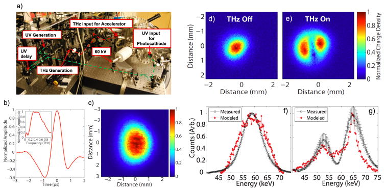
A proof-of-principle THz-acceleration experiment based on laser-generated single-cycle THz pulses at 0.45 THz has been performed recently [26]. Fig. 3(a) shows the setup. The single-cycle THz pulse with a center frequency of about 450 GHz (see Fig. 3(b) and (c)) was generated with a sub-ps Yb:KYW regenerative amplifier by optical rectification in cryogenically cooled LiNbO3 using the tilted-pulse-front technique. Of the generated 10 μJ THz energy, about 1 μJ was ultimately coupled into a dielectrically loaded metal waveguide optimized for accelerating sub-relativistic electron bunches provided by a 60-keV DC electron gun. Fig. 3(d) and (e) shows the electron beam on a multi-channel plate (MCP) after a deflecting dipole magnet without and with accelerating THz pulse. The electron bunches are generated by a UV-photocathode in the electron gun. The generated electron pulses have spread beyond one ps when reaching the THz-powered waveguide and therefore both acceleration and deceleration of electrons occur, which leads to a split beam on the MCP when the THz acceleration is on. Fig. 3(f) and (g) shows the measured and simulated electron energy spectra for both cases. From the measured electron energy spectra, we infer that the 1-μJ single-cycle THz pulse readily leads to a 7 keV electron beam energy modulation.
Fig. 3.
(a) THz LINAC and source with the THz acceleration chamber and accompanying power supplies, chillers and pumps on a portable optical cart. (b) THz acceleration pulse generated by optical rectification in cryogenically cooled LiNbO3. (c) THz beam profile. (d) Electron beam profile without THz pulse. (e) Electron beam profile with THz pulse on. (f) and (g) comparison between measured (black) and modelled (red) electron spectra without and with THz acceleration pulse [26].
The current goal is to achieve >5% percent of optical-to-THz generation efficiency. Close to a 1-Joule laser pulse will be necessary for generating the required 20 mJ THz pulse energy in the waveguide including transport losses. Such energy is also necessary for providing an optical undulator with an equivalent undulator parameter close to 1 as desired for efficient X-ray generation. Thus we aim for a few-ps 1-J laser capable of operating ultimately at up to 1 kHz pulse repetition rate. Cryogenically cooled Yb-based amplifiers are the laser technology of choice for this application. Cryogenic Yb:YAG amplifiers with 1 J of pulse energy operating at 100 Hz repetition rate have already been demonstrated [29]. We have recently demonstrated a compact composite thin-disk cryogenic Yb:YAG amplifier with up to 160 mJ of pulse energy at 100 Hz repetition rate [40] and scaling of this technology to the 1-J level at 1 kHz repetition rate is currently in progress.
The development of the proposed AXSIS source will of course face challenges and detailed start-to-end simulations are necessary to support the desired design principles and specifications outlined in this paper as well as to develop the machine. Such simulations are well on its way but surpass greatly the page limit set for this paper and therefore we defer them to a forthcoming article. However, back off the envelope simulations as well as first simulations indicate that the AXSIS source produces already as a low risk inverse Compton scattering source, i.e. no bunching of electrons is needed, 106 X-ray photons per shot at 12 keV within an opening angle of 1 mrad and at 1 kHz repetition rate. When operating in the regime where micro bunching is achieved either due to the use of an electron crystal generated in the gun or in combination with partial bunching due to FEL action, 108 to 109 photons per shot at 12 keV within an opening angle of less than 100 μrad and at 1 kHz repetition rate seem possible.
3. Attosecond serial crystallography
Very recently, our collaborators and we have introduced a new paradigm of femtosecond serial nano crystallography for collecting protein diffraction data from sub-micron crystals with ~10 to 100-fs pulses from X-ray FELs [28,7,8]. Our method combines two major advances: Firstly, the short exposure allows for diffraction patterns of the crystals being collected before the destruction occurs, even though the degree of ionization and subsequent Coulomb explosion is extreme [27,5]. “Damage-free” data have been collected at doses exceeding 3 GGy, from room-temperature samples. This exceeds 3000 times the tolerable dose for conventional crystallography at room temperature and is 100 times higher than the tolerable dose for cryogenically cooled samples. Secondly, we merge data from hundreds of thousands of micron or sub-micron crystals that flow continuously across the X-ray beam as a liquid suspension in a micro-jet [10,35]. Each pulse gives rise to a unique snapshot diffraction pattern of a randomly-oriented crystal. Diffraction data are combined for each indexed Bragg spot by Monte Carlo integration, [21]. This merging of terabytes of data [36] accumulates signals from many individual crystallites. We achieve a full 3D dataset that can be analyzed by conventional single-crystal phasing techniques, including anomalous diffraction [32]. Both of these advances allow us to use protein crystal sizes that would be prohibitively small for conventional studies, even with the brightest synchrotron sources. Consequently, we overcome the major bottleneck of structural biology, which is the requirement for large well-diffracting crystals that can take years of effort to achieve. Furthermore reactions can be induced in the small crystals by light or rapid mixing “on the fly”, which allows for time resolved data to be collected [23,34,4].
The method of serial crystallography with X-ray FEL pulses has already been applied to some of the most difficult protein complexes, including membrane protein complexes such as G-protein coupled receptors [19,24,39], and Photosystem I and II (see Fig. 4) [23,8]. Since the sample is refreshed on every pulse, this method is also ideal for time-resolved measurements of irreversible reactions [4] with an inherently high temporal resolution.
Fig. 4.
(left) 3D structure factors of Photosystem II generated by indexing and merging tens of thousands of single-shot diffraction patterns collected at LCLS. [from [23] with permission] (center) Structure of Photosystem II at 5 Å resolution, from LCLS diffraction data [from [23] with permission]; (right) Detail of a single-shot diffraction pattern of Photosystem I showing fringes due to the sub-micron finite size of the crystal ([8] with modifications).
In the AXSIS project, we will push the technique of serial crystallography to a new frontier and overcome limitations of current fs FELs, using attosecond pulses from the C-ICS source. Although fs pulses from FELs are indeed short enough to collect X-ray diffraction data before the molecule is destroyed [5], the ionization and destruction of electronic states in molecules is particularly severe [31]. As shown by electron spectroscopy and fluorescence [31,38], core electrons are rapidly displaced and “hollow atoms” are created. On large molecules and crystals the fastest loss is due to impact ionization due to photoelectrons liberated and Auger decay which requires attosecond pulses to outrun [33]. By bringing the technique from the X-ray FEL to a compact laboratory-scale X-ray source we vastly extend its availability. Attosecond pulses from the C-ICS source, while having fewer photons than the large scale XFEL (yet matching the peak power of those sources), are spectrally better defined and fully longitudinal coherent and will thereby provide X-ray diffraction patterns that can be more easily interpreted and merged, giving further gains in accuracy and reducing sample consumption. Furthermore, the electronic structure is not disturbed by the attosecond pulses, which allows for truly damage-free X-ray spectroscopy to be performed. A new advanced CMOS pixel array detector will be built to match the 1-kHz repetition rate of the source and to meet the dynamic range requirements. We will develop an X-ray emission and absorption spectroscopy (XES, XAS) setup [2,3,9], which will be combined with X-ray diffraction for simultaneous collection of XES/XAS spectra and diffraction data at 1-kHz repetition rate.
The groundbreaking experimental capabilities of attosecond X-ray diffraction and spectroscopy will be used to realize one of the grandest dreams of chemists and biochemists: the production of molecular movies of the formation of the excited states and transition states of molecules at the atomic level. By adding optical and X-ray spectroscopies we will gain new insights in spectroscopic measurements by giving a real-space accompaniment to changes in electronic states. Using this capability we will unravel the mechanisms of ultrafast light absorption and excitation energy transfer in important biological processes such as photosynthesis at the relevant spatial and temporal time scales.
Acknowledgments
This work has been supported by the European Research Council under the European Union Seventh Framework Program (FP/2007-2013)/ERC Grant Agreement no. 609920, the excellence cluster ‘The Hamburg Centre for Ultrafast Imaging - Structure, Dynamics and Control of Matter at the Atomic Scale under Grant agreement no. EXC 1074, Air Force Office of Scientific Research through contract FA9550-12-1-0499. Drs. Wu and Yahaghi, as well as R. Huang acknowledge support by Research Fellowships from the Alexander von Humboldt Foundation and a NDSEG Graduate Research Fellowship, respectively.
References
- 1.Allaria E, et al. Nat Phot. 2012;6:699. [Google Scholar]
- 2.Alonso-Mori R, et al. Proc Nat Acad Sci USA. 2012;109:19103–19107. doi: 10.1073/pnas.1211384109. [DOI] [PMC free article] [PubMed] [Google Scholar]
- 3.Alonso-Mori R, et al. Rev Sci Instrum. 2012;83:073114. doi: 10.1063/1.4737630. [DOI] [PMC free article] [PubMed] [Google Scholar]
- 4.Aquila A, et al. Opt Express. 2012;20:2706–2716. doi: 10.1364/OE.20.002706. [DOI] [PMC free article] [PubMed] [Google Scholar]
- 5.Barty A, et al. Nat Photon. 2012;6:35–40. doi: 10.1038/nphoton.2011.297. [DOI] [PMC free article] [PubMed] [Google Scholar]
- 6.Barends T, et al. J Synchrotron Radiat. 2015;22(Pt. 3):644–652. doi: 10.1107/S1600577515005184. [DOI] [PubMed] [Google Scholar]
- 7.Boutet S, et al. Science. 2012;337:362–364. doi: 10.1126/science.1217737. [DOI] [PMC free article] [PubMed] [Google Scholar]
- 8.Chapman HN, et al. Nature. 2011;470:73–77. doi: 10.1038/nature09750. [DOI] [PMC free article] [PubMed] [Google Scholar]
- 9.Davis KM, et al. J Phys Chem Lett. 2012;3:1858–1864. doi: 10.1021/jz3006223. [DOI] [PMC free article] [PubMed] [Google Scholar]
- 10.DePonte D, et al. J Phys D. 2008;41:195505. [Google Scholar]
- 11.Fülöp JA, et al. Opt Express. 2011;19:15090. doi: 10.1364/OE.19.015090. [DOI] [PubMed] [Google Scholar]
- 12.Fülöp JA, et al. Opt Lett. 2012;37:557. doi: 10.1364/OL.37.000557. [DOI] [PubMed] [Google Scholar]
- 13.Hau-Riege SP. Phys Rev Lett. 2012;108:238101. doi: 10.1103/PhysRevLett.108.238101. [DOI] [PubMed] [Google Scholar]
- 14.Hommelhoff P, et al. Phys Rev Lett. 2006;97:247402. doi: 10.1103/PhysRevLett.97.247402. [DOI] [PubMed] [Google Scholar]
- 15.Hobbs R, et al. Nanotechnology. 2014;25:465304–465401. doi: 10.1088/0957-4484/25/46/465304. [DOI] [PubMed] [Google Scholar]
- 16.Huang SW, et al. Opt Lett. 2013;38:796–798. doi: 10.1364/OL.38.000796. [DOI] [PubMed] [Google Scholar]
- 17.Huang SW, et al. Opt Lett. 2012;37:557. [Google Scholar]
- 18.Jordan P, et al. Nature. 2001;411:909–917. doi: 10.1038/35082000. [DOI] [PubMed] [Google Scholar]
- 19.Kang Y, et al. Nature. 2015;523:561–567. doi: 10.1038/nature14656. [DOI] [PMC free article] [PubMed] [Google Scholar]
- 20.Keathley PD, et al. Ann Phys. 2013;525:144–150. [Google Scholar]
- 21.Kirian R, et al. Opt Express. 2010;18:5713–5723. doi: 10.1364/OE.18.005713. [DOI] [PMC free article] [PubMed] [Google Scholar]
- 22.Kirian R, et al. Acta Crystallogr A. 2011;67(131–140):2011. doi: 10.1107/S0108767310050981. [DOI] [PMC free article] [PubMed] [Google Scholar]
- 23.Kupitz C, et al. Nature. 2014;513:261–265. doi: 10.1038/nature13453. [DOI] [PMC free article] [PubMed] [Google Scholar]
- 24.Liu W, et al. Science. 2013;342:1521–1524. doi: 10.1126/science.1244142. [DOI] [PMC free article] [PubMed] [Google Scholar]
- 25.Marinelli A. private communication.
- 26.Nanni EA, et al. Nat Commun. 2015;6:8486. doi: 10.1038/ncomms9486. [DOI] [PMC free article] [PubMed] [Google Scholar]
- 27.Neutze R, et al. Nature. 2000;406:753–757. [Google Scholar]
- 28.Redecke L, et al. Science. 2012;29 [Google Scholar]
- 29.Rocca J, et al. Opt Lett. 2012;37:557. [Google Scholar]
- 30.Ropers C, et al. Phys Rev Lett. 2007;98:043907. doi: 10.1103/PhysRevLett.98.043907. [DOI] [PubMed] [Google Scholar]
- 31.Rudek B, et al. Nat Photon. 2012;6:858–865. [Google Scholar]
- 32.Son SK, et al. Phys Rev Lett. 2011;107:218102. doi: 10.1103/PhysRevLett.107.218102. [DOI] [PubMed] [Google Scholar]
- 33.Son SK, et al. Phys Rev A. 2011;83:033402. [Google Scholar]
- 34.Tenboer, et al. Science. 2014;346:1242–1246. doi: 10.1126/science.1259357. [DOI] [PMC free article] [PubMed] [Google Scholar]
- 35.Weierstall U, et al. Rev Sci Instrum. 2012;83:035108. doi: 10.1063/1.3693040. [DOI] [PubMed] [Google Scholar]
- 36.White TA, et al. J Appl Cryst. 2012;45:335–341. [Google Scholar]
- 37.Ye H, et al. In: Ultrafast Phenomena XIX. Yamanouchi K, Cundiff S, de Vivie-Riedle R, Kuwata-Gonokami M, DiMauro L, editors. Springer; 2015. p. 663. [Google Scholar]
- 38.Young L, et al. Nature. 2010;466:56–61. doi: 10.1038/nature09177. [DOI] [PubMed] [Google Scholar]
- 39.Zhang H, et al. Cell. 2015;161:833–844. doi: 10.1016/j.cell.2015.04.011. [DOI] [PMC free article] [PubMed] [Google Scholar]
- 40.Zapata L, et al. Opt Lett. 2015;40:2610. doi: 10.1364/OL.40.002610. [DOI] [PubMed] [Google Scholar]
- 41.Wong LJ, et al. Opt Express. 2013;21:9792–9806. doi: 10.1364/OE.21.009792. [DOI] [PubMed] [Google Scholar]
- 42.Carbajo S, et al. Opt Lett. 2015;24:5762. doi: 10.1364/OL.40.005762. [DOI] [PubMed] [Google Scholar]


