Abstract
Despite the recent successes of using immune modulatory antibodies in cancer patients, autoimmune pathologies resulting from the activation of self-reactive T cells preclude the dose escalations necessary to fully exploit their therapeutic potential. To reduce the observed and expected toxicities associated with immune modulation, here we describe a clinically feasible and broadly applicable approach to limit immune costimulation to the disseminated tumor lesions of the patient whereby an agonistic 4-1BB oligonucleotide aptamer is targeted to the tumor stroma by conjugation to an aptamer that binds to a broadly expressed stromal product, vascular endothelial growth factor (VEGF). The approach was predicated on the premise that by targeting the costimulatory ligands to products secreted into the tumor stroma the T cells will be costimulated prior to their engagement of the MHC/peptide complex on the tumor cell, thereby obviating the need to target the costimulatory ligands to non-internalizing cell-surface products expressed on the tumor cells. Underscoring the potency of stroma-targeted costimulation and the broad spectrum of tumors secreting VEGF, in preclinical murine tumor models systemic administration of the VEGF-targeted 4-1BB aptamer conjugates engendered potent antitumor immunity against multiple unrelated tumors in subcutaneous, post-surgical lung metastasis, methylcholantrene-induced fibrosarcoma, and oncogene-induced autochthonous glioma models, and exhibited a superior therapeutic index compared to non-targeted administration of an agonistic 4-1BB antibody or 4-1BB aptamer.
Keywords: Cancer immunotherapy, oligonucleotide aptamers, 4-1BB costimulation, Tumor stroma, Vascular endothelial growth factor
INTRODUCTION
Antigen-activated T cells express stimulatory and inhibitory receptors that regulate their fate and ultimately control the outcome of the immune response. 4-1BB (CD137), is a major costimulatory receptor promoting the survival and expansion of activated CD8+ T cells and their differentiation into memory cells(1). Underscoring the lack of optimal 4-1BB costimulation at the tumor site, intratumoral administration of 4-1BB ligands (4-1BBL) as 4-1BB-Fc fusion protein (2) or adenoviral vector encoded 4-1BBL (3), as well as systemic administration of agonistic anti-4-1BB antibodies (4) or soluble 4-1BB ligands (5) enhanced tumor immunity and inhibited tumor growth. Systemic anti-4-1BB antibody therapy synergized with vaccination (6, 7) and other immune modalities (3, 8–12) to inhibit tumor growth in mice. Enhancing 4-1BB costimulation, therefore, represents a potentially useful modality to potentiate protective immunity in cancer patients. Notwithstanding, systemic administration of agonistic 4-1BB antibodies to mice was accompanied by immune anomalies, notably polyclonal activation of CD8+ T cells and a “cytokine storm” consisting predominantly of IFNγ, TNF and type-I IFNs that affected the function of organs such as liver, spleen and bone marrow (9, 13, 14).
The recent FDA approval of Ipilumimab, a blocking anti-CTLA-4 Ab, for the treatment of advanced melanoma has provided formal validation for using immune-potentiating drugs in the treatment of cancer. Nonetheless, treatment of cancer patients with Ipilumimab, and with other immune-potentiating antibodies like anti-PD-L1 or anti-PD-1 that have shown promise in clinical trials, was associated with autoimmune pathologies, including grade 3 or 4 toxicities (15). Clinical trials using an agonistic anti-4-1BB Ab in advanced cancers were associated with high frequencies of objective responses, yet adverse effects became significant at the highest dose used causing liver toxicity that resulted in two fatalities (16). Arguably, reducing the toxicity of immune-potentiating drugs without compromising their antitumor activity will be paramount for exploiting their clinical potential to the fullest extent.
We have shown previously that an agonistic 4-1BB oligonucleotide aptamer conjugated to a prostate-specific membrane antigen (PSMA)-binding aptamer is efficiently targeted to PSMA-expressing tumors in vivo and potentiates vaccine-induced protective immunity (17). Given that most receptors engaged by their ligand, including PSMA, are internalized (18), and since the tumor-targeted 4-1BB costimulatory ligands need to be displayed on the cell surface to engage the 4-1BB-expressing tumor-infiltrating T cells, tumor cells were engineered to express a mutant PSMA containing a small deletion in the cytoplasmic domain to prevent its internalization upon aptamer engagement. Since this is not clinically feasible, the need to identify tumor-specific surface products that do not internalize upon interaction with the bispecific aptamers significantly reduces the clinical applicability of this approach.
In general, the professional or nonprofessional (i.e. tumor) cell is costimulated concurrently with antigen presentation by the same cell expressing both the costimulatory ligand and the MHC/peptide complex. In this study we tested the hypothesis that by targeting the costimulatory ligands to products secreted into the tumor stroma, the tumor-infiltrating T cells will be costimulated prior to their engagement and presentation of the MHC/peptide complex by the tumor cell, thereby obviating the need to target the costimulatory ligands to non-internalizing cell-surface products expressed on the tumor cells. In addition, since unlike tumor cell-expressed products such as PSMA, Her2, or EGFR, stroma-secreted products, like vascular endothelial growth factor (VEGF), osteopontin (OPN), or metalloproteases, are secreted by many tumors of distinct origins, tumor-stroma-targeted costimulation would be more broadly applicable.
MATERIALS AND METHODS
Construction of aptamer conjugates
A 2′-fluoro-pyrimidine modified dimeric 4-1BB RNA aptamer transcribed in vitro from a DNA template described in reference (19) extended at the 3′ end with a linker sequence 5′-UCCCGCUAUAAGUGUGCAUGAGAAC-3′ was annealed to either a VEGF (20) or OPN (21) chemically synthesized (IDT, Colarville, IA) aptamer via a complementary linker sequence engineered at their 3 ends. Equimolar amounts of 4-1BB and either VEGF or OPN aptamers were mixed, heated to 75°C, and cooled to room temperature. Annealing efficiency, monitored by agarose gel electrophoresis was > 80%. To prevent conjugation of the two aptamers, they were annealed separately with a 2-fold excess of complimentary linker sequence before tail vein injection. 32P-labeled 4-1BB dimer was generated by transcription in the presence of αP32-ATP (PerkinElmer, Boston, MA).
In vitro Costimulation Assay
CD8+ T cells were isolated from the spleens of Balb/C mice using a Miltenyi CD8+ T-cell isolation kit (Auburn, CA). Briefly, 106 cells/mL were plated in a 96-well plate at 200 μL per well in the presence or absence of suboptimal concentration of CD3e antibody (250 ng/mL). 18 hours later, antibody (1 μg/mL) or aptamer (100 pmole/mL) was added. 48 hours later, media was supplemented with 1 μCi/mL of 3H-thymidine. 6 hours later cells were harvested and counted using a scintillation counter.
Histology and immunohistochemistry (IHC)
4T1 and 4T07 subcutaneously established tumors were resected and embedded in paraffin. Non-specific immunoreactivity in slide-mounted tissue sections was blocked with Serum Blocking Reagent D (R&D Systems, Minneapolis MN), and incubated with goat polyclonal anti-mouse VEGF at 1:20 (R&D Systems, Minneapolis MN) at 4°c overnight, washed with PBS, and incubated with biotinylated anti-Goat secondary antibody (R&D Systems, Minneapolis MN) for 40 minutes, followed by HSS-HRP (R&D Systems, Minneapolis MN) for 30 minutes. Slides were washed with PBS, and then incubated with an HRP-reactive DAB chromogen (R&D Systems, Minneapolis MN) for 7 minutes. Slides were rinsed in H20, incubated in CAT hematoxylin (Biocare Medical, Concord CA) at 1:5 for 3 minutes and Tacha’s Bluing Agent (Biocare Medical, Concord CA) at 1:5 for 3 minutes, followed by rinses with H20. Slides were dehydrated in increasing concentrations of alcohol (70%, 90%, 100%; 3 minutes each), briefly washed in xylene, and coverslipped using mounting medium (Richard-Allan Scientific, Kalamazoo MI). Negative control slides for VEGF IHC were prepared by completing the above protocol in the absence of primary antibody.
Tumor immunotherapy studies
The facilities at the University of Miami Division of Veterinary Resources are fully accredited by the Association for Assessment and Accreditation of Laboratory Animal Care and USDA. An OLAW assurance is on file ensuring that humane animal care and use practices, as outlined in the “Guide for the Care and Use of Laboratory Animals “ are as followed. 5–6 week old female C57BL/6 (H-2b) and Balb/c (H-2d) mice were purchased from The Jackson Laboratory (Bar Harbor, ME) and used within 1–3 weeks.
Subcutaneous tumor models
4T1 breast carcinoma model
Balb/c mice were injected subcutaneously in the right flank with 1.0×10⁴ 4T1 tumor cells, and immunized with a mixture of irradiated (6000 rad) B7-1 and MHC class II-expressing 4T1 cells (22) injected into the opposite flanks at days 7, 10, and 13. 18 hours after each injection, 100 pmoles of aptamer conjugates were administered by tail vein injection.
B16.F10 melanoma model
C57BL/6 mice were injected subcutaneously in the right flank with 1.0×10⁵ B16.F10 cells and immunized with 106 irradiated (6000 rad) GM-CSF-expressing B16.F10 cells (GVAX) (23) injected into the opposite flanks at days 4, 7, and 10. 18 hours after each injection, 100 pmoles of aptamer conjugates were administered by tail vein injection.
Mice were sacrificed when tumor diameter exceeded 1.2 cm or mice exhibited signs of morbidity. Experiment was terminated when 2 or more mice were sacrificed in the “no treatment” group. 4T1, 4T07, and B16.F10 parental, B7-1- and MHC class II-expressing cells were tested and validated to be mycoplasma-free. B7-1 and MHC class II expressions were validated by IHC; no other authentication assays were performed.
4T1 breast carcinoma post-surgical metastasis model
Balb/c mice were injected with 1.0×10⁴ 4T1 cells into the abdominal mammary fat pad. At Day 11, two days after local tumors became palpable, mice were anesthetized and primary tumors were surgically removed (24). Mice were vaccinated with the irradiated B7-1 and MHC class II-expressing 4T1 cells at days 13, 16 and 19; 18 hours after each vaccination 100 pmoles of aptamer conjugates were administered as described above. Mice were sacrificed when they showed signs of morbidity.
MCA carcinogenesis model
Balb/c mice were injected subcutaneously with 400 μg of 3-methylcholanthrene in castor oil. As indicated, 100 pmoles of VEGF-4-1BB conjugates were administered once weekly for a total of 4 injections. Mice were sacrificed when tumor diameter exceeded 1.2 cm or mice exhibited signs of morbidity.
Oncogene-induced high-grade glioma model
The immune competent RCAS/Ntv-a murine model in which intracerebral high-grade gliomas are induced by expressing PDGFB and STAT3 from glioneuronal precursors has been described previously (25, 26). Briefly, to transfer genes via RCAS vectors, DF-1 producer cells transfected with a particular RCAS vector (1 × 10⁵ DF-1 cells in 2 μl of PBS) were injected into the frontal lobes of day 1 or 2 Ntv-a mice at the coronal suture. Twenty-one days after introducing the glioma-inducing transgenes, mice were randomly assigned to the treatment or control group. Mice were sacrificed 90 days after injection or sooner if they demonstrated morbidity related to tumor burden. DF-1 parental and transfected cells (25, 26), were tested and validated to be mycoplasma-free. Transgene expression was validated by IHC/flow cytometry; no other authentication was performed.
Statistical analysis
Mouse survival, defined as the time (in days) the mouse was born to the date of death, was estimated using the method of Kaplan and Meier. Cox regression model was employed to explore the survival difference between individual treatment groups using statistical software R V-3.0.2. For other continuous measurements, unpaired, two-tailed student’s t-tests were performed between individual treatment groups using GraphPad Prism V-5.00 (GraphPad Software Inc. La Jolla, CA). P values less than or equal to 0.05 were considered statistically significant.
RESULTS
Potentiation of vaccine-induced antitumor immunity in subcutaneously implanted tumor-bearing mice with VEGF-4-1BB aptamer conjugates
To target costimulation to the tumor stroma we conjugated an agonistic dimeric 4-1BB aptamer (27) to a VEGF aptamer selected against human VEGF (20) (Supplementary Fig. 1). VEGF is secreted by progressing tumor lesions in many types of cancers (28). We have confirmed that the VEGF aptamer also binds to murine VEGF and the VEGF-4-1BB conjugate binds to both 4-1BB and VEGF (Fig. 1a). Figure 1b shows that the VEGF-4-1BB aptamer conjugates retained 4-1BB costimulatory activity.
Figure 1. Characterization of VEGF-4-1BB aptamer conjugates.
A. Binding of VEGF-4-1BB aptamer conjugates to filter-immobilized targets was measured using a double-filter binding assay. 32P-labeled 4-1BB aptamer, VEGF aptamer, or VEGF-4-1BB aptamer conjugates were passed through nitrocellulose filters immobilized with decreasing amounts of murine 4-1BB protein (m4-1BB), murine VEGF protein (mVEGF), or IgG, except for the rightmost lane that contained no protein. Bound radioactivity was visualized by exposure to X-ray sensitive film. B. 4-1BB costimulation of polyclonally activated CD8+ T cells. CD8+ T cells were activated with anti-CD3 antibody and incubated with an agonistic 4-1BB antibody or IgG control, 4-1BB aptamer, VEGF-4-1BB, or control aptamers in which the 4-1BB aptamer was replaced with a scrambled aptamer (Scram). Proliferation was measured 48 hours later by 3H-thymidine incorporation. Statistical analysis: 4-1BB antibody versus isotype antibody, p=0.01; 4-1BB aptamer versus scrambled aptamer, p=0.028, VEGF-4-1BB aptamer conjugate versus VEGF-scrambled aptamer conjugate, p=0.069.
To test whether the VEGF-4-1BB aptamer conjugate can enhance vaccine-induced antitumor immunity, mice were implanted subcutaneously with either 4T1 breast carcinoma cells and 7 days later vaccinated with a mixture of irradiated MHC class II- and B7-1-transfected 4T1 cells (22) (Fig. 2a), or with B16.F10 melanoma cells and 5 days later vaccinated with GM-CSF-expressing irradiated B16.F10 cells (23) (Fig 2b). 18 hours after vaccination, aptamer conjugates were administered intravenously via the tail vein. Vaccination and conjugate administration were repeated twice at three-day intervals. Mice were monitored for tumor growth and the experiment was terminated when the cohort of mice that received only tumor cells had to be sacrificed. As shown in Fig. 2, using experimental conditions whereby vaccination of tumor-bearing mice had minimal to no impact on tumor growth – simulating a scenario in which tumor vaccination as monotherapy is largely ineffective – co-treatment with the VEGF-4-1BB aptamer conjugates inhibited tumor growth. Treatment with conjugates alone had a small effect that did not reach statistical significance in either model. Vaccination with a mixture of VEGF and 4-1BB aptamers had no effect, ruling out the possibility that tumor inhibition was an additive contribution of the two aptamers. This was to be expected given that the mice were administered with 300 pmoles of (monovalent) aptamer conjugates that were 5- to 10-fold less than unconjugated 4-1BB aptamer (27) and 20- to 50-fold less than (bivalent) VEGF antibody (29–31) used to inhibit tumor growth in mice.
Figure 2. Treatment of tumor-bearing mice with VEGF-4-1BB aptamer conjugates potentiate vaccine-induced protective antitumor immunity.
A. Balb/c mice were implanted subcutaneously with 4T1 tumor cells, vaccinated with irradiated B7-1 and MHC class II-expressing 4T1 cells at days 7, 10 and 13 (arrows) and treated with VEGF-4-1BB aptamer conjugates or with a mixture of VEGF and 4-1BB aptamers 18 hours after each vaccination (8 mice per group). Statistical analysis: ** p < 0.01. (Days 25 and 27, p=0.0089 and 0.0027, respectively). B. C57BL/6 mice were implanted subcutaneously with B16.F10 melanoma cells, vaccinated with GVAX at days 5, 8 and 11 (arrows), and treated with VEGF-4-1BB aptamer conjugates or with a mixture of VEGF and 4-1BB aptamers 18 hours after each vaccination (8 mice per group). Statistical analysis: * p<0.05. (Days 19 and 21, p= 0.0433 and 0.0403, respectively).
VEGF-dependent tumor-targeted 4-1BB costimulation
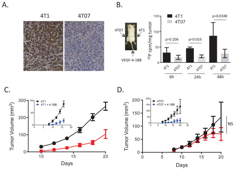
To determine whether inhibition of tumor growth was a result of VEGF-dependent tumor-targeted 4-1BB costimulation, we compared the homing and antitumor activities of the VEGF-4-1BB aptamer conjugates in 4T1 and 4T07 tumor-bearing mice. 4T1 and 4T07 are two sub-cell lines derived from a thioguanine-resistant breast carcinoma tumor (32). VEGF expression in subcutaneously implanted tumors was measured by immunohistochemistry. As shown in Figure 3a, the metastatic 4T1 tumors expressed higher levels of VEGF than the nonmetastatic 4T07 tumors. Mice were then implanted contralaterally in opposite flanks with 4T1 and 4T07 cells and 21 days later 32P-labeled VEGF-4-1BB aptamer conjugates were administered via the tail vein. As shown in Figure 3b, the VEGF-41BB aptamer conjugates accumulated preferentially in the VEGFhigh 4T1 tumors. Consistent with the preferential homing of the VEGF-4-1BB conjugates to 4T1 cells, the VEGF-4-1BB aptamer conjugates inhibited 4T1 (Fig. 3c), but not, or to a much lesser extent, 4T07 (Fig 3d) tumor growth.
Figure 3. VEGF-mediated tumor-targeting of 4-1BB aptamers.
A. VEGF expression in 4T1 and 4T07 tumors. 4T1 or 4T07 tumor cells were injected subcutaneously into Balb/c mice and when tumors reached about 0.5 cm diameter they were resected and stained for VEGF expression as described in Methods. B. Homing of systemically administered VEGF-4-1BB aptamer conjugates to VEGF-expressing tumors. Balb/c mice were implanted in opposite flanks with 1.0x10⁴ 4T1 (VEGFhigh) and 5x10⁴ 4T07 (VEGFlow) tumor cells and at day 21 when tumors became readily palpable mice were administered with 32P-labeled VEGF-4-1BB conjugates via the tail vein. As described in the Methods, only the dimeric 4-1BB was radioactively labeled. At indicated times mice were sacrificed, tumors excised, and radioactivity was determined by scintillation counting. Statistical significance 4T1 versus 4T07: 6h, p=0.2059; 24h, p=0.016; 48h, p=0.0348.
C and D. VEGF-4-1BB aptamer conjugates inhibit VEGFhigh 4T1, but not VEGFlow 4T07 cells. Balb/c mice were injected subcutaneously with 4T1 (C) or 4T07 (D) tumor cells and 4 days later administered with 100 pmoles of VEGF-4-1BB aptamer conjugates three times 3 days apart and tumor growth was monitored. Insert, mice were administered with 500 pmoles of unconjugated 4-1BB aptamer. (5 mice per group). Statistical significance: Panel C, days 16 and 18, p<0.001; day 20, p=0.0145, panel D, ns=not statistically significant.
Unlike the experiment shown in Figure 2a, in this experiment mice were not vaccinated, yet treatment with conjugates alone inhibited 4T1 tumor growth. The reason is that conditions used in this experiment were less stringent, starting treatment at day 4 rather than day 7, post tumor implantation. The lack of inhibition of 4T07 tumor growth was not due to its resistance to 4-1BB costimulation because a 5-fold higher dose of unconjugated 4-1BB aptamer inhibited both 4T07 and 4T1 tumor growth to a similar extent. The small inhibition of 4T07 tumor growth in mice treated with the aptamer conjugate, which did not reach statistical significance, could be due therefore to the non-targeted 4-1BB costimulatory effect of the conjugates administered at a 5-fold lower dose, and/or the low level of VEGF expressed in the 4T07 tumors (Fig. 3a). Supplementary Figure 2 shows the biodistribution of the systemically administered VEGF-4-1BB aptamer conjugates in various tissues and organs of the mouse. Since the VEGF-targeted aptamer conjugates do not become cell-associated, the tested tissues could not be perfused. Thus, the relatively high levels of aptamer conjugates in the various tissues and organs, especially in the blood-rich spleen and liver, are likely a reflection of their presence in the circulation.
To assess the generality of tumor stroma-targeted costimulation the dimeric 4-1BB aptamers were conjugated to an osteopontin (OPN)-binding aptamer that was selected for binding to both murine and human orthologs (21) (Supplementary Fig. 1). OPN, like VEGF, is secreted by progressing tumor lesions in many types of cancer (33). As shown in Supplementary Figure 3a, 4T1 tumors secrete higher levels of OPN than 4T07 tumors. 32P-labeled OPN-4-1BB aptamer conjugates injected into mice that were co-implanted with both 4T1 and 4T07 tumor cells accumulated preferentially in the OPNhigh 4T1 tumors (Supplementary Fig. 3b), and inhibited the growth of 4T1 tumor but not, or to a lesser extent, the growth of 4T07 tumor cells (Supplementary Fig. 3c). In a more stringent experimental setting, the OPN-4-1BB aptamer conjugates potentiated an otherwise ineffective vaccine-induced immune response resulting in reduced tumor growth (Supplementary Fig. 3d).
Superior therapeutic index of VEGF-4-1BB aptamer conjugates compared to 4-1BB antibody
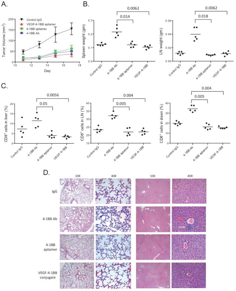
To determine whether tumor stroma-targeting of 4-1BB aptamers enhances the therapeutic index of 4-1BB costimulation, we compared treatment with VEGF-4-1BB aptamer conjugates to that of unconjugated 4-1BB aptamer and to anti-4-1BB Ab. As shown in Figure 4a, on a molar basis the VEGF-4-1BB aptamer conjugates were as effective as a 6-fold higher dose of unconjugated 4-1BB aptamer or anti-4-1B Ab at inhibiting tumor growth. Antibody, but not unconjugated 4-1BB aptamer or VEGF-4-1BB conjugate, treatment was associated with increased spleen, lymph node, lung, and liver weights (Fig. 4b and data not shown) resulting mostly from increased CD8+ T-cell numbers (Fig. 4c) and notably, extensive infiltration of leukocytes in the lung and liver (Fig. 4d), as was described previously (9, 13, 14). Interestingly, as we have shown (17), treatment with an equimolar dose of 4-1BB aptamer that elicits a comparable antitumor response to that of treatment with the 4-1BB antibody did not elicit CD8+ T-cell hyperplasia (Fig. 4c) or leukocytic infiltration (Fig. 4d). The experiments shown in Figure 4, therefore, suggest that the therapeutic index of the VEGF-targeted 4-1BB aptamer could be significantly higher than that of the non-targeted anti-4-1BB Ab, and that non-targeted 4-1BB aptamer exhibits an intermediate therapeutic index.
Figure 4. Therapeutic index of VEGF-4-1BB aptamer conjugates.
A. 4T1 tumor cells were injected subcutaneously into Balb/c mice and 4 days later administered via the tail vein with 150 pmoles of VEGF-4-1BB aptamer conjugates or with 800 pmoles of either unconjugated 4-1BB aptamer, anti-4-1BB Ab, or IgG control Ab. Aptamer and antibody administration was repeated two additional times at 3-day intervals as described in Methods, and tumor growth was monitored (5 mice per group). B. Mice shown in panel A were sacrificed at day 17, spleens, and lymph nodes weights were measured and C. CD8+ T cells were isolated from the spleens, lymph nodes, and livers, and were counted. The difference between the 4-1BB Ab group and all other treated groups in panels B and C was statistically significant (p< 0.001). There was no statistical difference between the control IgG, 4-1BB aptamer and VEGF-4-1BB aptamer conjugate groups. D. Tissue sections from the lung and liver were stained with hematoxylin and eosin and visualized by light microscopy.
Inhibition of tumor growth by VEGF-4-1BB aptamer conjugates in a post-surgical metastasis model
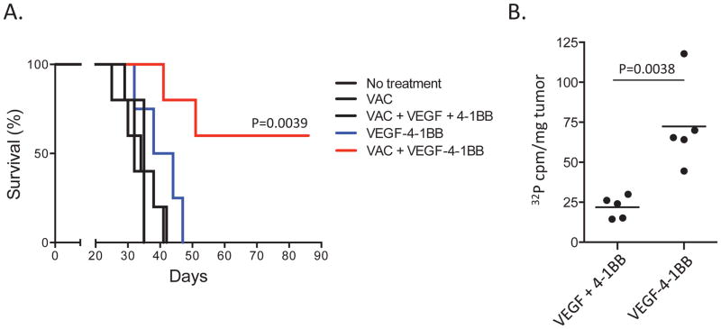
Given the limitations of subcutaneously implanted tumor models, we evaluated the therapeutic potential of stroma-targeted 4-1BB costimulation using increasingly relevant murine tumor models. In the experiment shown in Figure 5 we used a post-surgical metastasis model to simulate treatment of residual metastatic disease. In this model 4T1 breast carcinoma cells were implanted orthotopically into the abdominal mammary fat pad, and when they became palpable, 10–11 days later, tumors were surgically resected. 4T1 growth parallels highly invasive human metastatic stage IV breast cancer metastasizing sequentially to the lung, lymph nodes, liver, and brain (34, 35). Two days after surgical resections mice were subjected to three vaccinations and conjugate treatment as described in the legend to Figure 2, and monitored for survival. As shown in Figure 5a, vaccination or administration of VEGF-4-1BB aptamer conjugates did not improve the survival of treated mice, which presented extensive metastasis in the lung at the time of sacrifice. On the other hand, mice that were vaccinated and treated with the VEGF-4-1BB aptamer conjugates survived longer, 3 out of 5 mice surviving long-term showing no signs of metastasis in the lung at the time of sacrifice (day 90). The targeted nature of the VEGF-4-1BB aptamer conjugates mechanism of action is consistent with the observation that a mixture of VEGF and 4-1BB aptamers was ineffective (Fig 5a), and that accumulation of 32P-labeled 4-1BB aptamers in the lung was enhanced by conjugation to the VEGF aptamer (Fig. 5b).
Figure 5. 4T1 breast carcinoma post-surgical metastasis model.
A. 4T1 tumor cells were injected into the abdominal mammary fat pad. At day 11, 1–2 days after tumors became palpable, tumors were surgically excised with the mice under anesthesia. Two days later mice were vaccinated with irradiated B7-1- and MHC class II-expressing 4T1 cells and 18 hours later 100 pmoles of VEGF-4-1BB aptamer conjugates or a mixture of VEGF and 4-1BB aptamers were administered via the tail vein. Vaccination and aptamer conjugate treatment was repeated twice three days apart. Mice were sacrificed when they showed signs of morbidity. (6 mice per group). B. At day 32 before the onset of signs of discomfort and morbidity 4T1 implanted, surgically resected mice were injected with 32P-labeled VEGF-4-1BB aptamer conjugates or unconjugated mixtures of VEGF and 4-1BB aptamers via the tail vein. 24 hours later mice were sacrificed, lungs were removed, and radioactivity determined by scintillation counting.
Inhibition of tumor growth by VEGF-4-1BB aptamer conjugates in non-transplanted autochthonous tumor models
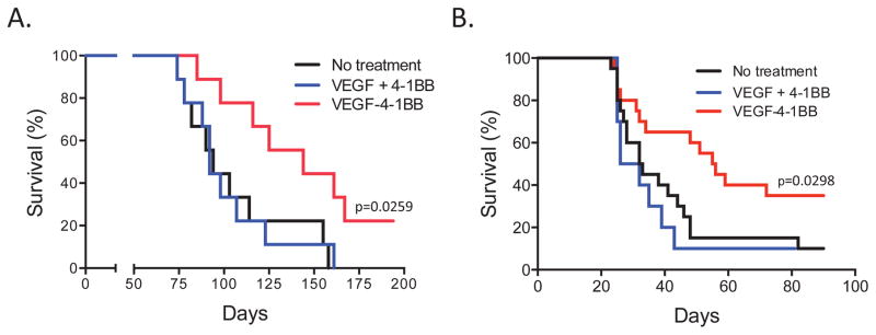
Given the limitations of modeling tumorigenesis with tissue culture-adapted tumor cell lines, we determined whether VEGF-targeted 4-1BB costimulation can control the growth of tumors that develop in situ from normal precursors. To this end we used a carcinogen-induced tumor model whereby mice injected with 3-methylcholanthrene (MCA) develop fibrosarcomas over the course of several months, by and large recapitulating the naturally-occurring multi-step carcinogenesis process (36). Starting at about day 70 when tumors are readily palpable, mice were treated with either VEGF-4-1BB conjugates or a mixture of VEGF and 4-1BB aptamers. As shown in Figure 6a, mice treated with the conjugates, but not with the non-conjugated aptamers, exhibited a significant survival advantage compared to untreated mice, with 2 of 9 mice surviving long-term.
Figure 6.
Autochthonous murine tumor models. A. Methylcholanthrene-induced fibrosarcoma model. C57BL/6 mice were injected subcutaneously with 400 mg of methylcholanthrene (MCA) in castor oil. When tumors became palpable, at days 71, 78, 85 and 92, mice were injected with 100 pmoles of VEGF-4-1BB aptamer conjugates or a mixture of VEGF and aptamers via the tail vein. Mice were sacrificed when mice exhibited signs of morbidity or when tumor diameter reached 1.2 cm, whichever came first (9 mice per group.) B. Oncogene-induced high-grade glioma model. Tumor-bearing Ntv-a mice were injected via the tail vein with 150 pmoles of VEGF-4-1BB aptamer conjugates or with a mixture of VEGF and 4-1BB aptamers starting at day 21 for a total of 6 injections every 3 or 4 days. Mice were sacrificed when they showed signs of morbidity (10–20 mice per group.) Statistical significance: VEGF-4-1BB aptamer conjugate versus a mixture ofVEGF and 4-1BB aptamers, p=0.0298.
As a second model we used an oncogene-induced high-grade glioma model whereby tumors are induced endogenously by the expression of PDGF-B and STAT3 in glioneuronal precursor cells of newborn mice (25, 26). This model was also used for assessing the ability of the aptamer conjugates to cross the blood brain barrier that may exist in tumor-bearing mice. As shown in Figure 6B, treatment of the tumor-bearing mice with the VEGF-targeted 4-1BB aptamer conjugates, but not with a mixture of VEGF and 4-1BB aptamers, led to the enhanced survival of the aptamer conjugate-treated mice. The therapeutic effect of the VEGF-4-1BB conjugates in this model was comparable to that of the previously reported treatment with an immune modulatory microRNA, miR124 (26).
DISCUSSION
Dose-limiting toxicities of cancer drugs, reflecting their limited tumor-specificity, are a major hurdle in developing effective treatments for cancer. Predicted by animal studies, systemic administration of immune modulatory antibodies to cancer patients was also associated with dose-limiting autoimmune pathologies (15, 16). Arguably, enhancing the therapeutic index of immune-potentiating drugs, namely reducing their toxicity without compromising their antitumor activity, is paramount for exploiting their clinical potential to the fullest extent.
A major step toward addressing dose-limiting drug toxicity was heralded with the introduction of molecular targeted therapies. Targeted therapies act by blocking essential biochemical pathways or mutant proteins that are required for tumor cells to grow and survive. The success of Imatinib, a BCR-ABL kinase inhibitor that has shown striking responses in patients with chronic myelogenous leukemia, an inhibitor that blocks a specific mutant of the B-RAF serine/threonine kinase (V600E-B-RAF) and demonstrated dramatic efficacy in the treatment of metastatic melanoma, as well as other inhibitors targeting EGFR, KIT, HER2 and ALK, have by-and-large validated this novel paradigm of cancer therapeutics (37). Nonetheless, the Achilles heel of targeted therapy is that the target is often a cancer-driving mutation in a product that normally regulates cell behavior, which leads to increasing fragmentation of drug treatments among subtypes of cancer, and more importantly, the development of resistance even to the most dramatically effective therapies.
An alternative, mutually non-exclusive, way to reduce drug toxicity is to limit drug exposure to normal tissues by targeting the drug to the tumor cells of the patients. Monoclonal antibodies are currently the main platform for targeting ligands. For example, Phase I clinical trials in cancer patients with a CD19-CD3 bispecific antibody have underscored the therapeutic benefit of targeting T cells to cancer cells using low-dose therapy (38). Nonetheless, despite a growing list of therapeutic antibodies that have been approved for cancer therapy, only two antibody-targeted cytotoxic drug conjugates are currently licensed for cancer therapy (39).
The development of therapeutic antibody-based targeting ligands is hindered by several factors. Antibodies are cell-based products which increases the complexity and cost of manufacturing, and the regulatory approval process. Conjugation of antibodies to their therapeutic drug requires special skill sets and expensive instrumentation, and monoclonal antibodies, even when fully humanized, run the risk of eliciting neutralizing antibody response upon repeated administrations (40, 41). Unlike antibodies, the short oligonucleotide aptamers can be synthesized in a cell-free chemical process that is more cost-effective and require a less complex regulatory approval process. Conjugation to nucleic acid-based therapeutic agents by hybridization between short complementary sequences is straightforward, and the short oligonucleotides are less likely to stimulate neutralizing immunity. Recent studies have underscored the therapeutic potential of using aptamers as targeting-ligands to deliver therapeutic siRNAs and aptamers to tumor cells and T cells to eradicate tumors (42), to sensitize tumor cells to radiation therapy (43), to inhibit HIV replication (44, 45), and to potentiate tumor immunity (17, 19, 46).
In addition to activated CD8+ T cells, 4-1BB is also upregulated, though to a lesser extent, on activated CD4+ T cells, NK cells, endothelial cells, mature DC, and foxp3+ CD4+ regulatory T cells (Treg) (47, 48). Administration of anti-4-1BB Ab to mice led not only to CD8+ T-cell hyperplasia (14), but also to the deletion or inactivation of activated CD4+ T cells, NK cells, and B cells (14, 49) that could compromise protective immunity against tumors and infectious agents, and may have contributed to the autoimmune pathologies in the antibody-treated mice (9, 13, 14). The risks of non-targeted antibody administration have been underscored by the severe liver toxicities in patients treated with an agonistic 4-1BB antibody (16). In this study we used aptamers to target 4-1BB costimulation to tumor lesions to reduce its systemic and potentially harmful effects. We have shown that on a molar basis tumor stroma-targeted 4-1BB costimulation with bispecific aptamer was as effective at inhibiting tumor growth as a 6-fold higher dose of an agonistic 4-1BB Ab (Fig. 4a), and that the antibody, but not the aptamer conjugate, elicited CD8+ T-cell hyperplasia (Fig. 4c) and immune pathology (Fig. 4d). The therapeutic index of tumor-targeted 4-1BB costimulation was, therefore, significantly higher than that of the non-targeted antibody, the current gold standard of therapeutic costimulation. Whereas the observed VEGF-dependent accumulation of the VEGF conjugated 4-1BB aptamer in the tumor lesions is likely to be the underlying mechanism for the enhanced therapeutic index, given that VEGF concentrations can be elevated in tumor-bearing mice and cancer patients, an additional mechanism contributing to the enhanced bioactivity of the VEGF-4-1BB aptamer conjugates could be due to stabilization of the 4-1BB aptamer in the circulation
Interestingly, as we have shown previously (17), treatment with an equimolar dose of 4-1BB aptamer that elicits a comparable antitumor response to that of treatment with the 4-1BB antibody did not elicit either CD8+ T-cell hyperplasia or immune pathology, suggesting that even the non-targeted agonistic 4-1BB aptamer exhibits a superior therapeutic index compared to antibody. A possible explanation that will require additional studies is that the half-life of aptamers in the circulation, from a few hours to 1–2 days, is considerably less than that of antibodies that can persist for weeks and thereby contribute more extensively to the activation of autoreactive T cells. Given the severe hepatic toxicities in patients treated with a 4-1BB antibody, predicted by preclinical studies showing extensive inflammatory foci in the liver of antibody-treated mice (9, 13, 14) and Fig 4d, the observations summarized in Figure 4 suggest that tumor-targeted, and even non-targeted, aptamer-based immune modulatory drugs, not limited to 4-1BB costimulation, could offer certain advantages over antibody-based immune modulatory drugs.
Targeting the 4-1BB costimulatory ligand to the tumor stroma, instead of to the tumor cell obviates the limitation imposed by the internalization of most receptors upon ligation (18). Using the aptamer platform, we have shown that VEGF or OPN aptamer conjugated 4-1BB aptamers accumulated preferentially in tumors that express higher levels of VEGF or OPN and inhibit their growth but not that of tumors expressing low levels of VEGF or OPN (Fig. 3 and Supplementary Fig. 3). Another important advantage of targeting costimulation to the tumor stroma is that unlike tumor-specific receptors that exhibit restricted expression patterns, for example PSMA expressed mostly on prostate tumors or Her2 expressed on a subset of breast tumors, many stroma secreted products like, metaloproteases, VEGF, or OPN, are secreted by most if not all tumor lesions regardless of their origin. The generality of this approach was indicated in this study by showing that the VEGF-targeted 4-1BB ligand inhibited tumor growth in three unrelated tumor models, B16 melanoma (Fig. 2b), 4T1 breast carcinoma (Fig. 2a and 5a), and MCA-induced fibrosarcoma (Fig. 6).
To assess the therapeutic potential of stroma-targeted costimulation we measured the survival of tumor-bearing mice using increasingly relevant models for human cancer. The post-surgical lung metastasis model (Fig. 5), except for using an established tumor cell line, simulates a common scenario and critical aspect of cancer therapy, the treatment of minimal or residual metastatic disease. Complementing the post-surgical metastasis model, in the carcinogen- and oncogene-induced models (Fig. 6) tumors develop in situ from normal cells recapitulating the multistep carcinogenesis process. The MCA-treated (Fig 6A) and the Ntv-a (Fig 6B) mice are excellent models for carcinogen-induced skin cancer and high-grade glioma, respectively. The therapeutic impact of VEGF-targeted 4-1BB costimulation in the 4T1 post-surgical (Fig. 5a) and MCA-induced (Fig. 6a) tumor models appears to be significantly more pronounced than what has been reported in the literature. In the 4T1 post-surgical model, vaccination with irradiated cells ectopically expressing syngeneic MHC class II, B7-1 (used in our study), and the bacterial toxin SEB was shown to prolong survival but eventually all mice succumbed to disease (24). In the MCA model a proportion of mice could be cured only when treated with a combination of 3 or 4 immune stimulatory antibodies (12, 50), while no cure was observed when a combination of two antibodies, anti-4-1BB and either anti-CD40 or anti-DR5 antibodies, was used (12). By contrast, VEGF-4-1BB aptamer conjugates potentiated vaccination in the 4T1 post-surgical metastasis model leading to long-term survival of over 50% of the treated mice (Fig. 5a), while in the MCA model monotherapy with the VEGF-4-1BB aptamer conjugates was able to extend the survival of the treated mice with 2 of the 9 mice surviving long-term (Fig. 6a). In the high-grade glioma model the therapeutic effect of the VEGF-targeted 4-1BB aptamer conjugates (Fig. 6b) was comparable to that of treatment with the previously described immune modulatory miR124 (26). In both the MCA fibrosarcoma (Fig. 6a) and the high-grade glioma (Fig. 6b) models the therapeutic effect of monotherapy with the VEGF-4-1BB aptamer conjugates reflects the potentiation of a naturally occurring antitumor immune response. It is conceivable that combination with other forms of immune therapy such as vaccination or checkpoint blockade would further potentiate the antitumor effects shown in Fig. 6.
In summary, this study suggests that tumor stroma-targeted costimulation with bispecific aptamer ligands, not limited to 4-1BB costimulation, is a clinically feasible broadly applicable approach to modulate immune response expressed on tumor-infiltrating immune cells that obviates the current limitations of using non-targeted administration of monoclonal antibodies.
Supplementary Material
Acknowledgments
Financial support. Bequest from the Dodson Estate and the Sylvester Comprehensive Cancer Center, Miller School of Medicine, University of Miami, and a grant (KG090348) from the Susan G. Komen for the Cure of Breast Cancer Foundation (EG)
GRANT SUPPORT
Funding was provided by a bequest from the Dodson Estate and the Sylvester Comprehensive Cancer Center (Miller School of Medicine, University of Miami), the Susan G. Komen for the Cure of Breast Cancer Foundation (KG090348), the Dr. Marnie Rose Foundation (ABH), and the National Institutes of Health CA120813, P50 CA127001 (ABH) and K08 NS070928 (GR).
Abbreviations
- FDA
federal drug agency
- PSMA
prostate specific membrane antigen
- VEGF
vascular endothelial growth factor
- OPN
osteopontin
- GM-CSF
granulocyte macrophage colony stimulatory factor
- GVAX
GM-CSF expressing irradiated B16 melanoma tumor cell vaccine
- Ab
antibody
- NK
natural killer
- Treg
foxp3+CD4+ regulatory T cell
- MCA
methylcholanthrene
- SEB
staphylococcal enterotoxin B
- IHC
immunohistochemistry
- IF
immunofluorescence
- IFN
interferon
Footnotes
Conflict of interest. Authors declare no conflict of interest.
AUTHORS CONTRIBUTIONS
B.S. developed the bi-specific aptamer conjugates and analyzed them in the transplantable tumor models. A.B. was responsible for developing the post-surgical metastasis model. R.B. was responsible for the MCA-induced carcinogenesis model, A.W. was responsible for the immunohistochemistry experiments, A.L. was responsible for large scale manufacturing of the aptamer conjugate and the in vitro binding and function studies. LK was responsible for the immunotherapy studies in the Ntv-a mice, GR prepared the RCAS vectors and maintains the colony, ZH was responsible for statistical analysis, AH oversaw the Ntv-a immunotherapy studies and analyzed the results, E.G. provided coordination, oversight and guidance.
References
- 1.Wang C, Lin GH, McPherson AJ, Watts TH. Immune regulation by 4-1BB and 4-1BBL: complexities and challenges. Immunol Rev. 2009;229:192–215. doi: 10.1111/j.1600-065X.2009.00765.x. [DOI] [PubMed] [Google Scholar]
- 2.Liu S, Breiter DR, Zheng G, Chen A. Enhanced antitumor responses elicited by combinatorial protein transfer of chemotactic and costimulatory molecules. J Immunol. 2007;178:3301–6. doi: 10.4049/jimmunol.178.5.3301. [DOI] [PubMed] [Google Scholar]
- 3.Martinet O, Ermekova V, Qiao JQ, Sauter B, Mandeli J, Chen L, Chen SH. Immunomodulatory gene therapy with interleukin 12 and 4–1BB ligand: long- term remission of liver metastases in a mouse model. J Natl Cancer Inst. 2000;92:931–6. doi: 10.1093/jnci/92.11.931. [DOI] [PubMed] [Google Scholar]
- 4.Melero I, Shuford WW, Newby SA, Aruffo A, Ledbetter JA, Hellstrom KE, et al. Monoclonal antibodies against the 4-1BB T-cell activation molecule eradicate established tumors. Nat Med. 1997;3:682–5. doi: 10.1038/nm0697-682. [DOI] [PubMed] [Google Scholar]
- 5.Xu DP, Sauter BV, Huang TG, Meseck M, Woo SL, Chen SH. The systemic administration of Ig-4-1BB ligand in combination with IL-12 gene transfer eradicates hepatic colon carcinoma. Gene Ther. 2005;12:1526–1533. doi: 10.1038/sj.gt.3302556. [DOI] [PubMed] [Google Scholar]
- 6.Ito F, Li Q, Shreiner AB, Okuyama R, Jure-Kunkel MN, Teitz-Tennenbaum S, Chang AE. Anti-CD137 monoclonal antibody administration augments the antitumor efficacy of dendritic cell-based vaccines. Cancer Res. 2004;64:8411–9. doi: 10.1158/0008-5472.CAN-04-0590. [DOI] [PubMed] [Google Scholar]
- 7.Wilcox RA, Flies DB, Zhu G, Johnson AJ, Tamada K, Chapoval AI, et al. Provision of antigen and CD137 signaling breaks immunological ignorance, promoting regression of poorly immunogenic tumors. J Clin Invest. 2002;109:651–9. doi: 10.1172/JCI14184. [DOI] [PMC free article] [PubMed] [Google Scholar]
- 8.Hirano F, Kaneko K, Tamura H, Dong H, Wang S, Ichikawa M, et al. Blockade of B7-H1 and PD-1 by monoclonal antibodies potentiates cancer therapeutic immunity. Cancer Res. 2005;65:1089–96. [PubMed] [Google Scholar]
- 9.Kocak E, Lute, Chang X, May KF, Jr, Exten KR, Zhang H, et al. Combination therapy with anti-CTL antigen-4 and anti-4-1BB antibodies enhances cancer immunity and reduces autoimmunity. Cancer Res. 2006;66:7276–84. doi: 10.1158/0008-5472.CAN-05-2128. [DOI] [PubMed] [Google Scholar]
- 10.Houot R, Goldstein MJ, Kohrt HE, Myklebust JH, Alizadeh AA, Lin JT, et al. Therapeutic effect of CD137 immunomodulation in lymphoma and its enhancement by Treg depletion. Blood. 2009;114:3431–8. doi: 10.1182/blood-2009-05-223958. [DOI] [PMC free article] [PubMed] [Google Scholar]
- 11.John LB, Howland LJ, Flynn JK, West AC, Devaud C, Duong CP, et al. Oncolytic virus and anti-4-1BB combination therapy elicits strong antitumor immunity against established cancer. Cancer Res. 2012;72:1651–60. doi: 10.1158/0008-5472.CAN-11-2788. [DOI] [PubMed] [Google Scholar]
- 12.Uno T, Takeda K, Kojima Y, Yoshizawa H, Akiba H, Mittler RS, et al. Eradication of established tumors in mice by a combination antibody-based therapy. Nat Med. 2006;12:693–8. doi: 10.1038/nm1405. [DOI] [PubMed] [Google Scholar]
- 13.Dubrot J, Milheiro F, Alfaro C, Palazon A, Martinez-Forero I, Perez-Gracia JL, et al. Treatment with anti-CD137 mAbs causes intense accumulations of liver T cells without selective antitumor immunotherapeutic effects in this organ. Cancer Immunol immunother. 2010;59:1223–33. doi: 10.1007/s00262-010-0846-9. [DOI] [PMC free article] [PubMed] [Google Scholar]
- 14.Niu L, Strahotin S, Hewes B, Zhang B, Zhang Y, Archer D, et al. Cytokine-mediated disruption of lymphocyte trafficking, hemopoiesis, and induction of lymphopenia, anemia, and thrombocytopenia in anti-CD137-treated mice. J Immunol. 2007;178:4194–213. doi: 10.4049/jimmunol.178.7.4194. [DOI] [PMC free article] [PubMed] [Google Scholar]
- 15.Topalian SL, Drake CG, Pardoll DM. Targeting the PD-1/B7-H1(PD-L1) pathway to activate anti-tumor immunity. Curr Opin Immunol. 2012;24:207–12. doi: 10.1016/j.coi.2011.12.009. [DOI] [PMC free article] [PubMed] [Google Scholar]
- 16.Melero I, Hirschhorn-Cymerman D, Morales-Kastresana A, Sanmamed MF, Wolchok JD. Agonist antibodies to TNFR molecules that costimulate T and NK cells. Clin Cancer Res. 2012;19:1044–53. doi: 10.1158/1078-0432.CCR-12-2065. [DOI] [PMC free article] [PubMed] [Google Scholar]
- 17.Pastor F, Kolonias D, McNamara JO, 2nd, Gilboa E. Targeting 4-1BB costimulation to disseminated tumor lesions with bi-specific oligonucleotide aptamers. Mol Ther. 2011;19:1878–1886. doi: 10.1038/mt.2011.145. [DOI] [PMC free article] [PubMed] [Google Scholar]
- 18.Doherty GJ, McMahon HT. Mechanisms of endocytosis. Annu Rev Biochem. 2009;78:857–902. doi: 10.1146/annurev.biochem.78.081307.110540. [DOI] [PubMed] [Google Scholar]
- 19.Berezhnoy A, Castro I, Levay A, Malek TR, Gilboa E. Aptamer-targeted inhibition of mTOR in T cell enhances antitumor immunity. J Clin Invest. 2014;124:188–97. doi: 10.1172/JCI69856. [DOI] [PMC free article] [PubMed] [Google Scholar]
- 20.Burmeister PE, Lewis SD, Silva RF, Preiss JR, Horwitz LR, Pendergrast PS, et al. Direct in vitro selection of a 2′-O-methyl aptamer to VEGF. Chem Biol. 2005;12:25–33. doi: 10.1016/j.chembiol.2004.10.017. [DOI] [PubMed] [Google Scholar]
- 21.Mi Z, Guo H, Russell MB, Liu Y, Sullenger BA, Kuo PC. RNA aptamer blockade of osteopontin inhibits growth and metastasis of MDA-MB231 breast cancer cells. Mol Ther. 2009;17:153–61. doi: 10.1038/mt.2008.235. [DOI] [PMC free article] [PubMed] [Google Scholar]
- 22.Baskar S, Glimcher L, Nabavi N, Jones RT, Ostrand-Rosenberg S. Major histocompatibility complex class II+B7-1+ tumor cells are potent vaccines for stimulating tumor rejection in tumor-bearing mice. J Exp Med. 1995;181:619–29. doi: 10.1084/jem.181.2.619. [DOI] [PMC free article] [PubMed] [Google Scholar]
- 23.Dranoff G, Jaffee E, Lazenby A, Golumbek P, Levitsky H, Brose K, et al. Vaccination with irradiated tumor cells engineered to secrete murine granulocyte-macrophage colony-stimulating factor stimulates potent, specific, and long-lasting anti-tumor immunity. Proc Natl Acad Sci U S A. 1993;90:3539–43. doi: 10.1073/pnas.90.8.3539. [DOI] [PMC free article] [PubMed] [Google Scholar]
- 24.Pulaski BA, Terman DS, Khan S, Muller E, Ostrand-Rosenberg S. Cooperativity of Staphylococcal aureus enterotoxin B superantigen, major histocompatibility complex class II, and CD80 for immunotherapy of advanced spontaneous metastases in a clinically relevant postoperative mouse breast cancer model. Cancer Res. 2000;60:2710–2715. [PubMed] [Google Scholar]
- 25.Doucette TA, Kong LY, Yang Y, Ferguson SD, Yang J, Wei J, et al. Signal transducer and activator of transcription 3 promotes angiogenesis and drives malignant progression in glioma. Neuro Oncol. 2012;14:1136–45. doi: 10.1093/neuonc/nos139. [DOI] [PMC free article] [PubMed] [Google Scholar]
- 26.Wei J, Wang F, Kong LY, Xu S, Doucette T, Ferguson SD, et al. miR-124 inhibits STAT3 signaling to enhance T cell-mediated immune clearance of glioma. Cancer Res. 2013;73:3913–26. doi: 10.1158/0008-5472.CAN-12-4318. [DOI] [PMC free article] [PubMed] [Google Scholar]
- 27.McNamara JO, Kolonias D, Pastor F, Mittler RS, Chen L, Giangrande PH, et al. Multivalent 4-1BB binding aptamers costimulate CD8 T cells and inhibit tumor growth in mice. J Clin Invest. 2008;118:376–86. doi: 10.1172/JCI33365. [DOI] [PMC free article] [PubMed] [Google Scholar]
- 28.Dvorak HF. Vascular permeability factor/vascular endothelial growth factor: a critical cytokine in tumor angiogenesis and a potential target for diagnosis and therapy. J Clin Oncol. 2002;20:4368–80. doi: 10.1200/JCO.2002.10.088. [DOI] [PubMed] [Google Scholar]
- 29.Bagri A, Berry L, Gunter B, Singh M, Kasman I, Damico LA, et al. Effects of anti-VEGF treatment duration on tumor growth, tumor regrowth, and treatment efficacy. Clin Cancer Res. 2010;16:3887–900. doi: 10.1158/1078-0432.CCR-09-3100. [DOI] [PubMed] [Google Scholar]
- 30.Kim KJ, Li B, Winer J, Armanini M, Gillett N, Phillips HS, Ferrara N. Inhibition of vascular endothelial growth factor-induced angiogenesis suppresses tumour growth in vivo. Nature. 1993;362:841–4. doi: 10.1038/362841a0. [DOI] [PubMed] [Google Scholar]
- 31.Shrimali RK, Yu Z, Theoret MR, Chinnasamy D, Restifo NP, Rosenberg SA. Antiangiogenic agents can increase lymphocyte infiltration into tumor and enhance the effectiveness of adoptive immunotherapy of cancer. Cancer Res. 2010;70:6171–80. doi: 10.1158/0008-5472.CAN-10-0153. [DOI] [PMC free article] [PubMed] [Google Scholar]
- 32.Heppner GH, Miller BE, Miller FR. Tumor subpopulation interactions in neoplasms. Biochim Biophys Acta. 1983;695:215–26. doi: 10.1016/0304-419x(83)90012-4. [DOI] [PubMed] [Google Scholar]
- 33.Wai PY, Kuo PC. The role of Osteopontin in tumor metastasis. J Surg Res. 2004;121:228–41. doi: 10.1016/j.jss.2004.03.028. [DOI] [PubMed] [Google Scholar]
- 34.Aslakson CJ, Miller FR. Selective events in the metastatic process defined by analysis of the sequential dissemination of subpopulations of a mouse mammary tumor. Cancer Res. 1992;52:1399–405. [PubMed] [Google Scholar]
- 35.Pulaski BA, Ostrand-Rosenberg S. Reduction of established spontaneous mammary carcinoma metastases following immunotherapy with major histocompatibility complex class II and B7.1 cell-based tumor vaccines. Cancer Res. 1998;58:1486–93. [PubMed] [Google Scholar]
- 36.DiGiovanni J. Multistage carcinogenesis in mouse skin. Pharmacol Ther. 1992;54:63–128. doi: 10.1016/0163-7258(92)90051-z. [DOI] [PubMed] [Google Scholar]
- 37.Haber DA, Gray NS, Baselga J. The evolving war on cancer. Cell. 2011;145:19–24. doi: 10.1016/j.cell.2011.03.026. [DOI] [PubMed] [Google Scholar]
- 38.Bargou R, Leo E, Zugmaier G, Klinger M, Goebeler M, Knop S, et al. Tumor regression in cancer patients by very low doses of a T cell-engaging antibody. Science. 2008;321:974–7. doi: 10.1126/science.1158545. [DOI] [PubMed] [Google Scholar]
- 39.Sievers L, Senter PD. Antibody-drug conjugates in cancer therapy. Annu Rev Med. 2013;64:15–29. doi: 10.1146/annurev-med-050311-201823. [DOI] [PubMed] [Google Scholar]
- 40.Kuus-Reichel K, Grauer LS, Karavodin LM, Knott C, Krusemeier M, Kay NE. Will immunogenicity limit the use, efficacy, and future development of therapeutic monoclonal antibodies? Clin Diagn Lab Immunol. 1994;1:365–72. doi: 10.1128/cdli.1.4.365-372.1994. [DOI] [PMC free article] [PubMed] [Google Scholar]
- 41.Hwang WY, Foote J. Immunogenicity of engineered antibodies. Methods. 2005;36:3–10. doi: 10.1016/j.ymeth.2005.01.001. [DOI] [PubMed] [Google Scholar]
- 42.Dassie JP, Liu XY, Thomas GS, Whitaker RM, Thiel KW, Stockdale KR, et al. Systemic administration of optimized aptamer-siRNA chimeras promotes regression of PSMA-expressing tumors. Nat Biotechnol. 2009;27:839–49. doi: 10.1038/nbt.1560. [DOI] [PMC free article] [PubMed] [Google Scholar]
- 43.Ni X, Zhang Y, Ribas J, Chowdhury WH, Castanares M, Zhang Z, et al. Prostate-targeted radiosensitization via aptamer-shRNA chimeras in human tumor xenografts. J Clin Invest. 2011;121:2383–90. doi: 10.1172/JCI45109. [DOI] [PMC free article] [PubMed] [Google Scholar]
- 44.Neff CP, Zhou J, Remling L, Kuruvilla J, Zhang J, Li H, et al. An aptamer-siRNA chimera suppresses HIV-1 viral loads and protects from helper CD4(+) T cell decline in humanized mice. Sci Transl Med. 2011;3:66ra66. doi: 10.1126/scitranslmed.3001581. [DOI] [PMC free article] [PubMed] [Google Scholar]
- 45.Wheeler LA, Trifonova R, Vrbanac V, Basar E, McKernan S, Xu Z, et al. Inhibition of HIV transmission in human cervicovaginal explants and humanized mice using CD4 aptamer-siRNA chimeras. J Clin Invest. 2011;121:2401–12. doi: 10.1172/JCI45876. [DOI] [PMC free article] [PubMed] [Google Scholar]
- 46.Pastor F, Kolonias D, Giangrande PH, Gilboa E. Induction of tumor immunity by targeted inhibition of nonsense mediated mRNA decay. Nature. 2010;465:227–31. doi: 10.1038/nature08999. [DOI] [PMC free article] [PubMed] [Google Scholar]
- 47.Palazon A, Teijeira A, Martinez-Forero, Hervas-Stubbs S, Roncal C, Penuelas I, et al. Agonist anti-CD137 mAb act on tumor endothelial cells to enhance recruitment of activated T lymphocytes. Cancer Res. 2011;71:801–11. doi: 10.1158/0008-5472.CAN-10-1733. [DOI] [PubMed] [Google Scholar]
- 48.Vinay DS, Kwon BS. 4-1BB signaling beyond T cells. Cell Mol Immunol. 2011;8:281–4. doi: 10.1038/cmi.2010.82. [DOI] [PMC free article] [PubMed] [Google Scholar]
- 49.Vinay DS, Cha K, Kwon BS. Dual immunoregulatory pathways of 4-1BB signaling. J Mol Med. 2006;84:726–36. doi: 10.1007/s00109-006-0072-2. [DOI] [PubMed] [Google Scholar]
- 50.Takeda K, Kojima Y, Uno T, Hayakawa Y, Teng MW, Yoshizawa H, et al. Combination therapy of established tumors by antibodies targeting immune activating and suppressing molecules. J Immunol. 2010;184:5493–501. doi: 10.4049/jimmunol.0903033. [DOI] [PubMed] [Google Scholar]
Associated Data
This section collects any data citations, data availability statements, or supplementary materials included in this article.


