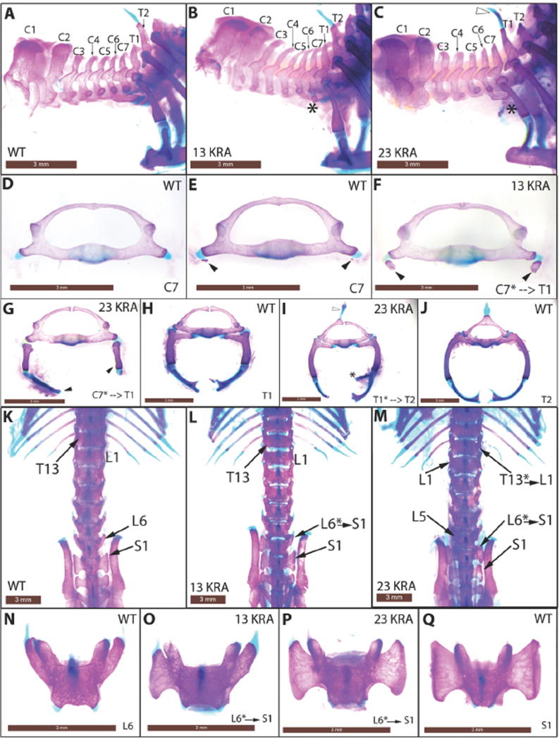
Fig. 3.
Anterior-to-posterior transformations in Cbx223KRA and Cbx213KRA homozygous mutant mice. (A–C) Lateral views of the cervical-thoracic boundary of the axial skeleton. Asterisks mark C7 rib anlage (B) or ectopic rib (C) that is indicative of C7-to-T1 transformation. Open arrowhead marks ectopic spinous process on T1 that characterizes T1-to-T2 transformation (C, also in I). (D–J) Anterior views of disarticulated C7 to T2 vertebrae. Black arrowheads mark rib anlages (E, F) or ectopic ribs (G) on C7. Asterisk marks a spurious second attachment site to the sternum (I) on T1. (K–M) Dorsal views of the thoracic-lumbar and lumbar-sacral transitions. Asterisks indicate positions of homeotic transformations. (N–Q) Dorsal views of disarticulated L6 and S1vertebrae. L6-to-S1 transformation is characterized by the sacral ala (large triangular surface) on L6 (O, unilateral left; P, bilateral). Scale bar = 3mm.

