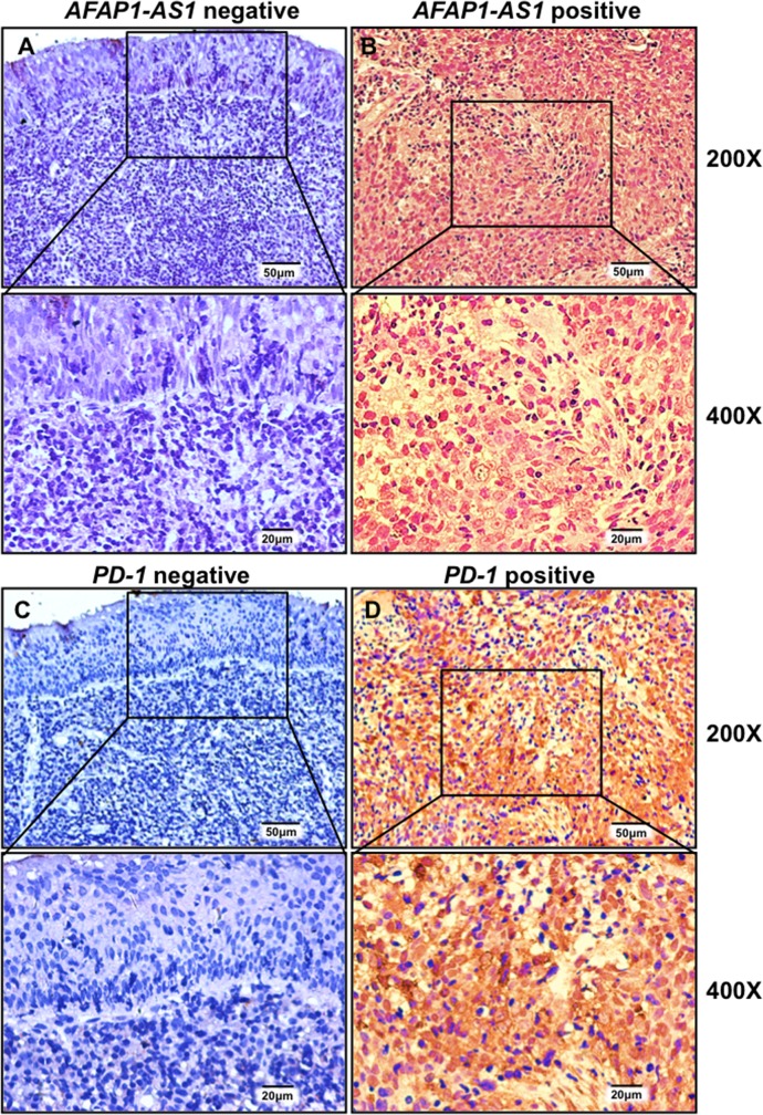
Figure 2. AFAP1-AS1 and PD-1 are highly and jointly expressed in infiltrating lymphocytes of NPC tissues.
Representative images of in situ hybridization for AFAP1-AS1 (A & B) and immunohistochemical staining for PD-1 (C & D) are shown. AFAP1-AS1 and PD-1 are negatively expressed in adjacent non-tumor NPE tissue (A & C) but highly expressed in infiltrating lymphocytes (B & D).

