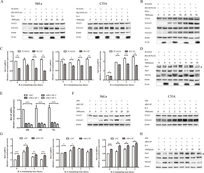
Figure 5. BLCAP inhibited IL-6 induced STAT3 activation.

(A) Empty PCAGGS vector or 3×FLAG-BLCAP plasmid was transfected into HeLa cells (left) and C33A cells (right) respectively. After 24h post-transfection, cells were treated with human IL-6 (10ng/ml for HeLa cells and 100ng/ml for C33A cells) and harvested at the indicated time. (B) PCAGGS or 3×FLAG-BLCAP was transfected into HeLa cells for 24 hours, cells were stimulated with human IFN-α (30ng/ml) for various lengths of time before harvested. (C and D) HeLa cells were transfected with PCAGGS and 3×FLAG-BLCAP for 24 hours, and treated with IL-6 (10ng/ml) for the indicated time before lysed. (E) SiNC and three types of siBLCAP were transfected into HeLa cells and harvested after 48-72 hours. (F) siNC or siBLCAP (siBLCACP-3) was transfected into HeLa (left) and C33A (right) cells for 72 hours. Cells were treated with IL-6 (10ng/ml for HeLa cells and 100ng/ml for C33A cells) for various length of time before lysed. (G and H) siNC or siBLCAP (siBLCACP-3) was transfected into HeLa cells for 72 hours. Cells were treated with IL-6 (10ng/ml) and harvested at the indicated time. All the harvested cells mentioned above were lysed and detected by realtime-PCR assay with different specific primers or Western blot analysis with the corresponding antibodies. β-actin was used as a loading control. The asterisk showed specific band. Data were shown by analysis of unpaired Student's t test, *P<0.05, **P<0.01, ***P<0.001.
