Figure 4.
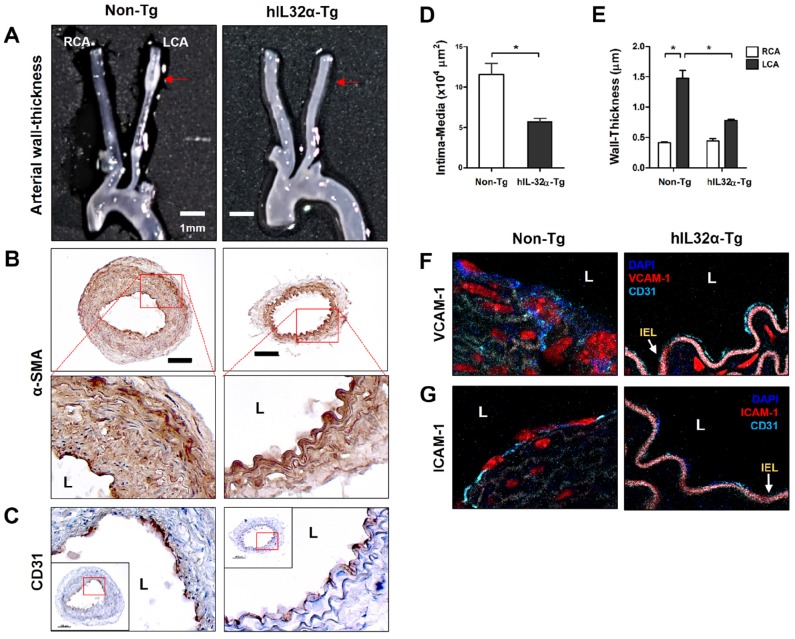
Arterial wall thickening is diminished in hIL-32α-Tg mice.(A-C) For the assessment of arterial wall thickening, hIL-32α-Tg and non-Tg littermate control mice (n = 6 each) were partially ligated and fed a high-fat diet for 4 weeks. (A) Aortic trees including the carotid arteries were dissected and examined by bright-field imaging. Scale bar, 1 mm. (B-C) Frozen sections prepared from the lesion area of LCA, denoted by red arrows in A, were stained for (B) alpha-smooth muscle actin (α-SMA), a vascular smooth muscle cell marker, and (C) CD31, an endothelium marker. The red rectangle indicates the magnified area shown in the lower panel. Scale bar, 100 μm. The thicknesses of the (D) intima media and (E) wall were quantified (data shown as mean ± SEM; n = 6, *p < 0.05 as determined by Student's t-test). Expression of (D) VCAM-1 and (E) ICAM-1 (red) in the intimal layer was determined by immunofluorescence double-stained with a specific endothelium marker CD31 (cyan). Auto-fluorescence (pink) shows internal elastic lamina (IEL). Nuclei were stained with DAPI (blue). L, lumen.

