

Abstract
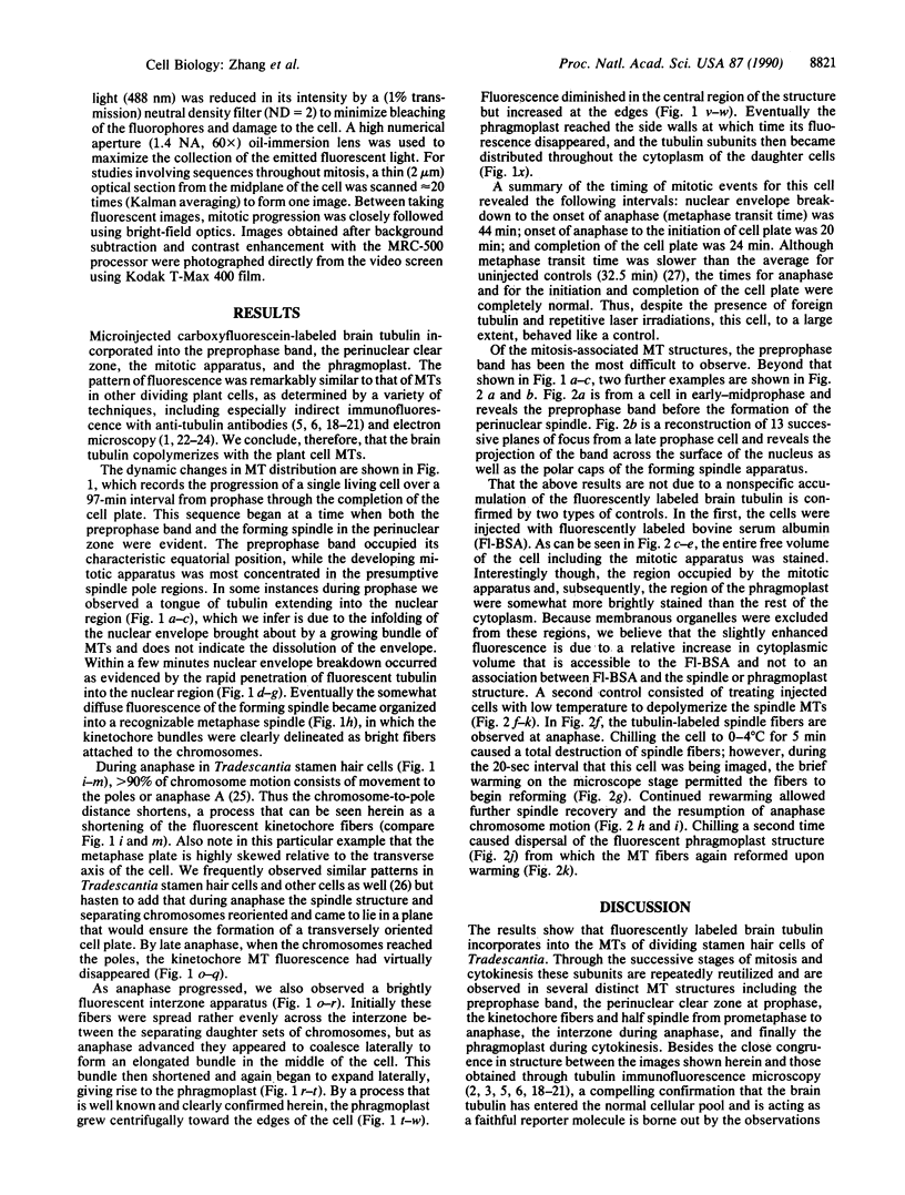
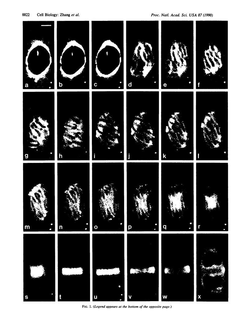
Carboxyfluorescein-labeled brain tubulin has been microinjected into stamen hair cells of Tradescantia, and its distribution during mitosis and cytokinesis was examined using confocal laser scanning fluorescence microscopy. The results show that brain tubulin incorporates into plant microtubules and is utilized throughout mitosis and cytokinesis. Microtubule structures that incorporate brain tubulin include the preprophase band, the perinuclear sheath at late prophase, the kinetochore fibers during prometaphase, metaphase, and anaphase, the interzone spindle during anaphase, and finally the phragmoplast during late anaphase and telophase. All of these microtubule-containing structures and, notably, their transitions from one to another have been observed in single live cells progressing through mitosis and cytokinesis.
Full text
PDF
Images in this article
Selected References
These references are in PubMed. This may not be the complete list of references from this article.
- Bajer A. S., Molè-Bajer J. Reorganization of microtubules in endosperm cells and cell fragments of the higher plant Haemanthus in vivo. J Cell Biol. 1986 Jan;102(1):263–281. doi: 10.1083/jcb.102.1.263. [DOI] [PMC free article] [PubMed] [Google Scholar]
- Gorbsky G. J., Sammak P. J., Borisy G. G. Chromosomes move poleward in anaphase along stationary microtubules that coordinately disassemble from their kinetochore ends. J Cell Biol. 1987 Jan;104(1):9–18. doi: 10.1083/jcb.104.1.9. [DOI] [PMC free article] [PubMed] [Google Scholar]
- Gunning B. E., Wick S. M. Preprophase bands, phragmoplasts, and spatial control of cytokinesis. J Cell Sci Suppl. 1985;2:157–179. doi: 10.1242/jcs.1985.supplement_2.9. [DOI] [PubMed] [Google Scholar]
- Hepler P. K. Calcium restriction prolongs metaphase in dividing Tradescantia stamen hair cells. J Cell Biol. 1985 May;100(5):1363–1368. doi: 10.1083/jcb.100.5.1363. [DOI] [PMC free article] [PubMed] [Google Scholar]
- Hepler P. K., Callaham D. A. Free calcium increases during anaphase in stamen hair cells of Tradescantia. J Cell Biol. 1987 Nov;105(5):2137–2143. doi: 10.1083/jcb.105.5.2137. [DOI] [PMC free article] [PubMed] [Google Scholar]
- Hepler P. K., Jackson W. T. Microtubules and early stages of cell-plate formation in the endosperm of Haemanthus katherinae Baker. J Cell Biol. 1968 Aug;38(2):437–446. doi: 10.1083/jcb.38.2.437. [DOI] [PMC free article] [PubMed] [Google Scholar]
- Hepler P. K., Palevitz B. A. Metabolic inhibitors block anaphase A in vivo. J Cell Biol. 1986 Jun;102(6):1995–2005. doi: 10.1083/jcb.102.6.1995. [DOI] [PMC free article] [PubMed] [Google Scholar]
- INOUE S., BAJER A. Birefringence in endosperm mitosis. Chromosoma. 1961;12:48–63. doi: 10.1007/BF00328913. [DOI] [PubMed] [Google Scholar]
- INOUE S. [Polarization optical studies of the mitotic spindle. I. The demonstration of spindle fibers in living cells]. Chromosoma. 1953;5(5):487–500. doi: 10.1007/BF01271498. [DOI] [PubMed] [Google Scholar]
- Inoué S. Cell division and the mitotic spindle. J Cell Biol. 1981 Dec;91(3 Pt 2):131s–147s. doi: 10.1083/jcb.91.3.131s. [DOI] [PMC free article] [PubMed] [Google Scholar]
- Keith C. H., Feramisco J. R., Shelanski M. Direct visualization of fluorescein-labeled microtubules in vitro and in microinjected fibroblasts. J Cell Biol. 1981 Jan;88(1):234–240. doi: 10.1083/jcb.88.1.234. [DOI] [PMC free article] [PubMed] [Google Scholar]
- Kreis T. E., Birchmeier W. Microinjection of fluorescently labeled proteins into living cells with emphasis on cytoskeletal proteins. Int Rev Cytol. 1982;75:209–214. doi: 10.1016/s0074-7696(08)61005-0. [DOI] [PubMed] [Google Scholar]
- Salmon E. D., Leslie R. J., Saxton W. M., Karow M. L., McIntosh J. R. Spindle microtubule dynamics in sea urchin embryos: analysis using a fluorescein-labeled tubulin and measurements of fluorescence redistribution after laser photobleaching. J Cell Biol. 1984 Dec;99(6):2165–2174. doi: 10.1083/jcb.99.6.2165. [DOI] [PMC free article] [PubMed] [Google Scholar]
- Sloboda R. D., Dentler W. L., Rosenbaum J. L. Microtubule-associated proteins and the stimulation of tubulin assembly in vitro. Biochemistry. 1976 Oct 5;15(20):4497–4505. doi: 10.1021/bi00665a026. [DOI] [PubMed] [Google Scholar]
- Taylor D. L., Wang Y. L. Molecular cytochemistry: incorporation of fluorescently labeled actin into living cells. Proc Natl Acad Sci U S A. 1978 Feb;75(2):857–861. doi: 10.1073/pnas.75.2.857. [DOI] [PMC free article] [PubMed] [Google Scholar]
- Vantard M., Levilliers N., Hill A. M., Adoutte A., Lambert A. M. Incorporation of Paramecium axonemal tubulin into higher plant cells reveals functional sites of microtubule assembly. Proc Natl Acad Sci U S A. 1990 Nov;87(22):8825–8829. doi: 10.1073/pnas.87.22.8825. [DOI] [PMC free article] [PubMed] [Google Scholar]
- Vigers G. P., Coue M., McIntosh J. R. Fluorescent microtubules break up under illumination. J Cell Biol. 1988 Sep;107(3):1011–1024. doi: 10.1083/jcb.107.3.1011. [DOI] [PMC free article] [PubMed] [Google Scholar]
- Wadsworth P., Salmon E. D. Analysis of the treadmilling model during metaphase of mitosis using fluorescence redistribution after photobleaching. J Cell Biol. 1986 Mar;102(3):1032–1038. doi: 10.1083/jcb.102.3.1032. [DOI] [PMC free article] [PubMed] [Google Scholar]
- White J. G., Amos W. B., Fordham M. An evaluation of confocal versus conventional imaging of biological structures by fluorescence light microscopy. J Cell Biol. 1987 Jul;105(1):41–48. doi: 10.1083/jcb.105.1.41. [DOI] [PMC free article] [PubMed] [Google Scholar]
- Wick S. M. Immunofluorescence microscopy of tubulin and microtubule arrays in plant cells. III. Transition between mitotic/cytokinetic and interphase microtubule arrays. Cell Biol Int Rep. 1985 Apr;9(4):357–371. doi: 10.1016/0309-1651(85)90031-1. [DOI] [PubMed] [Google Scholar]