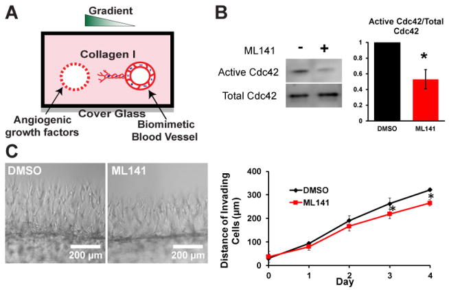
Figure 1.
Inhibition of Cdc42 in 3D biomimetic angiogenic model. (A) Schematic of our 3D biomimetic model of angiogenesis. A device is consisted of 2 channels fully embedded inside 2.5mg/ml collagen gel. (B) Cdc42 activity was reduced in half in the presence of 15 μM Cdc42 inhibitor ML141. (C) Representative phase images of sprouts guided by a gradient of angiogenic cocktail including MCP-1, VEGF, PMA, and S1P at Day 4 for control DMSO and Cdc42-inhibited devices. Average invading distance of invading cells into matrix was reduced in the presence of ML141 (N=4 individual experiments); * (p<0.05) indicates statistical significance.

