

Abstract
The integration of different noble metal nanostructures, which exhibit desirable plasmonic and/or electrocatalytic properties, with electrospun polymer nanofibers, which display unique mechanical and thermodynamic properties, yields novel hybrid nanoscale systems of synergistic properties and functions. This review summarizes recent advances on how to incorporate noble metal nanoparticles into electrospun polymer nanofibers and illustrates how such integration paves the way towards chemical sensing applications with improved sensitivity, stability, flexibility, compatibility, and selectivity. It is expected that further development of this field will eventually make a wide impact on many areas of research.
Keywords: Electrospinning, Polymer nanofibers, Noble metal nanoparticles, Sensing
Review
Background
The rapid development of nanoscience and nanotechnology has led to a wide variety of practical applications, including air filtration, wound dressings, drug delivery, detection, energy production, and food packaging [1–10]. Nanomaterials often have physical and chemical properties that are very different from the same materials at larger scales. Many different strategies have been developed for the synthesis and construction of nanostructured materials [11–13]. Based on dimensionality, nanomaterials may be classified into four categories: zero-dimensional (0D), one-dimensional (1D), two-dimensional (2D), and three-dimensional (3D). 1D nanomaterials such as nanowires, nanorods, and nanotubes have been widely investigated in the last few decades. Among the aforementioned materials, 1D nanofibers have attracted tremendous attention due to their unique structural and physical properties such as small diameters, large surface area per unit mass, small pore size, and flexibility in surface functionalities [14, 15]. There are many processing techniques that have been utilized to produce 1D nanofibers such as template synthesis [16], self-assembly [17], and electrospinning [18, 19]. Among these methods, electrospinning appears to be the most versatile and simplest one for preparing nanofibers [15]. It is notable that by adjusting the parameters of the polymer solution or the electrospinning setup, most of the known polymers such as polyacrylonitrile (PAN) [20, 21], polyvinylidene fluoride (PVdF) [18], and polyvinylalcohol (PVA) [22] can be successfully electrospun into ultrafine fibers. Therefore, due to the significant simplicity and versatility of electrospinning, electrospun polymer nanofibers have garnered substantial attention in recent years, particularly in the field of chemical sensors.
The field of plasmonics that deals with light-matter interactions between adsorbed molecules and noble metal structures at nanoscale dimensions has recently emerged as a rapidly growing area of interest, as evidenced by the explosive growth in various fields including surface-enhanced Raman scattering (SERS) [23–25], surface-enhanced infrared absorption spectroscopy [26, 27], surface-enhanced fluorescence spectroscopy [28–30], surface plasmon resonance spectroscopy [31–34], and plasmonic colorimetry [35]. The fascinating optical properties of plasmonic nanostructures are dominated by collective oscillations of the conduction band electrons in the noble metal (e.g., Au, Ag, and Pt) nanostructures known as surface plasmons. The quest for simple methods to fabricate reproducible plasmonic nanostructures has spurred much interest in a variety of scientific disciplines; however, it has remained a big challenge to hierarchically assemble individual noble metal nanostructures with desirable long-range order at predefined sites on a substrate. Templated synthesis and assembly of nanoscale plasmonic building blocks to form rationally designed architectures have emerged as an overarching strategy for addressing this challenge [36–38]. Electrospun polymer nanofibers have been shown to be one of the most promising templates to pack noble metal nanostructures with great precision. The controlled incorporation of noble metal nanostructures with desired plasmonic properties into electrospun polymer nanofibers paves the way towards sensing applications with improved sensitivity, stability, flexibility, compatibility, and selectivity.
This review highlights recent advances in integrating electrospun polymer nanofibers with noble metal nanoparticles and their applications for chemical sensing. We summarize the following: (1) the basic setup and process parameters for electrospinning, (2) different strategies for the synthesis of Au or Ag nanostructures, (3) preparation of electrospun polymer nanofibers decorated with Au or Ag nanoparticles, and (4) examples of chemical sensing applications of electrospun polymer nanofibers decorated with Au or Ag nanoparticles.
Electrospinning: Basic Set-up and Process Parameters
The electrospinning system generally consists of four main parts: a direct current power supply with high voltage, a syringe that contains polymer solution, a metallic needle with a blunt tip, and a grounded conductive collector, as shown in Fig. 1. During the electrospinning process, the polymer solution in the syringe will be pumped out through the metallic needle tip at a specific rate. A high voltage is applied to create charges on the surfaces of the polymer droplet forming a Taylor cone, and when the repulsive electrostatic force is sufficiently strong to overcome the surface tension of the polymer droplet, the polymer droplet will be elongated into a conical shape [39]. Subsequently, the polymer jets will undergo an elongation process, during which the polymer will be stretched and the polymer solution solvent will evaporate, leaving the long and thin polymer nanofibers collected on the grounded conductive collector.
Fig. 1.
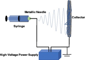
The basic laboratory setup for electrospinning
One of the great advantages of electrospinning is that by changing the parameters during the electrospinning process, the morphology of the electrospun nanofibers can be easily controlled. These parameters include polymer concentration, solution viscosity, solution conductivity, flow rate, applied voltage, the working distance between the collector and tip of the needle, and air humidity [12]. Polymer concentration is an important parameter as it determines the morphology of the electrospun nanofibers, because the surface tension can be dominant as the polymer concentration decreases, which will lead to polymer bead formation [39]. In addition, solution viscosity is another critical parameter, which determines if the polymer can be elestrospun into nanofibers or not. The solution viscosity is highly dependent on the polymer concentration and the molecular weight of the polymer used for the electrospinning. In principle, a polymer with higher molecular weight has, on average, longer molecular chains, and it will form more entanglements leading to a higher viscosity of the polymer solution. Therefore, for a solution made with high molecular weight polymers, even though the polymer concentration is low, it still can produce a uniform jet due to a sufficient level of solution viscosity. Conversely, if the molecular weight is too low, an appropriate polymer solution viscosity cannot be guaranteed even with a high polymer concentration and the polymer tends to form a bead structure on the collector [40]. Comparatively, the processing conditions such as applied voltage and flow rate also play a significant role in nanofiber formation during electrospinning. For applied voltage, it has been proven that varied applied voltage will not change the nanofiber morphology dramatically. According to the past work, both larger and smaller fiber diameters can be obtained when a higher voltage is applied [40].
Synthesis and Assembly of Au Or Ag Nanostructures
During the last few decades, great advances have been made in the synthesis of Ag and Au nanostructures with different sizes and shapes. It is worth noting that different nanostructures can give rise to significantly different optical, electronic, magnetic, or chemical properties, which may be suitable for different applications. Generally, based on the different mechanisms, the reductive approaches to Au or Ag nanostructures can be approximately classified into chemical and physical methods. Typically, the way to obtain Au or Ag nanostructures is to mix Au or Ag precursors with a reducing agent and/or a colloidal stabilizer, and nanostructured Au or Ag with different sizes and shapes can be generated under specific conditions. AgNO3 and HAuCl4 are the most commonly used precursors for Ag and Au nanostructure synthesis, and various reducing agents such as sodium borohydride, alcohols, sodium citrate, and poly(vinyl pyrrolidone) (PVP) can reduce Ag/Au ions into Ag/Au atoms with exceptional control over their sizes and shapes. It has been proved that the plasmon resonance frequencies of the Au or Ag nanoparticles depend on their sizes. For example, Xia and coworkers have synthesized Ag nanocubes ranging from 60 to 100 nm and compared their SERS with respect to both size and shape (sharp vs. truncated) [39]. It demonstrates that larger particles (90 and 100 nm) were found to have higher SERS efficiencies (90 and 100 nm), which is primarily attributed to the overlap between the laser source and plasmon resonance band. Additionally, particles with shaper corners also gave more intense SERS signals than their truncated counterparts.
Synthesis of Au Nanostructures
Based on Turkevich’s research in 1951, HAuCl4 could be reduced in a water solution in the presence of citrate, which has been one of the most commonly used methods for Au nanoparticle synthesis [41]. By changing the amount of citrate, the mean size of the Au nanoparticles can be easily manipulated and citrate plays a role as a nucleating agent and a growth agent at the same time [41]. It has been proven that the citrate reduction method can produce relatively narrow size distributions of the Au nanoparticles. Subsequent studies demonstrated that the mechanism of the control on different Au nanoparticle sizes as a function of the amount of citrate is intimately related to the pH values, because different pH values will determine the formation process of the Au nanoparticles [42].
In 1994, Brust and Schiffrin made a great contribution to the Au nanostructure synthesis by inventing a two-phase synthetic strategy. In this approach, AuCl4 − was transferred from aqueous solution to toluene using tetraoctylammonium bromide as the phase-transfer reagent and strong thiol−gold interactions were utilized to protect AuNPs with thiol ligands. Au clusters with a size range between 1 and 3 nm (Fig. 2) were obtained through the reduction reaction by sodium borohydride (NaBH4) in the presence of dodecanethiol [41]. As NaBH4 was added into the organic phase, the color of the solution turned into deep brown immediately. Several parameters including gold/thiol ratio, temperature, and reduction rate can be varied to control the size of the resulting Au nanoparticles. For example, larger thiol/gold mole ratios led to Au nanoparticles with smaller average core sizes [43]. Different ligands were utilized to form monolayer-protected gold clusters and the ratio between thiol and AuCl4 − could be adjusted in the synthesis to control the size of the AuNPs. Seed-mediated growth, developed by Jana et al. has also shown great promise for generating Au nanoparticles with controlled and monodispersed particle size [44–47]. In a typical process, high-quality seeds are required and then the cylindrical Au nanostructures are grown in multiple steps. In the seed-mediated growth approach, the yield of Au nanostructures is relatively low and high-quality seeds are necessary [48].
Fig. 2.

TEM pictures of the thiol derivatized gold nanoparticles at a low and b high magnification [147]. Reprinted with permission from [147]. Copyright {2010} Royal Society of Chemistry
Various polymers have been reported for the stabilization of Au nanoparticles, which include PVP, poly(ethylene glycol) (PEG), PVA, poly(vinyl methyl ether) (PVME), chitosan, and polyethyleneimine (PEI) [49–57]. Different polymers exhibit different formation processes for Au nanostructures; for example, the reduction between gold ions and PVP may involve a solid–liquid (S–L) mechanism and the nitrogen and oxygen atom heterocyclic ring can contribute to the reducing ability of the PVP [56]. In the reduction reaction, PVP plays the roles of both a reducing agent and a steric stabilizer; therefore, by varying the concentration or ratio between PVP and Au ions, different Au nanostructures with different shapes and sizes can be achieved (as shown in Fig. 3).
Fig. 3.
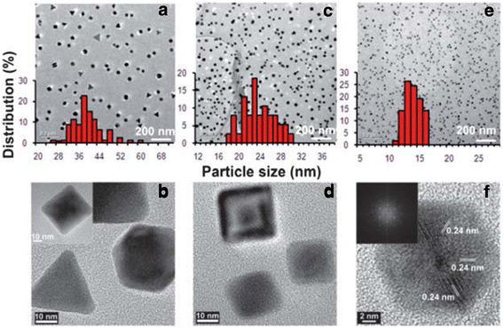
TEM images and histograms of AuNPs from AuNPs–PVP nanocomposite films with weight ratios of HAuCl4 to PVP, [HAuCl4/PVP] = 1:1.5 (a, b), 1:2 (c, d), and 1:4 (e, f) [88]. Reprinted with permission from ref. [67]. Copyright {2010} Royal Society of Chemistry
In addition to the chemical synthesis strategies of Au nanostructures, several physical methods have also been used to improve the quality of the Au nanostructures, including photochemistry (UV, Near-IR), sonochemistry, radiolysis, thermolysis, and microwave irradiation [58–65]. In the microwave irradiation synthesis process, the addition of different amounts of oleic acid not only increases the growth rate but also controls the morphology of the resulting Au nanostructures as shown in Fig. 4 [65]. In addition, oleylamine could also be added as the reducing agent and the nucleated Au functions as the catalyst to initiate the reaction between oleic acid and oleylamine to form dioleamide, which plays a role as the capping agent for the as-prepared Au nanoparticles.
Fig. 4.

TEM images of the gold nanoparticles prepared in i 60, ii 70, iii 80, and iv 90% oleic acid [65]. Reprinted with permission from ref. [65]. Copyright {2010} American Chemical Society
Synthesis of Ag Nanostructures
Use of citrate as the reducing agent for Ag colloid synthesis in an aqueous solution has been discovered for decades. Typically, a set amount of sodium citrate solution is added into a boiling aqueous solution of AgNO3 and the Ag nanocrystals will be obtained after keeping the system boiling for 1 h. During the reaction, the citrate ions serve as both a reducing agent and a stabilizer and they can complex with the silver seeds, thereby influence the particle growth, leading to formation of larger clusters of silver [66]. Furthermore, by varying the pH of the solution or the concentration of citrate ions, different protonation states associated with citrate ion can be achieved, resulting in different growth mechanisms and morphologies of Ag clusters [13].
Another commonly used method for synthesizing Ag nanostructures is the polyol process, which can lead to the formation of Ag nanostructures with a wide variety of sizes and shapes [67–71]. In the polyol reduction process, the nucleation, growth process and the resultant Ag nanostructure morphology are sensitive to reaction conditions, such as temperature, reagent concentration, and presence of trace ions [13]. In a typical polyol reduction process, a Ag precursor with a capping agent is injected into a preheated polyol such as ethylene glycol; 1,2-propylene glycol; or 1,5-pentanediol, which plays a dual role as a solvent and a reducing agent [13, 66]. The exact mechanism during the polyol reduction process is still largely unknown due to its complex nature, and one of the possible reactions is as follows:
| 1 |
By using a spectroscopic method, the formation of glycolaldehyde (GA) has been confirmed and it is the intermediate product of ethylene glycol and a stronger reductant that can effectively reduce AgNO3 into Ag [72]. This may also explain why the polyol process is highly dependent on the reaction temperature [13]. During the polyol reaction, Ag atoms initially form small clusters and later grow into stable and bigger clusters. Finally, Ag nanostructures with different shapes and sizes will be formed after continuous growth.
Moreover, silver nitrate, in the presence of aldehyde-containing compounds (or sugar, e.g., glucose), can form Tollen’s reagent and subsequently transform into elemental Ag through the reduction reaction:
| 2 |
This reaction is also called the silver mirror reaction, which will produce a shiny mirror coating on the inner surface of a reaction container [13]. However, no shape control can be achieved through this reaction, which limits its use in synthesizing Ag nanostructures.
Seed-mediated growth, which uses nanocrystals as seeds for further growth, has attracted a lot of attention and become another popular synthetic approach for Ag nanostructures. Basically, there are two main steps involved: the seed nucleation and the growth of the nanostructures. These two steps are essentially separate, enabling great control over the final morphology of Ag nanostructures [13, 67]. For Ag nanostructures synthesized by this method, the final shape of the nanostructure not only depends on the initial seed but is also governed by the growth rates of different crystallographic facets [13]. It has been found that the growth rates of specific facets are significantly influenced by the capping agent. For example, when used as the capping agent, citrate has been shown to bind more strongly to {111} than {100} crystal facets, tending to form nanoplates. However, for PVP, it binds more strongly to {100} than {111} crystal facets and can thereby reduce the growth rate along the [73] direction, leading to different morphology formation. These studies demonstrate that by changing the reaction conditions including capping agents and seed types,. binding strengths with different facets can be simply manipulated, leading to precise control over the morphology of the Ag nanostructures [13].
A long time ago, people found that a silver precursor (e.g., AgNO3) could interact with light leading to the formation of elemental silver. Therefore, in the presence of appropriate chemical species, Ag nanostructures can be formed under laser irradiation of a sample of Ag colloids [13]. Early studies found when ultrafast (femto- or nanosecond) laser pulses were applied to Ag nanostructures, these nanostructures would melt and tend to form rough spheres due to the low surface energy and thermodynamic stability of this shape [13, 74, 75]. Inspired by these early studies, later investigations revealed that light excitation could also be utilized to grow or modify nanostructures in a controllable fashion and the size and shape of the resultant nanostructures are found to be dependent on applied laser wavelength and power [76–79]. Recent studies indicate that citrate, oxygen, and light are necessary for the reaction. The mechanism behind the light-mediated synthesis is as follows: the Ag seeds that absorb/scatter light weakly reduce dioxygen and release Ag+ into solution; in the presence of light and Ag+, citrate will degrade into acetoacetate, and the resulting electrons are transferred into the Ag nanostructure, accelerating the rate of silver deposition on the surface [13, 78, 80]. By increasing irradiation light intensity, the photochemical process can be significantly enhanced which increases the photoreaction rate and the yield of Ag nanoprisms [78].
Electrospun Polymer Nanofibers Decorated with Noble Metal Nanoparticles
Ag and Au nanostructures have proven to be versatile platforms for various application such as plasmonics, biomedical research, sensing, and catalysis [81–86]. Taking advantage of the flexibility, uniform distribution, controllable morphology, and free-standing properties of electrospun polymer nanofibers, the combination of Au or Ag nanostructures with polymer nanofibers have great potential to improve the reusability and widen the current applications. For example, it is notable that encapsulation of Ag nanoparticles into polymer fiber matrix can efficiently prevent the sulfuration on the surface of Ag nanoparticles. The addition of Au or Ag nanostructures into electrospun nanofibers can also change the nanofiber morphology. Kim and coworkers synthesized Au NP/PEO composites and found that there was a 50-nm increase of fiber diameter after addition of Au NPs to PEO (poly (ethylene oxide)) [87].
Preparation of Electrospun Polymer Nanofibers Decorated with Ag Nanoparticles
Based on the sequence of the reduction process of Ag+ to Ag nanostructure, the preparation of electrospun polymer nanofibers decorated with Ag nanoparticles can be classified into two different methods. In the first method, the Ag nanostructure with different morphologies is either prepared first or the Ag precursor is reduced into Ag nanostructures inside the polymer precursor solution. If the reduction reaction is conducted in a separate solution, the as-prepared Ag nanostructures will be separated and added into the polymer precursor solution subsequently. In this way, because the Ag nanostructure reduction takes place before the electrospun nanofiber formation, it does not require a solvent that can dissolve and stabilize the Ag precursor. In addition, it is not necessary for the polymer to be able to reduce the Ag precursor and it means suitable polymers can be utilized for the composite without limitation. Furthermore, since it is easier to control the Ag nanostructure morphology in a dependent reduction process, the separate synthesis processes permit Ag nanostructure/electrospun nanofiber composites with more Ag nanostructure morphologies.
The other method to prepare electrospun polymer nanofibers decorated with Ag nanoparticles involves first dissolving the Ag precursor into the polymer precursor solution or attaching it onto the surface of the electrospun polymer nanofibers, followed by a reduction process which transforms the Ag precursor into Ag nanostructures. This method is also called the in situ growth of plasmonic nanoparticles. Generally, to perform the reduction reaction, one of the approaches is to utilize a reducing polymer or mixed polymer that contains a reducing polymer as the electrospun nanofiber precursor such as chitosan and PVP [88, 89–91]. The exact mechanism of how the PVP reduces Ag precursor into Ag nanoparticles is still not fully understood, and it has been hypothesized that the aldehyde functional groups, resulting from the oxidation of the hydroxyl end group, might reduce the metal ions in a similar way to that of Tollen’s reagent [92]. Furthermore, it is worth pointing out that the metal formation capacity is highly dependent on the molecule weight of PVP when the same mass of polymer is used [88]. Other approaches to reduce Ag+ in or outside the polymer nanofibers include heating, UV irradiation, microwave irradiation, or hydrogen reduction [93–97]. Leonard et al. prepared tourmaline nanoparticles/polyurethane nanofiber composite and decorated with silver nitrite on the surface [98]. After the irradiation treatment for 4 h, silver nitrate was reduced into Ag nanoparticles which exhibited a wire-like structure on the surface of the composites.
Preparation of Electrospun Polymer Nanofibers Decorated with Au Nanoparticles
Similar to the strategies to encapsulate Ag nanostructures, most researchers demonstrate that electrospun polymer nanofibers decorated with Au nanoparticles could firstly be synthesized using a regular Au nanostructure method such as citrate reduction and seed-mediated approach and then disperse the as-prepared Au nanostructures into the electrospinning polymer precursor solution [73, 99–102]. For some specific applications, Au nanostructures are required to decorate on the surface of the nanofibers and Au nanostructures are found to be attracted by some specific functional groups on the polymers. By adjusting the Ag or Au solution pH, Dong et al. found that one of the three COONa groups from the surface-bound citrate on the NPs would become COOH, which could bridge the amide group on the surface of the nylon 6 fibers through two intermolecular hydrogen bonds and bond the Ag or Au NPs on the surface of the nylon 6 nanofibers as shown in Fig. 5 [103].
Fig. 5.
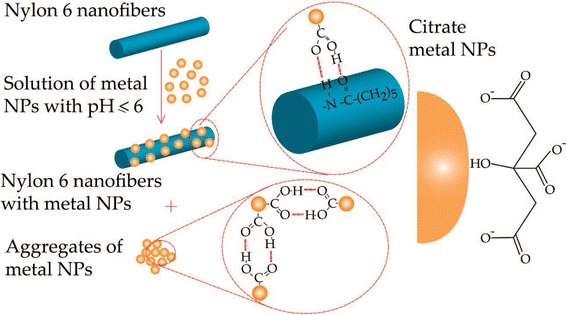
Postulated mechanism of pH-induced assembly of metal nanoparticles on the surface of nylon 6 nanofibers [103]. Reprinted with permission from ref. [103]. Copyright {2008} American Chemical Society
Some polymers contain functional groups on their backbones that can be easily modified with other materials such as 3-mercaptopropyltrimethoxysilane (MPTES) to provide stronger binding sites to attract the Au nanoparticles [99]. In addition, some polymers can be used to stabilize Au nanostructures and function as the electron donor in the reduction process of Au3+ to Au0. Pucci et al. found that under irradiation, the RCH2OH in the PVA with available α hydrogen atoms could be transformed into RCHO while releasing H+ and e− [52]. Subsequently, the produced e− might be trapped by the Au3+ to produce Au0 making PVA additives more efficient photo-reduction reactions [104].
Sensing Applications of Electrospun Polymer Nanofibers Decorated with Noble Metal Nanoparticles
Some recent examples of sensing applications based on electrospun polymer nanofibers decorated with noble metal nanoparticle (e.g., Au and Ag NPs) are illustrated in Table 1. From Table 1, it is seen that the metal particle/nanofiber composites have many advantages such as simplicity, high sensitivity, and high selectivity in detecting various biological and chemical specimens. Through electrospinning, the metal particle/nanofiber composites can be easily fabricated with a high surface area, which can provide easy access for the detection molecules leading to excellent activities for SERS. Therefore, many metal particle/nanofiber composites show low limit of detection. In addition, based on the results in Table 1, it can be concluded that the density and size of metal particles have an important impact on the SERS activity/sensitivity because enhancement of Raman signals result from the presence of hot spots between/among metal particles.
Table 1.
Examples of sensing applications of electrospun polymer nanofibers decorated with noble metal nanoparticles
| Detection mode | Nanofiber materials | Au or Ag nanostructures | Limit of detection | Reference |
|---|---|---|---|---|
| SERS | Cellulose | Ag NPs | 1 ppm thiabendazole | [118] |
| SERS | PVA | Au nanorods with Ag nanowires | 10−4 M 3,3'diethylthiatricarbocyanine iodide | [117] |
| SERS | PVA | Au nanorods | 10−4 M 3,3'diethylthiatricarbocyanine iodide | [100] |
| SERS | PVA | Ag NPs | 1 μM 4-mercaptobenzoic acid (4-MBA) | [116] |
| SERS | PVA | Au and Ag NPs | 4-MBA (2 mM) and thiophenol (1 mM) | [123] |
| SERS | PAA/PVA | Au NPs | 10−8 Rhodamine 6G (R6G) and 10−9 4-Aminothiophenol (4-ATP) | [115] |
| SERS | Poly(2-vinyl pyridine) | Au nanorods | 1 mM 1,4-benzenedithiol | [73] |
| SERS | PVP | Ag nanowires | 5 mg/mL 4,4'-bipyridine | [122] |
| SERS | PAN | Ag nanoparticles | 10−4 p-Aminothiophenol | [148] |
| SERS | PAN | Ag NPs | 10 ppb R6G | [114] |
| SERS | Silica | Au and Ag NPs | 1 mM MBA | [121] |
| SERS | Chitosan | Ag NPs | 1 μM R6G and 0.001 mg/mL d-glucose | [106] |
| SERS | PMMA | Au NPs | 0.1 nM malachite green isothiocyanate | [119] |
| SERS | PMMA | Ag NPs | 1 mM 4-MBA | [149] |
| Electrochemical | Bacteria cellulose | Au NPs | 1 μM H2O2 | [150] |
| Electrochemical | PAN | Ag−Pt Bimetallic NPs | 0.11 μM dopamine (DA) | [151] |
| Electrochemical | PVA | Au NPs | 0.5 μM H2O2 | [99] |
| Electrochemical | PVA/poly(ethyleneimine)/glucose oxidase | Au NPs | 0.9 μM glucose | [146] |
Electrospun Polymer Nanofibers Decorated with Noble Metal Nanoparticles for Chemical Sensing Based on SERS
Surface-enhanced Raman scattering (SERS) has emerged as one of the most promising and powerful analytical tools for probing single molecules, ions, biomolecules, and for cell studies [105–111]. Since the mid-1980s, more researchers began to focus on the exploration of promising analytical applications of SERS instead of the fundamental understanding of the phenomenon [112]. Organized Au or Ag nanostructures have attracted tremendous attention due to their signal-amplifying function as SERS substrates, which have been attributed to a local electromagnetic field enhancement induced by the metallic nanostructures. The SERS enhancement factor (ratio between the Raman signals from a given number of molecules in the presence and in the absence of the nanostructure) is closely related to the size and shape of the nanostructures that give rise to the effect [113]. Typically, the Au, Ag, or AuAg-mixed nanostructures are arranged on rigid materials as the SERS substrate and these methods are either complicated and time consuming in synthesis processes or require strict synthetic conditions.
Recently, a flexible substrate fabricated by combining electrospun nanofibers with Au, Ag, or AuAg-mixed nanostructures has become popular due to their excellent SERS performance and, compared with the rigid substrate, these flexible structures are adaptable to a rough substrate in terms of wrapping and bending [106, 114]. These metal/nanofiber composites demonstrated a 3D structure, which can provide high density of “hot spots”, which refers to the regions of highly enhanced local electromagnetic field [115]. In addition, the polymer outside the nanostructures can protect them from the surrounding environment especially for Ag nanostructures, which gives the composite long lifetime and high sensitivity [116].
Different polymers or ceramic nanofibers, such as PVA [100, 116, 117], cellulose [118], poly(methyl methacrylate) (PMMA) [119], chitosan [106], poly (acrylic acid) (PAA)/PVA [120], and silica [121] have been utilized to combine with different Ag or Au nanostructures to fabricate the flexible substrate for SERS. PVA is a nontoxic, biocompatible polymer, which has good electrospinability, and it is a popular material for electrospinning. When it is used as the supporting material for Ag nanostructures for SERS, it functions not only as the host matrix but also as an organic additive inducing the aggregation of individual Ag nanostructures [116]. In a typical process, the Au or Ag nanostructures are produced in specific morphologies first and these nanostructures are added into the polymer solution as the precursor solution. He et al. synthesized nearly monodispersed Ag NPs via a microwave-assisted method and then these as-prepared silver dimers and aggregates were mixed into a 7% aqueous PVA solution for electrospinning [116]. In order to reduce the specific surface area and the surface Gibbs free energy of individual nanofibers, Ag NPs were self-assembled inside the PVA nanofibers. The assembly of Ag dimers or aligned aggregates within PVA nanofibers was confirmed using transmission electron microscopy (TEM) and X-ray photoelectron spectroscopy (XPS) analyses. Moreover, Ag NPs tended to form a linear chain-like structure along the axial direction of fibers (Fig. 6) because when a high voltage was applied to the solution, Ag NPs became positively charged on one side and negatively charged on the other, leading to a self-alignment by electrostatic attraction in the direction of the electric field [116].
Fig. 6.
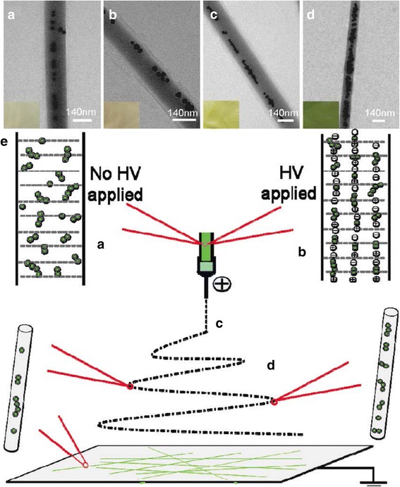
a–d Typical TEM image of Ag/PVA nanofibers with the molar ratio of PVA/Ag 530:1 (a), 530:2 (b), 530:3 (c), and 530:4 (d). The increase of the molar ratio of Ag/PVA in the Ag/PVA solution led to stronger aggregation state and a larger distribution in the sizes of the aggregated Ag NPs. e Schematic representation of the formation of chain-like arrays of Ag NP aggregates within PVA nanofibers [116]. Reprinted with permission from ref. [116]. Copyright {2009} American Chemical Society
As the amount of Ag NPs increased in the PVA nanofibers, the enhancement factor did not increase accordingly, which indicated that different morphology of Ag NP aggregation had a great influence on the enhancement effects of SERS [116]. When 4-mercaptobenzoic acid (4-MBA) was used as a probing molecule to study the Raman enhancement effects, the Ag/PVA nanofibers showed excellent detection reproducibility (i.e., the average relative standard deviation values of the major Raman peak were less than 0.07). Taking advantage of the same nanoparticle alignment in the polymer nanofibers, Ag nanowires (NWs) were also synthesized and electrospun into PVA nanofibers [122]. The Ag NW/PVA nanofibers showed similar morphology and the NWs were “frozen-up” within the polymer fibers. In addition, the electrospun Ag NW/PVA nanofibers were arranged into different structures and stronger SERS intensities were obtained from the arranged samples [122]. Besides Ag nanostructures, Au nanostructures were also encapsulated into the PVA electrospun nanofibers as SERS substrates [100]. Zhang et al. used a seed-mediated surfactant-directed approach to synthesize Au nanorods (AuNRs), and these Au NRs exhibited good alignment along the axial direction of the nanofibers, which demonstrates that electrospinning is a powerful tool to assemble anisotropic nanorods on a large scale [100]. Ag and Au nanostructures can be co-assembled into the PVA nanofibers [117, 123]. Different SERS effects can be obtained by varying the Au/Ag ratio and the excitation wavelength due to the different activities of Au and Ag nanostructures under different wavelengths [124]. In spite of the different morphologies of Ag and Au nanostructures, both Au/PVA and Ag/PVA composites showed excellent SERS performance.
Electrospun Polymer Nanofibers Decorated with Noble Metal Nanoparticles for Chemical Sensing Based on Electrochemical Techniques
Nowadays, metal nanoparticles (such as Au, Ag, Cu, and Ni) have become widely utilized in electrochemical sensing applications, which can be attributed to their rich electronic properties, high surface area, and excellent chemical stability [125, 126]. Au NPs can decrease the overpotentials of many electroanalytical reactions and maintain the reversibility of redox reactions [41, 127]. The Au NP platform can be used for detection of different kinds of analytes including small molecules such as glucose [128, 129], dopamine [130–133], bisphenol A [134], toxic chemicals and drugs such as mercury [135–138], antimony [139], and hydrogen peroxide [140]. Au NPs hold great promise as substrates for designing electrochemical biosensors, which benefit from their ability to provide a stable immobilization of biomolecules retaining their bioactivity, ease of use in chemical synthesis, narrow size distribution, and their convenient labeling of biomolecules [141–143]. Furthermore, both Ag and Au NPs have good biocompatibility and large surface area which can help adsorb biomolecules strongly and play an important role in the immobilization of biomolecules [144]. Accordingly, combining Au or Ag NPs with large-surface-area polymer nanofibers, which provide a large loading capacity for nanoparticles, can further enhance the sensitivity of the sensors [145].
Sapountzi et al. decorated the PVA/poly(ethyleneimine) (PEI)/glucose oxidase nanofibers with Au NPs to further improve the conductivity of the mat and used these composites as electrochemical biosensors [146]. However, both PVA and PEI are water soluble polymers and it may weaken the stability of the composite. Therefore, the researchers conducted a post-electrospinning cross-linking step by exposing the NFs to glutaraldehyde (GA) vapors and the morphology of the fibers was still well retained, suggesting a successful chemical cross-linking reaction induced by GA vapors [146]. The same treatment was also performed by other researchers and the cross-linked PVA nanofiber mat maintained its morphology even after being soaked in water for 15 days [99]. After obtaining the water soluble PVA nanofiber mat, 3-mercaptopropyltrimethoxysilanes (MPTES) were first modified on the surface of electrospun PVA nanofibers. Then, the modified PVA nanofiber mat was immersed into the as-prepared Au NPs aqueous solutions and Au NPs were strongly bonded onto the surface of the modified PVA nanofibers due to the strong affinity between the thiol groups and Au NPs [99]. Au NPs were homogenously decorated on the surface of the modified PVA nanofibers for different Au NP concentrations, leading to highly sensitive detection of H2O2 and the Au NPs/modified PVA also showed more advantages such as fast response, broad linear range, and low detection limit [99].
Conclusions
Extensive research has been carried out to study the properties and applications of both Au or Ag nanostructures and electrospun nanofiber materials in recent years. Taking advantage of the flexibility, large surface area, ease of production, and surface modification of the electrospun polymer nanofibers, the combination of Au/Ag nanostructures with nanofibers makes these composites versatile platforms for various applications in optics, antibacterial coatings, photovoltaics, and chemical and biological sensors etc. The adaptable functionalization of both electrospun nanofibers and Au or Ag nanostructures can lead to unique morphologies and structures for Au or Ag nanostructure/electrospun nanofiber composites, followed by more applications with enhanced performance.
Despite the increasing number of publications using electrospun polymer nanofibers decorated with noble metal nanoparticles for sensing applications, the field is in its infancy. The rational integration of noble metal nanoparticles to nanofiber matrices to achieve desirable plasmonic properties will bring unprecedented strategies for sensor development. Further investigations are required to better understand the morphology control, formation mechanism, and applications to specific applications. It is expected that further development of this field will eventually make a wide impact on many areas of research.
Acknowledgements
This work was supported in part by the U.S. NSF (DMR-1523617) and ARO: W911NF-13-0165. FY thanks Dr. Guy D. Griffin (Oak Ridge, TN) for proofreading the manuscript.
Abbreviations
- GA
Glutaraldehyde
- MPTES
3-Mercaptopropyltrimethoxysilane
- NWs
Nanowires
- PAA
Poly (acrylic acid)
- PAN
Polyacrylonitrile
- PEG
Poly(ethylene glycol)
- PEI
Polyethyleneimine
- PEO
Poly (ethylene oxide)
- PVA
Polyvinyl alcohol
- PVdF
Polyvinylidene fluoride
- PVME
Poly(vinyl methyl ether)
- PVP
Polyvinylpropelene
- SERS
Surface-enhanced Raman scattering
- TEM
Transmission electron microscopy
- XPS
X-ray photoelectron spectroscopy
Authors’ Contributions
FY jointly conceived the study with YT and BV. CC and FY conducted a thorough literature review on the topic. CC wrote the original draft of the manuscript. YT and FY reviewed and edited the manuscript. All authors reviewed the manuscript. All authors read and approved the final manuscript.
Authors’ Information
C. Chen is a postdoctoral fellow with the Department of Chemistry and Biochemistry at North Carolina Central University (NCCU). His research interests include integrated polymer and metallic nanoparticles for energy, sensing, and environmental applications. Y. Tang is an assistant professor of Physics at NCCU, and his research interests are in developing metallic nanoparticles and nanostructures for applications in optics and beyond. B. Vlahovic is a professor of Physic at NCCU, and his research interests include nanotechnologies, astronomies, and sensor development. F. Yan is an associate professor of chemistry at NCCU. His research is centered on developing novel plasmonic sensing strategies, which can be potentially exploited in environmental monitoring, forensic investigation, and biomedical diagnostics.
Competing Interests
The authors declare that they have no competing interests.
Publisher’s Note
Springer Nature remains neutral with regard to jurisdictional claims in published maps and institutional affiliations.
References
- 1.Zhu, C. Chen, Y. Lu, Y. Ge, H. Jiang, K. Fu, X. Zhang, Nitrogen-doped carbon nanofibers derived from polyacrylonitrile for use as anode material in sodium-ion batteries, Carbon N. Y. 94 (2015) 189–195. doi:10.1016/j.carbon.2015.06.076
- 2.Chen C, Li G, Lu Y, Zhu J, Jiang M, Hu Y, Cao L, Zhang X. Chemical vapor deposited MoS2/electrospun carbon nanofiber composite as anode material for high-performance sodium-ion batteries. Electrochim Acta c. 2016 [Google Scholar]
- 3.Jing X, Mi HY, Peng J, Peng XF, Turng LS. Electrospun aligned poly(propylene carbonate) microfibers with chitosan nanofibers as tissue engineering scaffolds. Carbohydr Polym. 2015;117:941–949. doi: 10.1016/j.carbpol.2014.10.025. [DOI] [PubMed] [Google Scholar]
- 4.Wen P, Zhu D-H, Wu H, Zong M-H, Jing Y-R, Han S-Y. Encapsulation of cinnamon essential oil in electrospun nanofibrous film for active food packaging. Food Control. 2016;59:366–376. doi: 10.1016/j.foodcont.2015.06.005. [DOI] [Google Scholar]
- 5.GhavamiNejad A, Rajan Unnithan A, Ramachandra Kurup Sasikala A, Samarikhalaj M, Thomas RG, Jeong YY, Nasseri S, Murugesan P, Wu D, Hee Park C, Kim CS. Mussel-inspired electrospun nanofibers functionalized with size-controlled silver nanoparticles for wound dressing application. ACS Appl Mater Interfaces. 2015;7:12176–12183. doi: 10.1021/acsami.5b02542. [DOI] [PubMed] [Google Scholar]
- 6.Zhang R, Liu C, Hsu PC, Zhang C, Liu N, Zhang J, Lee HR, Lu Y, Qiu Y, Chu S, Cui Y. Nanofiber air filters with high-temperature stability for efficient PM2.5 removal from the pollution sources. Nano Lett. 2016;16:3642–3649. doi: 10.1021/acs.nanolett.6b00771. [DOI] [PubMed] [Google Scholar]
- 7.Jiang S, Hou H, Agarwal S, Greiner A. Polyimide nanofibers by “Green” electrospinning via aqueous solution for filtration applications. ACS Sustain. Chem. Eng. 2016;4:4797–4804. doi: 10.1021/acssuschemeng.6b01031. [DOI] [Google Scholar]
- 8.Xu J, Liu C, Hsu PC, Liu K, Zhang R, Liu Y, Cui Y. Roll-to-roll transfer of electrospun nanofiber film for high-efficiency transparent air filter. Nano Lett. 2016;16:1270–1275. doi: 10.1021/acs.nanolett.5b04596. [DOI] [PubMed] [Google Scholar]
- 9.Liu C, Hsu P-C, Lee H-W, Ye M, Zheng G, Liu N, Li W, Cui Y. Transparent air filter for high-efficiency PM2.5 capture. Nat Commun. 2015;6:6205. doi: 10.1038/ncomms7205. [DOI] [PubMed] [Google Scholar]
- 10.Chen C, Fu K, Lu Y, Zhu J, Xue, L, Hu Y, Zhang X (2015) Tin antimony alloy-filled porous carbon nanofiber composite for use as anode in sodium-ion batteries, RSC Adv 39: 30793–30800. doi:10.1039/C5RA01729G.
- 11.Liang HW, Liu S, Yu SH. Controlled synthesis of one-dimensional inorganic nanostructures using pre-existing one-dimensional nanostructures as templates. Adv Mater. 2010;22:3925–3937. doi: 10.1002/adma.200904391. [DOI] [PubMed] [Google Scholar]
- 12.Zhang C-L, Yu S-H. Nanoparticles meet electrospinning: recent advances and future prospects. Chem Soc Rev. 2014;43:4423–48. doi: 10.1039/c3cs60426h. [DOI] [PubMed] [Google Scholar]
- 13.Rycenga M, Cobley CM, Zeng J, Li W, Moran CH, Zhang Q, Qin D, Xia Y. Controlling the synthesis and assembly of silver nanostructures for plasmonic applications. Chem Rev. 2011;111:3669–3712. doi: 10.1021/cr100275d. [DOI] [PMC free article] [PubMed] [Google Scholar]
- 14.Palmqvist AEC. Synthesis of ordered mesoporous materials using surfactant liquid crystals or micellar solutions. Curr Opin Colloid Interface Sci. 2003;8:145–155. doi: 10.1016/S1359-0294(03)00020-7. [DOI] [Google Scholar]
- 15.Huang Z, Zhang Y, Kotaki M, Ramakrishna S (2003) A review on polymer nanofibers by electrospinning and their applications in nanocomposites. Compos Sci Technol. http://www.sciencedirect.com/science/article/pii/S0266353803001787.
- 16.Feng L, Li S, Li H, Zhai J, Song Y, Jiang L, Zhu D. Super-hydrophobic surface of aligned polyacrylonitrile nanofibers. Angew. Chem. Int. Ed. 2002;41:1221–1223. doi: 10.1002/1521-3773(20020402)41:7<1221::AID-ANIE1221>3.0.CO;2-G. [DOI] [PubMed] [Google Scholar]
- 17.Whitesides GM, Grzybowski B. Self-assembly at all scales. Science. 2002;295:2418–21. doi: 10.1126/science.1070821. [DOI] [PubMed] [Google Scholar]
- 18.Yanilmaz M, Chen C, Zhang X. Fabrication and characterization of SiO2/PVDF composite nanofiber-coated PP nonwoven separators for lithium-ion batteries. J. Polym. Sci. B Polym. Phys. 2013;51:1719–1726. doi: 10.1002/polb.23387. [DOI] [Google Scholar]
- 19.Ge Y, Jiang H, Fu K, Zhang C, Zhu J, Chen C, Lu Y, Qiu Y, Zhang X. Copper-doped Li4Ti5O12/carbon nanofiber composites as anode for high-performance sodium-ion batteries. J Power Sources. 2014;272:860–865. doi: 10.1016/j.jpowsour.2014.08.131. [DOI] [Google Scholar]
- 20.Li S, Chen C, Fu K, White R, Zhao C, Bradford PD, Zhang X. Nanosized Ge@CNF, Ge@C@CNF and Ge@CNF@C composites via chemical vapour deposition method for use in advanced lithium-ion batteries. J Power Sources. 2014;253:366–372. doi: 10.1016/j.jpowsour.2013.12.017. [DOI] [Google Scholar]
- 21.Li S, Chen C, Fu K, Xue L, Zhao C, Zhang S, Hu Y, Zhou L, Zhang X. Comparison of Si/C, Ge/C and Sn/C composite nanofiber anodes used in advanced lithium-ion batteries. Solid State Ion. 2014;254:17–26. doi: 10.1016/j.ssi.2013.10.063. [DOI] [Google Scholar]
- 22.Koski A, Yim K, Shivkumar S. Effect of molecular weight on fibrous PVA produced by electrospinning. Mater Lett. 2004;58:493–497. doi: 10.1016/S0167-577X(03)00532-9. [DOI] [Google Scholar]
- 23.Zhang C, Jiang SZ, Yang C, Li CH, Huo YY, Liu XY, Liu AH, Wei Q, Gao SS, Gao XG, Man BY. Gold@silver bimetal nanoparticles/pyramidal silicon 3D substrate with high reproducibility for high-performance SERS. Sci Rep. 2016;6:25243. doi: 10.1038/srep25243. [DOI] [PMC free article] [PubMed] [Google Scholar]
- 24.Sivanesan A, Witkowska E, Adamkiewicz W, Dziewit Ł, Kamińska A, Waluk J. Nanostructured silver-gold bimetallic SERS substrates for selective identification of bacteria in human blood. Analyst. 2014;139:1037–43. doi: 10.1039/c3an01924a. [DOI] [PubMed] [Google Scholar]
- 25.Dong X, Zhou J, Liu X, Lin D, Zha L. Preparation of monodisperse bimetallic nanorods with gold nanorod core and silver shell and their plasmonic property and SERS efficiency. J. Raman Spectrosc. 2014;45:431–437. doi: 10.1002/jrs.4495. [DOI] [Google Scholar]
- 26.Perry DA, Razer TM, Primm KM, Chen T, Shamburger JB, Golden JW, Owen AR, Price AS, Borchers RL, Parker WR. Surface-enhanced infrared absorption and density functional theory study of dihydroxybenzene isomer adsorption on silver nanostructures. J Phys Chem C. 2013;117:8170–8179. doi: 10.1021/jp3121462. [DOI] [Google Scholar]
- 27.Brown LV, Yang X, Zhao K, Zheng BY, Nordlander P, Halas NJ. Fan-shaped gold nanoantennas above reflective substrates for surface-enhanced infrared absorption (SEIRA) Nano Lett. 2015;15:1272–1280. doi: 10.1021/nl504455s. [DOI] [PubMed] [Google Scholar]
- 28.Zhang Z, Yang P, Xu H, Zheng H (2013) Surface enhanced fluorescence and Raman scattering by gold nanoparticle dimers and trimers. J Appl Phys 113:033102. doi:10.1063/1.4776227.
- 29.Shtoyko T, Raut S, Rich RM, Sronce RJ, Fudala R, Mason RN, Akopova I, Gryczynski Z, Gryczynski I. Preparation of plasmonic platforms of silver wires on gold mirrors and their application to surface enhanced fluorescence. ACS Appl Mater Interfaces. 2014;6:18780–18787. doi: 10.1021/am504431j. [DOI] [PMC free article] [PubMed] [Google Scholar]
- 30.Damm S, Lordan F, Murphy A, McMillen M, Pollard R, Rice JH. Application of AAO matrix in aligned gold nanorod array substrates for surface-enhanced fluorescence and Raman scattering. Plasmonics. 2014;9:1371–1376. doi: 10.1007/s11468-014-9751-y. [DOI] [Google Scholar]
- 31.Zeng S, Baillargeat D, Ho H-P, Yong K-T. Nanomaterials enhanced surface plasmon resonance for biological and chemical sensing applications. Chem Soc Rev. 2014;43:3426–52. doi: 10.1039/c3cs60479a. [DOI] [PubMed] [Google Scholar]
- 32.Juvé V, Cardinal MF, Lombardi A, Crut A, Maioli P, Pérez-Juste J, Liz-Marzán LM, Del Fatti N, Vallée F. Size-dependent surface plasmon resonance broadening in nonspherical nanoparticles: single gold nanorods. Nano Lett. 2013;13:2234–2240. doi: 10.1021/nl400777y. [DOI] [PubMed] [Google Scholar]
- 33.Huang YF, Zhang M, Bin Zhao L, Feng JM, Wu DY, Ren B, Tian ZQ. Activation of oxygen on gold and silver nanoparticles assisted by surface plasmon resonances. Angew. Chem. Int. Ed. 2014;53:2353–2357. doi: 10.1002/anie.201310097. [DOI] [PubMed] [Google Scholar]
- 34.Cao J, Sun T, Grattan KTV. Gold nanorod-based localized surface plasmon resonance biosensors: a review. Sensors Actuators B Chem. 2014;195:332–351. doi: 10.1016/j.snb.2014.01.056. [DOI] [Google Scholar]
- 35.Guo Y, Wu J, Li J, Ju H. A plasmonic colorimetric strategy for biosensing through enzyme guided growth of silver nanoparticles on gold nanostars. Biosens Bioelectron. 2016;78:267–273. doi: 10.1016/j.bios.2015.11.056. [DOI] [PubMed] [Google Scholar]
- 36.Zhang L, Liu T, Liu K, Han L, Yin Y, Gao C. Gold nanoframes by nonepitaxial growth of Au on AgI nanocrystals for surface-enhanced Raman spectroscopy. Nano Lett. 2015;15:4448–4454. doi: 10.1021/acs.nanolett.5b01544. [DOI] [PubMed] [Google Scholar]
- 37.Li C, Jamison AC, Rittikulsittichai S, Lee T, Lee TR (2014) In situ growth of hollow gold–silver nanoshells within porous silica offers tunable plasmonic extinctions and enhanced colloidal stability. ACS Appl Mater Interfaces 6:19943–19950. doi:10.1021/am505424w. [DOI] [PubMed]
- 38.Gao X, Lu F, Dong B, Liu Y, Gao Y, Zheng L. Facile synthesis of gold and gold-based alloy nanowire networks using wormlike micelles as soft templates. Chem Commun. 2015;51:843–846. doi: 10.1039/C4CC08549C. [DOI] [PubMed] [Google Scholar]
- 39.Agarwal S, Greiner A, Wendorff JH. Functional materials by electrospinning of polymers. Prog Polym Sci. 2013;38:963–991. doi: 10.1016/j.progpolymsci.2013.02.001. [DOI] [Google Scholar]
- 40.Tan SH, Inai R, Kotaki M, Ramakrishna S. Systematic parameter study for ultra-fine fiber fabrication via electrospinning process. Polymer (Guildf) 2005;46:6128–6134. doi: 10.1016/j.polymer.2005.05.068. [DOI] [Google Scholar]
- 41.Saha K, Agasti SS, Kim C, Li X, Rotello VM. Gold nanoparticles in chemical and biological sensing. Chem Rev. 2012;112:2739–2779. doi: 10.1021/cr2001178. [DOI] [PMC free article] [PubMed] [Google Scholar]
- 42.Ji X, Song X, Li J, Bai Y, Yang W, Peng X. Size control of gold nanocrystals in citrate reduction: the third role of citrate. J Am Chem Soc. 2007;129:13939–13948. doi: 10.1021/ja074447k. [DOI] [PubMed] [Google Scholar]
- 43.Daniel MCM, Astruc D. Gold nanoparticles: assembly, supramolecular chemistry, quantum-size related properties and applications toward biology, catalysis and nanotechnology. Chem Rev. 2004;104:293–346. doi: 10.1021/cr030698+. [DOI] [PubMed] [Google Scholar]
- 44.Jana NR, Gearheart L, Murphy CJ. Seeding growth for size control of 5-40 nm diameter gold nanoparticles. Langmuir. 2001;17:6782–6786. doi: 10.1021/la0104323. [DOI] [Google Scholar]
- 45.Jana NR, Jana NR, Gearheart L, Gearheart L, Murphy CJ, Murphy CJ. Wet chemical synthesis of high aspect ratio cylindrical gold nanorods. J. Phys. Chem. B. 2001;105:4065–4067. doi: 10.1021/jp0107964. [DOI] [Google Scholar]
- 46.Johnson CJ, Dujardin E, Davis SA, Murphy CJ, Mann S. Growth and form of gold nanorods prepared by seed-mediated, surfactant-directed synthesis. J Mater Chem. 2002;12:1765–1770. doi: 10.1039/b200953f. [DOI] [Google Scholar]
- 47.Gole A, Murphy CJ (2004) Seed-mediated synthesis of gold nanorods: Role of the size and naure of the seed. Chem Mater 16:3633–3640. doi:10.1021/cm0492336
- 48.Nik B, El Sayed A. Preparation and growth mechanism of gold nanorods (NRs) using seed—mediated growth method. Chem Mater. 2003;15:1957–1962. doi: 10.1021/cm020732l. [DOI] [Google Scholar]
- 49.Tracy JB, Kalyuzhny G, Crowe MC, Balasubramanian R, Choi JP, Murray RW. Poly(ethylene glycol) ligands for high-resolution nanoparticle mass spectrometry. J Am Chem Soc. 2007;129:6706–6707. doi: 10.1021/ja071042r. [DOI] [PubMed] [Google Scholar]
- 50.Susumu K, Mei BC, Mattoussi H. Multifunctional ligands based on dihydrolipoic acid and polyethylene glycol to promote biocompatibility of quantum dots. Nat Protoc. 2009;4:424–436. doi: 10.1038/nprot.2008.247. [DOI] [PubMed] [Google Scholar]
- 51.Sun X, Dong S, Wang E. One-step synthesis and characterization of polyelectrolyte-protected gold nanoparticles through a thermal process. Polymer (Guildf) 2004;45:2181–2184. doi: 10.1016/j.polymer.2004.01.010. [DOI] [Google Scholar]
- 52.Pucci A, Bernabò M, Elvati P, Meza LI, Galembeck F, de Paula Leite CA, Tirelli N, Ruggeri G. Photoinduced formation of gold nanoparticles into vinyl alcohol based polymers. J Mater Chem. 2006;16:1058. doi: 10.1039/B511198F. [DOI] [Google Scholar]
- 53.Oh E, Susumu K, Goswami R, Mattoussi H. One-phase synthesis of water-soluble gold nanoparticles with control over size and surface functionalities. Langmuir. 2010;26:7604–7613. doi: 10.1021/la904438s. [DOI] [PubMed] [Google Scholar]
- 54.Krishna Rao KSV, Ramasubba Reddy P, Lee YI, Kim C. Synthesis and characterization of chitosan-PEG-Ag nanocomposites for antimicrobial application. Carbohydr Polym. 2012;87:920–925. doi: 10.1016/j.carbpol.2011.07.028. [DOI] [PubMed] [Google Scholar]
- 55.Harnish B, Robinson JT, Pei Z, Ramström O, Yan M. UV-cross-linked poly(vinylpyridine) thin films as reversibly responsive surfaces. Chem Mater. 2005;17:4092–4096. doi: 10.1021/cm050144i. [DOI] [Google Scholar]
- 56.Carotenuto G. Synthesis and characterization of poly(N-vinylpyrrolidone) filled by monodispersed silver clusters with controlled size. Appl Organomet Chem. 2001;15:344–351. doi: 10.1002/aoc.165. [DOI] [Google Scholar]
- 57.Pal T, Sau TK, Jana NR. Reversible formation and dissolution of silver nanoparticles in aqueous surfactant media. Langmuir. 1997;13:1481–1485. doi: 10.1021/la960834o. [DOI] [Google Scholar]
- 58.Pol VG, Gedanken A, Calderon-Moreno J. Deposition of gold nanoparticles on silica spheres: a sonochemical approach. Chem Mater. 2003;15:1111–1118. doi: 10.1021/cm021013+. [DOI] [Google Scholar]
- 59.Gachard E, Remita H, Khatouri J, Keita B, Nadjo L, Belloni J. Radiation-induced and chemical formation of gold clusters. New J Chem. 1998;22:1257–1265. doi: 10.1039/a804445g. [DOI] [Google Scholar]
- 60.Chen W, Cai WP, Liang CH, Zhang LD. Synthesis of gold nanoparticles dispersed within pores of mesoporous silica induced by ultrasonic irradiation and its characterization. Mater Res Bull. 2001;36:335–342. doi: 10.1016/S0025-5408(01)00497-4. [DOI] [Google Scholar]
- 61.Moessner S, Spatz JP, Moeller M, Aberle T, Schmidt J, Burchard W. Solution behavior of poly(styrene)- Macromolecules. 2000;33:4791–4798. doi: 10.1021/ma992006i. [DOI] [Google Scholar]
- 62.Okitsu K, Yue A, Tanabe S, Matsumoto H, Yobiko Y. Formation of colloidal gold nanoparticles in an ultrasonic field: control of rate of gold(III) reduction and size of formed gold particles. Langmuir. 2001;17:7717–7720. doi: 10.1021/la010414l. [DOI] [Google Scholar]
- 63.Zhou Y, Wang CY, Zhu YR, Chen ZY. A novel ultraviolet irradiation technique for shape-controlled synthesis of gold nanoparticles at room temperature. Chem Mater. 1999;11:2310–2312. doi: 10.1021/cm990315h. [DOI] [Google Scholar]
- 64.Mallick K, Wang ZL, Pal T. Seed-mediated successive growth of gold particles accomplished by UV irradiation: a photochemical approach for size-controlled synthesis. J Photochem Photobiol A Chem. 2001;140:75–80. doi: 10.1016/S1010-6030(01)00389-6. [DOI] [Google Scholar]
- 65.Mohamed MB, Abouzeid KM, Abdelsayed V, Aljarash AA, El-Shall MS. Growth mechanism of anisotropic gold nanocrystals via microwave synthesis: formation of dioleamide by gold nanocatalysis. ACS Nano. 2010;4:2766–2772. doi: 10.1021/nn9016179. [DOI] [PubMed] [Google Scholar]
- 66.Pillai ZS, Kamat PV. What factors control the size and shape of silver nanoparticles in the citrate ion reduction method? J. Phys. Chem. B. 2004;108:945–951. doi: 10.1021/jp037018r. [DOI] [Google Scholar]
- 67.Xia Y, Xiong Y, Lim B, Skrabalak SE. Shape-controlled synthesis of metal nanocrystals: simple chemistry meets complex physics? Angew. Chem. Int. Ed. 2009;48:60–103. doi: 10.1002/anie.200802248. [DOI] [PMC free article] [PubMed] [Google Scholar]
- 68.Wiley B, Herricks T, Sun Y, Xia Y. Polyol synthesis of silver nanoparticles: use of chloride and oxygen to promote the formation of single-crystal, truncated cubes and tetrahedrons. Nano Lett. 2004;4:1733–1739. doi: 10.1021/nl048912c. [DOI] [Google Scholar]
- 69.Wiley BJ, Wang Z, Wei J, Yin Y, Cobden DH, Xia Y. Synthesis and electrical characterization of silver nanobeams. Nano Lett. 2006;6:2273–2278. doi: 10.1021/nl061705n. [DOI] [PubMed] [Google Scholar]
- 70.Sun Y, Mayers B, Herricks T, Xia Y. Polyol synthesis of uniform silver nanowires: a plausible growth mechanism and the supporting evidence. Nano Lett. 2003;3:955–960. doi: 10.1021/nl034312m. [DOI] [Google Scholar]
- 71.Tao A, Sinsermsuksakul P, Yang P. Polyhedral silver nanocrystals with distinct scattering signatures. Angew. Chem. Int. Ed. 2006;45:4597–4601. doi: 10.1002/anie.200601277. [DOI] [PubMed] [Google Scholar]
- 72.Skrabalak SE, Wiley BJ, Kim M, Formo EV, Xia Y. On the polyol synthesis of silver nanostructures: glycolaldehyde as a reducing agent. Nano Lett. 2008;8:2077–2081. doi: 10.1021/nl800910d. [DOI] [PubMed] [Google Scholar]
- 73.Lee CH, Tian L, Abbas A, Kattumenu R, Singamaneni S. Directed assembly of gold nanorods using aligned electrospun polymer nanofibers for highly efficient SERS substrates. Nanotechnology. 2011;22:275311. doi: 10.1088/0957-4484/22/27/275311. [DOI] [PubMed] [Google Scholar]
- 74.Kawasaki M, Hori M. Laser-induced conversion of noble metal-island films to dense monolayers of spherical nanoparticles. J. Phys. Chem. B. 2003;107:6760–6765. doi: 10.1021/jp034768s. [DOI] [Google Scholar]
- 75.Tsuji T, Kakita T, Tsuji M. Preparation of nano-size particles of silver with femtosecond laser ablation in water. Appl Surf Sci. 2003;206:314–320. doi: 10.1016/S0169-4332(02)01230-8. [DOI] [Google Scholar]
- 76.Zhang J, Li S, Wu J, Schatz GC, Mirkin CA. Plasmon-mediated synthesis of silver triangular bipyramids. Angew. Chem. Int. Ed. 2009;48:7787–7791. doi: 10.1002/anie.200903380. [DOI] [PMC free article] [PubMed] [Google Scholar]
- 77.Jin R, Cao Y, Mirkin CA, Kelly KL, Schatz GC, Zheng JG (2001) Photoinduced conversion of silver nanospheres to nanoprisms. Science 294:1901-1903. doi:10.1126/science.1066541. [DOI] [PubMed]
- 78.Xue C, Métraux GS, Millstone JE, Mirkin CA. Mechanistic study of photomediated triangular silver nanoprism growth. J Am Chem Soc. 2008;130:8337–8344. doi: 10.1021/ja8005258. [DOI] [PMC free article] [PubMed] [Google Scholar]
- 79.Zheng X, Xu W, Corredor C, Xu S, An J, Zhao B, Lombardi JR. Laser-induced growth of monodisperse silver nanoparticles with tunable surface plasmon resonance properties and a wavelength self-limiting effect. J Phys Chem C. 2007;111:14962–14967. doi: 10.1021/jp074583b. [DOI] [Google Scholar]
- 80.Wu X, Redmond PL, Liu H, Chen Y, Steigerwald M, Brus L. Photovoltage mechanism for room light conversion of citrate stabilized silver nanocrystal seeds to large nanoprisms. J Am Chem Soc. 2008;130:9500–9506. doi: 10.1021/ja8018669. [DOI] [PubMed] [Google Scholar]
- 81.Shang L, Chen H, Dong S. Electrochemical preparation of silver nanostructure on the planar surface for application in metal-enhanced fluorescence. J Phys Chem C. 2007;111:10780–10784. doi: 10.1021/jp068713c. [DOI] [Google Scholar]
- 82.Le Ru EC, Meyer M, Etchegoin PG. Proof of single-molecule sensitivity in surface enhanced Raman scattering (SERS) by means of a two-analyte technique. J. Phys. Chem. B. 2006;110:1944–1948. doi: 10.1021/jp054732v. [DOI] [PubMed] [Google Scholar]
- 83.Pakizeh T, Käll M. Unidirectional ultracompact optical nanoantennas. Nano Lett. 2009;9:2343–2349. doi: 10.1021/nl900786u. [DOI] [PubMed] [Google Scholar]
- 84.Fantuzzi, G, Pengo P, Gomila R, Ballester P, Hunter CA, Itm CNR, Padova U, Uk S, Multivalent recognition of bis- and tris-Zn-porphyrins by N -methylimidazole functionalized gold nanoparticles. Chem Comm 8:1004–1005 [DOI] [PubMed]
- 85.Devilez A, Stout B, Bonod N. Compact metallo-dielectric optical antenna for ultra directional and enhanced radiative emission. ACS Nano. 2010;4:3390–3396. doi: 10.1021/nn100348d. [DOI] [PubMed] [Google Scholar]
- 86.Blackie EJ, Le Ru EC, Etchegoin PG. Single-molecule surface-enhanced raman spectroscopy of nonresonant molecules. J Am Chem Soc. 2009;131:14466–14472. doi: 10.1021/ja905319w. [DOI] [PubMed] [Google Scholar]
- 87.Kim G-M, Wutzler A, Radusch H-J, Michler GH, Simon P, Sperling RA, Parak WJ. One-dimensional arrangement of gold nanoparticles by electrospinning. Chem Mater. 2005;17:4949–4957. doi: 10.1021/cm0508120. [DOI] [Google Scholar]
- 88.Jeon SH, Xu P, Zhang B, Mack NH, Tsai H, Chiang LY, Wang HL. Polymer-assisted preparation of metal nanoparticles with controlled size and morphology. J Mater Chem. 2011;21:2550–2554. doi: 10.1039/C0JM02340J. [DOI] [Google Scholar]
- 89.Patakfalvi R, Virányi Z, Dékány I. Kinetics of silver nanoparticle growth in aqueous polymer solutions. Colloid Polym Sci. 2004;283:299–305. doi: 10.1007/s00396-004-1138-8. [DOI] [Google Scholar]
- 90.Laudenslager MJ, Schiffman JD, Schauer CL. Carboxymethyl chitosan as a matrix material for platinum, gold, and silver nanoparticles. Biomacromolecules. 2008;9:2682–2685. doi: 10.1021/bm800835e. [DOI] [PubMed] [Google Scholar]
- 91.Zhao Y, Zhou Y, Wu X, Wang L, Xu L, Wei S. A facile method for electrospinning of Ag nanoparticles/poly (vinyl alcohol)/carboxymethyl-chitosan nanofibers. Appl Surf Sci. 2012;258:8867–8873. doi: 10.1016/j.apsusc.2012.05.106. [DOI] [Google Scholar]
- 92.Washio I, Xiong Y, Yin Y, Xia Y. Reduction by the end groups of poly(vinyl pyrrolidone): a new and versatile route to the kinetically controlled synthesis of Ag triangular nanoplates. Adv Mater. 2006;18:1745–1749. doi: 10.1002/adma.200600675. [DOI] [Google Scholar]
- 93.Nguyen TH, Lee KH, Lee BT. Fabrication of Ag nanoparticles dispersed in PVA nanowire mats by microwave irradiation and electro-spinning. Mater Sci Eng C. 2010;30:944–950. doi: 10.1016/j.msec.2010.04.012. [DOI] [Google Scholar]
- 94.Sengupta R, Chakraborty S, Bandyopadhyay S, Dasgupta S, Mukhopadhyay R, Auddy K, Deuri AS. A short review on rubber/clay nanocomposites with emphasis on mechanical properties. Engineering. 2007;47:21–25. [Google Scholar]
- 95.Hiep NT, Khon HC, Niem VVT, Toi VV, Quyen TN, Hai ND, Anh MNT (2016) Microwave-assisted synthesis of chitosan/polyvinyl alcohol silver nanoparticles gel for wound dressing applications. Int J Polym Sci 2016:1–11. doi:10.1155/2016/1584046
- 96.Hang AT, Tae B, Park JS. Non-woven mats of poly(vinyl alcohol)/chitosan blends containing silver nanoparticles: fabrication and characterization. Carbohydr Polym. 2010;82:472–479. doi: 10.1016/j.carbpol.2010.05.016. [DOI] [Google Scholar]
- 97.Dong G, Xiao X, Liu X, Qian B, Liao Y, Wang C, Chen D, Qiu J. Functional Ag porous films prepared by electrospinning. Appl Surf Sci. 2009;255:7623–7626. doi: 10.1016/j.apsusc.2009.04.039. [DOI] [Google Scholar]
- 98.Tijing LD, Amarjargal A, Jiang Z, Ruelo MTG, Park CH, Pant HR, Kim DW, Lee DH, Kim CS. Antibacterial tourmaline nanoparticles/polyurethane hybrid mat decorated with silver nanoparticles prepared by electrospinning and UV photoreduction. Curr Appl Phys. 2013;13:205–210. doi: 10.1016/j.cap.2012.07.011. [DOI] [Google Scholar]
- 99.Wang J, Bin Yao H, He D, Zhang CL, Yu SH. Facile fabrication of gold nanoparticles-poly(vinyl alcohol) electrospun water-stable nanofibrous mats: efficient substrate materials for biosensors. ACS Appl Mater Interfaces. 2012;4:1963–1971. doi: 10.1021/am300391j. [DOI] [PubMed] [Google Scholar]
- 100.Zhang CL, Lv KP, Cong HP, Yu SH. Controlled assemblies of gold nanorods in PVA nanofiber matrix as flexible free-standing SERS substrates by electrospinning. Small. 2012;8:647–653. doi: 10.1002/smll.201290030. [DOI] [PubMed] [Google Scholar]
- 101.Zhang H, Hu Z, Ma Z, Gecevičius M, Dong G, Zhou S, Qiu J. Anisotropically enhanced nonlinear optical properties of ensembles of gold nanorods electrospun in polymer nanofiber film. ACS Appl Mater Interfaces. 2016;8:2048–2053. doi: 10.1021/acsami.5b10411. [DOI] [PubMed] [Google Scholar]
- 102.Bai J, Li Y, Yang S, Du J, Wang S, Zheng J, Wang Y, Yang Q, Chen X, Jing X. A simple and effective route for the preparation of poly(vinylalcohol) (PVA) nanofibers containing gold nanoparticles by electrospinning method. Solid State Commun. 2007;141:292–295. doi: 10.1016/j.ssc.2006.10.024. [DOI] [Google Scholar]
- 103.Dong H, Wang D, Sun G, Hinestroza JP (2008) Assembly of metal nanoparticles on electrospun nylon 6 nanofibers by control of interfacial hydrogen-bonding interactions. Chem Mater 20:6627–6632. doi:10.1021/cm801077p.
- 104.Tanahashi I, Kanno H. Temperature dependence of photoinduced Au particle formation in polyvinyl alcohol films. Appl Phys Lett. 2000;77:3358. doi: 10.1063/1.1328092. [DOI] [Google Scholar]
- 105.Rodríguez-Lorenzo L, Alvarez-Puebla RA, Pastoriza-Santos I, Mazzucco S, Stéphan O, Kociak M, Liz-Marzán LM, de Abajo FJ G. Zeptomol detection through controlled ultrasensitive surface-enhanced raman scattering. J Am Chem Soc. 2009;131:4616–4618. doi: 10.1021/ja809418t. [DOI] [PubMed] [Google Scholar]
- 106.Severyukhina AN, Parakhonskiy BV, Prikhozhdenko ES, Gorin DA, Sukhorukov GB, Möhwald H, Yashchenok AM. Nanoplasmonic chitosan nanofibers as effective SERS substrate for detection of small molecules. ACS Appl Mater Interfaces. 2015;7:15466–15473. doi: 10.1021/acsami.5b03696. [DOI] [PubMed] [Google Scholar]
- 107.Yashchenok A, Masic A, Gorin D, Shim BS, Kotov NA, Fratzl P, Mohwald H, Skirtach A. Nanoengineered colloidal probes for raman-based detection of biomolecules inside living cells. Small. 2013;9:351–356. doi: 10.1002/smll.201201494. [DOI] [PubMed] [Google Scholar]
- 108.Xu LJ, Zong C, Zheng XS, Hu P, Feng JM, Ren B. Label-free detection of native proteins by surface-enhanced Raman spectroscopy using iodide-modified nanoparticles. Anal Chem. 2014;86:2238–2245. doi: 10.1021/ac403974n. [DOI] [PubMed] [Google Scholar]
- 109.Li J, Chen L, Lou T, Wang Y. Highly sensitive SERS detection of As3+ ions in aqueous media using glutathione functionalized silver nanoparticles. ACS Appl Mater Interfaces. 2011;3:3936–3941. doi: 10.1021/am200810x. [DOI] [PubMed] [Google Scholar]
- 110.Khlebtsov BN, Khanadeev VA, Panfilova EV, Bratashov DN, Khlebtsov NG. Gold nanoisland films as reproducible SERS substrates for highly sensitive detection of fungicides. ACS Appl Mater Interfaces. 2015;7:6518–6529. doi: 10.1021/acsami.5b01652. [DOI] [PubMed] [Google Scholar]
- 111.Drescher D, Büchner T, McNaughton D, Kneipp J. SERS reveals the specific interaction of silver and gold nanoparticles with hemoglobin and red blood cell components. Phys Chem Chem Phys. 2013;15:5364–73. doi: 10.1039/c3cp43883j. [DOI] [PubMed] [Google Scholar]
- 112.Vo‐Dinh T, Yan F, MB Wabuyele WB (2005) Surface‐enhanced Raman scattering for medical diagnostics and biological imaging. J Raman Spectrosc 36:640-647. doi:10.1002/jrs.1348.
- 113.Fan M, Andrade GFS, Brolo AG. A review on the fabrication of substrates for surface enhanced Raman spectroscopy and their applications in analytical chemistry. Anal Chim Acta. 2011;693:7–25. doi: 10.1016/j.aca.2011.03.002. [DOI] [PubMed] [Google Scholar]
- 114.Zhang L, Gong X, Bao Y, Zhao Y, Xi M, Jiang C, Fong H. Electrospun nanofibrous membranes surface-decorated with silver nanoparticles as flexible and active/sensitive substrates for surface-enhanced Raman scattering. Langmuir. 2012;28:14433–14440. doi: 10.1021/la302779q. [DOI] [PubMed] [Google Scholar]
- 115.Yang T, Ma J, Zhen S, Huang C (2016) Electrostatic assemblies of well-dispersed AgNPs on the surface of electrospun nanofibers as highly active SERS substrates for wide-range pH sensing. ACS Appl Mater Interfaces 8:14802–14811. doi:10.1021/acsami.6b03720. [DOI] [PubMed]
- 116.He D, Hu B, Yao Q, Wang K, Yu S. Large-scale synthesis of flexible free-standing SERS substrates with high sensitivity: electrospun PVA nanofibers embedded with controlled alignment of silver nanoparticles. ACS Nano. 2009;3:3993–4002. doi: 10.1021/nn900812f. [DOI] [PubMed] [Google Scholar]
- 117.Zhang C-L, Lv K-P, Huang H-T, Cong H-P, Yu S-H. Co-assembly of Au nanorods with Ag nanowires within polymer nanofiber matrix for enhanced SERS property by electrospinning. Nanoscale. 2012;4:5348. doi: 10.1039/c2nr30736g. [DOI] [PubMed] [Google Scholar]
- 118.Liou P, Nayigiziki FX, Kong F, Mustapha A, Lin M. Cellulose nanofibers coated with silver nanoparticles as a SERS platform for detection of pesticides in apples. Carbohydr Polym. 2017;157:643–650. doi: 10.1016/j.carbpol.2016.10.031. [DOI] [PubMed] [Google Scholar]
- 119.Zhong L, Yin J, Zheng Y, Liu Q, Cheng X, Luo F (2014) Self-assembly of Au nanoparticles on PMMA template as flexible, transparent, and highly active SERS substrates. Anal Chem 86:6262–6267. doi:10.1021/ac404224f. [DOI] [PubMed]
- 120.Liu Z, Yan Z, Jia L, Song P, Mei L, Bai L, Liu Y. Gold nanoparticle decorated electrospun nanofibers: a 3D reproducible and sensitive SERS substrate. Appl Surf Sci. 2017;403:29–34. doi: 10.1016/j.apsusc.2017.01.157. [DOI] [Google Scholar]
- 121.Zhang S, Ni W, Kou X, Yeung MH, Sun L, Wang J, Yan C. Formation of gold and silver nanoparticle arrays and thin shells on mesostructured silica nanofibers. Adv Funct Mater. 2007;17:3258–3266. doi: 10.1002/adfm.200700366. [DOI] [Google Scholar]
- 122.Zhang CL, Lv KP, Hu NY, Yu L, Ren XF, Liu SL, Yu SH. Macroscopic-scale alignment of ultralong Ag nanowires in polymer nanofiber mat and their hierarchical structures by magnetic-field-assisted electrospinning. Small. 2012;8:2936–2940. doi: 10.1002/smll.201201353. [DOI] [PubMed] [Google Scholar]
- 123.Wu H, Lin D, Pan W (2010) High performance surface-enhanced Raman scattering substrate combining low dimensional and hierarchical nanostructures. Langmuir 26:6865–6868. doi:10.1021/la1000649 [DOI] [PubMed]
- 124.Li X, Cao M, Zhang H, Zhou L, Cheng S, Yao JL, Fan LJ. Surface-enhanced Raman scattering-active substrates of electrospun polyvinyl alcohol/gold-silver nanofibers. J Colloid Interface Sci. 2012;382:28–35. doi: 10.1016/j.jcis.2012.05.048. [DOI] [PubMed] [Google Scholar]
- 125.Li D, Lv P, Zhu J, Lu Y, Chen C, Zhang X, Wei Q. Nicu alloy nanoparticle-loaded carbon nanofibers for phenolic biosensor applications. Sensors (Switzerland) 2015;15:29419–29433. doi: 10.3390/s151129419. [DOI] [PMC free article] [PubMed] [Google Scholar]
- 126.Wang B, Akiba U, Anzai J (2017) Recent progress in nanomaterial-based electrochemical biosensors for cancer biomarkers: A review. Molecules 22:1048, 1-20. doi:10.3390/molecules22071048. [DOI] [PMC free article] [PubMed]
- 127.Li Y, Schluesener HJ, Xu S. Gold nanoparticle-based biosensors. Gold Bull. 2010;43:29–41. doi: 10.1007/BF03214964. [DOI] [Google Scholar]
- 128.Chen M, Diao G. Electrochemical study of mono-6-thio-β-cyclodextrin/ferrocene capped on gold nanoparticles: characterization and application to the design of glucose amperometric biosensor. Talanta. 2009;80:815–820. doi: 10.1016/j.talanta.2009.07.068. [DOI] [PubMed] [Google Scholar]
- 129.Li Y, Song YY, Yang C, Xia XH. Hydrogen bubble dynamic template synthesis of porous gold for nonenzymatic electrochemical detection of glucose. Electrochem Commun. 2007;9:981–988. doi: 10.1016/j.elecom.2006.11.035. [DOI] [Google Scholar]
- 130.Li J, Lin X. Simultaneous determination of dopamine and serotonin on gold nanocluster/overoxidized-polypyrrole composite modified glassy carbon electrode. Sensors Actuators, B Chem. 2007;124:486–493. doi: 10.1016/j.snb.2007.01.021. [DOI] [Google Scholar]
- 131.Raj CR, Okajima T, Ohsaka T. Gold nanoparticle arrays for the voltammetric sensing of dopamine. J Electroanal Chem. 2003;543:127–133. doi: 10.1016/S0022-0728(02)01481-X. [DOI] [Google Scholar]
- 132.Li M, Gao F, Yang P, Wang L, Fang B. Conveniently assembling dithiocarbamate and gold nanoparticles onto the gold electrode: a new type of electrochemical sensors for biomolecule detection. Surf Sci. 2008;602:151–155. doi: 10.1016/j.susc.2007.10.006. [DOI] [Google Scholar]
- 133.Zhang L, Jiang X. Attachment of gold nanoparticles to glassy carbon electrode and its application for the voltammetric resolution of ascorbic acid and dopamine. J Electroanal Chem. 2005;583:292–299. doi: 10.1016/j.jelechem.2005.06.014. [DOI] [Google Scholar]
- 134.Yin H, Zhou Y, Ai S, Han R, Tang T, Zhu L. Electrochemical behavior of bisphenol a at glassy carbon electrode modified with gold nanoparticles, silk fibroin, and PAMAM dendrimers. Microchim Acta. 2010;170:99–105. doi: 10.1007/s00604-010-0396-z. [DOI] [Google Scholar]
- 135.Domínguez-Renedo O, Alonso-Lomillo MA, Ferreira-Gonçalves L, Arcos-Martínez MJ. Development of urease based amperometric biosensors for the inhibitive determination of Hg (II) Talanta. 2009;79:1306–1310. doi: 10.1016/j.talanta.2009.05.043. [DOI] [PubMed] [Google Scholar]
- 136.Zhu Z, Su Y, Li J, Li D, Zhang J, Song S, Zhao Y, Li G, Fan C. Highly sensitive electrochemical sensor for mercury (II) ions by using a mercury-specific oligonucleotide probe and gold nanoparticle-based amplification. Work. 2009;81:7660–7666. doi: 10.1021/ac9010809. [DOI] [PubMed] [Google Scholar]
- 137.Xu H, Zeng L, Xing S, Shi G, Xian Y, Jin L. Microwave-radiated synthesis of gold nanoparticles/carbon nanotubes composites and its application to voltammetric detection of trace mercury(II) Electrochem Commun. 2008;10:1839–1843. doi: 10.1016/j.elecom.2008.09.030. [DOI] [Google Scholar]
- 138.Abollino O, Giacomino A, Malandrino M, Piscionieri G, Mentasti E. Determination of mercury by anodic stripping voltammetry with a gold nanoparticle-modified glassy carbon electrode. Electroanalysis. 2008;20:75–83. doi: 10.1002/elan.200704044. [DOI] [Google Scholar]
- 139.Domínguez Renedo O, Arcos Martínez MJ. Anodic stripping voltammetry of antimony using gold nanoparticle-modified carbon screen-printed electrodes. Anal Chim Acta. 2007;589:255–260. doi: 10.1016/j.aca.2007.02.069. [DOI] [PubMed] [Google Scholar]
- 140.Xu S, Peng B, Han X. A third-generation H2O2 biosensor based on horseradish peroxidase-labeled Au nanoparticles self-assembled to hollow porous polymeric nanopheres. Biosens Bioelectron. 2007;22:1807–1810. doi: 10.1016/j.bios.2006.07.008. [DOI] [PubMed] [Google Scholar]
- 141.Kerman K, Saito M, Tamiya E, Yamamura S, Takamura Y. Nanomaterial-based electrochemical biosensors for medical applications. TrAC - Trends Anal Chem. 2008;27:585–592. doi: 10.1016/j.trac.2008.05.004. [DOI] [Google Scholar]
- 142.Pingarrón JM, Yáñez-Sedeño P, González-Cortés A. Gold nanoparticle-based electrochemical biosensors. Electrochim Acta. 2008;53:5848–5866. doi: 10.1016/j.electacta.2008.03.005. [DOI] [Google Scholar]
- 143.Siangproh W, Dungchai W, Rattanarat P, Chailapakul O. Nanoparticle-based electrochemical detection in conventional and miniaturized systems and their bioanalytical applications: a review. Anal Chim Acta. 2011;690:10–25. doi: 10.1016/j.aca.2011.01.054. [DOI] [PubMed] [Google Scholar]
- 144.Luo X, Morrin A, Killard AJ, Smyth MR. Application of nanoparticles in electrochemical sensors and biosensors. Electroanalysis. 2006;18:319–326. doi: 10.1002/elan.200503415. [DOI] [Google Scholar]
- 145.Che X, Yuan R, Chai Y, Li J, Song Z, Wang J. Amperometric immunosensor for the determination of α-1-fetoprotein based on multiwalled carbon nanotube-silver nanoparticle composite. J Colloid Interface Sci. 2010;345:174–180. doi: 10.1016/j.jcis.2010.01.033. [DOI] [PubMed] [Google Scholar]
- 146.Sapountzi E, Braiek M, Vocanson F, Chateaux JF, Jaffrezic-Renault N, Lagarde F. Gold nanoparticles assembly on electrospun poly(vinyl alcohol)/poly(ethyleneimine)/glucose oxidase nanofibers for ultrasensitive electrochemical glucose biosensing. Sensors Actuators, B Chem. 2017;238:392–401. doi: 10.1016/j.snb.2016.07.062. [DOI] [Google Scholar]
- 147.M. Brust, M. Walker, D. Bethell, D.J. Schiffrin, R. Whyman, Synthesis of thiol-derivatised gold nanoparticles in, Chem. Commun. (1994) 801–802. doi:10.1039/c39940000801
- 148.Ren S, Dong L, Zhang X, Lei T, Ehrenhauser F, Song K, Li M, Sun X, Wu Q. Electrospun nanofibers made of silver nanoparticles, cellulose nanocrystals, and polyacrylonitrile as substrates for surface-enhanced Raman scattering. Materials (Basel) 2017;10:68. doi: 10.3390/ma10010068. [DOI] [PMC free article] [PubMed] [Google Scholar]
- 149.Bao Y, Lai C, Zhu Z, Fong H, Jiang C. SERS-active silver nanoparticles on electrospun nanofibers facilitated via oxygen plasma etching. RSC Adv. 2013;3:8998. doi: 10.1039/c3ra41322e. [DOI] [Google Scholar]
- 150.Zhang T, Wang W, Zhang D, Zhang X, Yurong M, Zhou Y, Qi L. Biotemplated synthesis of cold nanoparticle-bacteria cellulose nanofiber nanocomposites and their application in biosensing. Adv Funct Mater. 2010;20:1152–1160. doi: 10.1002/adfm.200902104. [DOI] [Google Scholar]
- 151.Huang Y, Miao YE, Ji S, Tjiu WW, Liu T. Electrospun carbon nanofibers decorated with Ag-Pt bimetallic nanoparticles for selective detection of dopamine. ACS Appl Mater Interfaces. 2014;6:12449–12456. doi: 10.1021/am502344p. [DOI] [PubMed] [Google Scholar]