A 47-year-old Japanese man had been suffering from acute posterior neck pain for the previous 2 days. He visited our outpatient department because he had limited neck mobility and was unable to sleep well due to this pain. The posterior neck pain became exacerbated when swallowing, and he also had dysphagia.
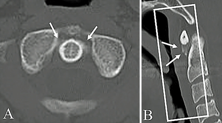
Computed tomography scans showed light calcification on the anterior surface of the axis below the atlas (Picture A, arrows), with edematous changes in the retropharyngeal space (Picture B, arrows inside the box). We diagnosed his condition as acute calcific prevertebral tendinitis, and we administered a non-steroidal anti-inflammatory drug. His symptoms thereafter disappeared 1 week later.
Picture.
Acute calcific prevertebral tendinitis is caused by an inflammatory reaction of the longus colli muscle due to the absorption process of hydroxyapatite which is deposited in the tendons of this muscle (1). Acute calcific prevertebral tendinitis is characterized by calcifications on the anterior surface of the odontoid process, as observed on either a simple radiograph or computed tomography scan (2).
The authors state that they have no Conflict of Interest (COI).
References
- 1.Hartley J. Acute cervical pain associated with retropharyngeal calcium deposit: a case report. J Bone Joint Surg Am 46: 1753-1754, 1964. [PubMed] [Google Scholar]
- 2.Eastwood JD, Hudgins PA, Malone D. Retropharyngeal effusion in acute calcific prevertebral tendinitis: diagnosis with CT and MR imaging. AJNR Am J Neuroradiol 19: 1789-1792, 1998. [PMC free article] [PubMed] [Google Scholar]


