Abstract
Right-sided aortic arch associated with Kommerell’s diverticulum is a rare congenital defect of aorta with complexity. The challenging surgical treatment calls for a direct and entire visibility of aorta and its branches which can be provided by the three-dimensional (3D) printing technique. A 42-year-old man with right-sided aortic arch, Kommerell’s diverticulum and an aberrant left subclavian artery underwent the surgery with the guiding assistance of 3D printed model. The satisfying outcome suggests the novel combination of 3D printing technique and surgical procedure a promising perspective on treating complex aortic defect.
Keywords: Right-sided aortic arch, Kommerell’s diverticulum, three-dimensional printing, precision treatment
Introduction
A right-sided aortic arch is a rare congenital defect of aorta, presenting in 0.05% to 0.1% of radiology series (1) and 0.04–0.1% of autopsy (2), which was first reported by Fioratti and Aglietti centuries ago (3). In the adult population, based on epidemiologic study, a right-sided aortic arch, presenting asymptomatic unless aneurysmal disease develops which may result in symptoms of tracheal and esophageal compression, is often associated with a Kommerell’s diverticulum, first reported by Dr. Kommerell in 1936 (4), which is known as the pseudoaneurysm usually occurs at the origination of an aberrant subclavian artery carrying a 19% to 53% risk of rupture or dissection (5). This condition is clinically relevant and may be life-threatening due to the mortality associated with rupture, the morbidity caused by compression of mediastinal structures, and the complexity of surgery. The variety of anomalies, the indirect visibility by means of usual imaging, including computed tomography (CT), ultrasound, magnetic resonance imaging (MRI), and the required precision of treatment reveal the utilization of the emerging three-dimensional (3D) printing technique, which are mostly presented in maxillofacial surgery, neurosurgery, orthopedics and congenital heart disease to date (6). In this case report, we induced a 3D printing model, deriving from multi-detector computed tomography (MDCT), to a patient suffering from right-sided aortic arch associated with Kommerell’s diverticulum at the origination of aberrant left subclavian artery for the diagnosis and preoperative planning.
Case report
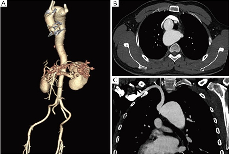
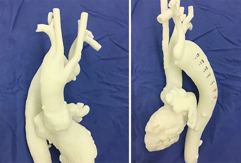
A 42-year-old man was admitted to ward of Cardiac surgery with presentation of chest pain after exercise for 1 year. Medical history revealed an outpatient record of aortic CT angiography (CTA) indicating right-sided aortic arch associated with an enlarged arch 1 and a half years ago when he was recommended to be followed up. The aortic CTA after admission diagnosed with right-sided aortic arch associated with an enlarged arch, of which the maximum cross-sectional area is about 38 mm × 52 mm, known as Kommerell’s diverticulum as well, at the origination of aberrant left subclavian artery passing behind the esophagus (Figure 1). The CTA of intracranial artery and vertebral artery showed no abnormity as well as other examinations. No difference was found between the blood pressures measured from the left and right arm. We developed a 3D printing model of the aorta and its branches at a level up to the internal carotid sinus and down to diaphragm based on the aortic CTA data before surgery to make an entire acquaintance to the complex anatomy of aorta as well as a detailed preoperative plan. In order to get a precise 3D printed model reflecting real situation, we used the dense CTA scanning at a slice distance of 1mm and resolution 512×512 pixels. The CTA images was exported in the format of standard DICOM and then imported to Chaos II (Meditool Enterprise Co., Ltd, China) for regions of interest segmentation and 3D surface reconstruction. Pangu V4.1 3D printer (Meditool Enterprise Co., Ltd, China) was used to print the 3D physical model. The building layer thickness is 0.1 mm and the laser spot diameter 80 µm (Figure 2).
Figure 1.
CTA before surgery. (A) 3D reconstruction of aorta; (B) cross section of Kommerell’s diverticulum; (C) coronal view of Kommerell’s diverticulum. CTA, CT angiography.
Figure 2.
3D model &3D model with measured mark.
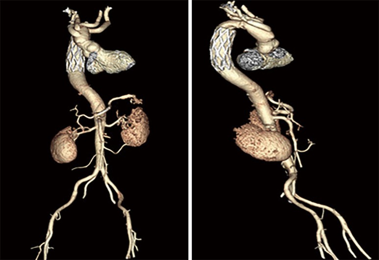
The operation, including replacement of the arch and insertion of frozen elephant trunk, was underwent at the third day after admission. A cannulation of right femoral artery was performed following the systemic heparinization in a supine position of the patient after anaesthesia. A standard median sternotomy was done. The great vessels were exposed and detected carefully, from which the enlarged arch, the Kommerell’s diverticulum and the four branches of arch were found and marked (Figure 3), almost the same as those showed in the 3D model. A standard right atrium cannulation was performed and the CPB as well as was established and initiated. Ante-grade and retro-grade coronary perfusion after aortic clamping were combined to maintain the cardioplegia as well as a better myocardial protection. Ante-grade cerebral perfusion was introduced through the cannulation in the left carotid artery. A frozen elephant trunk (MicroPort Cronus, 32 mm × 100 mm) was placed in the distal to the Kommerell’s diverticulum of descending aorta after the removal of partial ascending aorta, aortic arch, and Kommerell’s diverticulum. Then a four-branched graft (28/10/8/8 mm × 10 mm, Gelweave) was used to connect the elephant trunk, branches vessels and remaining ascending aorta (Figure 3). Aortic clamping was released following the exhaust after the accomplishment of all sutures. The heart came to resuscitation and the CPB procedure terminated progressively. The patient was transferred to intensive care unit after surgery. The postoperative process was uneventful without any complication such as bleeding, pneumonia, and incision infection. The aortic CTA after surgery showed sound patency, position, and shape of the graft, including the arch branches, and the trunk (Figure 4). The patient was discharged at the 11th day after surgery with no abnormity in all lab or clinical examinations.
Figure 3.
Exposure of arch branches & the connection of graft and great vessels. (A) Right subclavian artery; (B) right common carotid artery; (C) left common carotid artery; (D) left subclavian artery.
Figure 4.
3D reconstruction of aorta from CTA after surgery. CTA, CT angiography.
Discussion
Complex aortic pathologies involving the ascending aorta, the aortic arch, and the descending aorta which can be presented in various diseases such as aortic dissection, Marfan syndrome, extensive aneurysm, aortic coarctation remain a challenging issue in aortic surgery due to the complexity and indirect visibility of the aortic anatomy. Numerous methods including frozen elephant trunk, endovascular repair and hybrid procedures have been described for treatment of complex aortic disease to overcome the substantial morbidity and mortality associated with a classic open-surgery (7,8). A right-sided aortic arch associated with Kommerell’s diverticulum, possessing a 19% to 53% risk of rupture or dissection, being devastating to the outcome and definitely life-threatening, was actually one type of complex aortic diseases which should be medically corrected indicating by signs such as aortic aneurysm, clinical symptoms and aggravation in imaging during follow-up. Several opinions for treatment are available among which surgical treatment remains the most commonly described one for Kommerell’s diverticulum (4). The variety of aortic anatomies, indirect detection of usual imaging (CT, US, MRI, etc.) and required precision of surgery reveal the introduction of 3D printing models into diagnosis with more accuracy and detailed planning before surgery for patients suffering from the complex aortic defect.
3D printing applications, guiding treatment strategies, rehearsing fully endovascular and surgical procedures, benefiting medical teaching, advancing basic and clinical researches, enriching patient-physician communication, have been utilized and reported several times in cardiovascular surgery (9). While the combination of the emerging 3D printing technique and surgical treatment of right-sided aortic arch with Kommerell’s diverticulum described in this study is the first time being reported to the best of our knowledge. Compared to the usual imaging methods, 3D models provide a direct and entire visibility of the aberrant aorta, aortic branches and Kommerell’s diverticulum which is vital for surgeons to make a perioperative plan in detail. We measured exactly the diameter of the descending aorta distal to the Kommerell’s diverticulum at different levels, which is much more convenient compared to MDCT imaging, to select the frozen elephant trunk at a suitable size of 32 mm in diameter (Figure 2B). Furthermore, the 3D model has been used as a guideline in anatomy of the arch branches and Kommerell’s diverticulum during exposure making it a time-saving procedure. Moreover, the extensive comprehension of the aorta derived from the 3D model indicated the optimal section which should be removed for surgeons as part of preoperative planning.
3D printing models have shown an impressive potential to be incorporated into the precise treatment for many complex cardiovascular applications, akin to the role that 3D printed models play currently in planning and simulating surgical procedures for congenital heart diseases and complex aortic pathologies. From perspective of short-term goals, more cardiovascular applications should be available including all complex defects for medical utilization. Furthermore, the complex process of 3D printing needs to be simplified as well the cost less to make it an in-hospital procedure being done by physicians with none or few training even by artificial intelligence. From that of long-term goals, the research of materials used for printing in cardiovascular tissue engineering should be a highlighted issue.
Conclusions
In summary, the patient suffered from right-sided aortic arch associated with Kommerell’s diverticulum and an aberrant left subclavian artery underwent the precise treatment by mean of combining the surgical procedure including replacement of the arch and insertion of frozen elephant trunk and the 3D printing technique showed a good inpatient outcome after surgery in spite of the rarity and complexity of the defect. The assistance of 3D model, providing a well-designed plan before surgery and a detailed guidance for anatomy during surgery, as a perioperative guideline to some extent, plays a critical role in the surgical treatment of the patient. However, more cases and case-control studies should be performed. Data of follow-up are absent as well due to the timeliness of this report.
Acknowledgements
The authors acknowledge Meditool Enterprise Company (Ltd, China) for the selfless help in the fabrication of the 3D printed model.
Funding: This work was supported for funding of No. 14PJD008 from the Sponsored by Shanghai Pujiang Program.
Footnotes
Conflicts of Interest: The authors have no conflicts of interest to declare.
References
- 1.Shuford WH, Sybers RG, Gordon IJ, et al. Circumflex retroesophageal right aortic arch simulating mediastinal tumor or dissecting aneurysm. AJR Am J Roentgenol 1986;146:491-6. 10.2214/ajr.146.3.491 [DOI] [PubMed] [Google Scholar]
- 2.Hastreiter AR, D'Cruz IA, Cantez T, et al. Right-sided aorta. I. Occurrence of right aortic arch in various types of congenital heart disease. II. Right aortic arch, right descending aorta, and associated anomalies. Br Heart J 1966;28:722-39. 10.1136/hrt.28.6.722 [DOI] [PMC free article] [PubMed] [Google Scholar]
- 3.Fioratti F, Aglietti F. A case of human right aorta. Anatom Rec 1763;45:365. [Google Scholar]
- 4.Kommerell B. Verlagerung des Ösophagus durch eine abnorm verlaufende Arteria subclavia dextra (Arteria lusoria). Fortschr Geb Roentgenstrahlen 1936;54:590-5. [Google Scholar]
- 5.Idrees J, Keshavamurthy S, Subramanian S, et al. Hybrid repair of Kommerell diverticulum. J Thorac Cardiovasc Surg 2014;147:973-6. 10.1016/j.jtcvs.2013.02.063 [DOI] [PubMed] [Google Scholar]
- 6.Rengier F, Mehndiratta A, von Tengg-Kobligk H, et al. 3D printing based on imaging data: review of medical applications. Int J Comput Assist Radiol Surg 2010;5:335-41. 10.1007/s11548-010-0476-x [DOI] [PubMed] [Google Scholar]
- 7.Garg V, Ouzounian M, Peterson MD. Advances in aortic disease management: a year in review. Curr Opin Cardiol 2016;31:127-31. 10.1097/HCO.0000000000000267 [DOI] [PubMed] [Google Scholar]
- 8.Jakob H, Dohle D, Benedik J, et al. Long-term experience with the E-vita Open hybrid graft in complex thoracic aortic disease†. Eur J Cardiothorac Surg 2017;51:329-38. [DOI] [PubMed] [Google Scholar]
- 9.Sen-Chowdhry S, Jacoby D, Moon JC, et al. Update on hypertrophic cardiomyopathy and a guide to the guidelines. Nat Rev Cardiol 2016;13:651-75. 10.1038/nrcardio.2016.140 [DOI] [PubMed] [Google Scholar]


