

Dear Editor,
Fluorodeoxyglucose positron emission tomography-computed tomography (FDG PET-CT) scan has been a valuable tool in metabolically characterizing morphologically significant lesions based on glucose-transporter expression, thereby labeling them as “FDG avid” or “non-FDG avid.” In addition to this, in our case, it helped in providing favorable site for biopsy, which yielded the accurate diagnosis. It highlights the value of FDG PET-CT scan in posttransplant scenario, irrespective of the time interval following transplant procedure, wherein chances of malignancy are high.
A 46-year-old female presented with left-sided neck swelling for the past 1 month, increasing in size, associated with weakness and loss of appetite. Fine-needle aspiration from palpable left cervical nodal mass was suggestive of reactive hyperplasia. This was followed by biopsy and was inconclusive although histopathology raised the possibility of sarcoma.
Subsequently, patient was referred for whole-body F-18 FDG PET-CT scan, to assess for disease extent and recommend an appropriate site for biopsy. Maximum intensity projection image showed intense tracer uptake in the scalp and in bilateral neck region [Figure 1]. Axial CT and fused PET-CT images showed high grade metabolically active soft tissue thickening involving entire scalp and conglomerate nodal masses in bilateral level II and V-neck nodes. Biopsy under CT guidance was performed from this FDG avid hypermetabolic left neck nodal mass (which was different from the previous site). Histopathology showed diffuse sheet like the proliferation of somewhat cohesive cells, infiltrated by few small lymphocytes and occasional mast cells. The features were suggestive of sarcoma.
Figure 1.
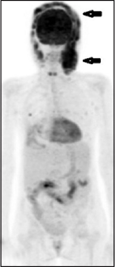
Maximum intensity projection image of whole body fluorodeoxyglucose positron emission tomography-computed tomography scan demonstrating intense uptake in scalp and neck region (black arrows)
Immunohistochemistry findings showed diffuse vimentin expression with a weak patchy expression of epithelial membrane antigen, CD99 had a moderate membrane expression. Thus, the diagnosis of epithelioid angiosarcoma was confirmed and the patient was started on chemotherapy.
The most frequent types of tumor postrenal transplantation are posttransplant lymphoproliferative disorders, squamous cell carcinoma and Kaposi's sarcoma.[1] Lymphoproliferative malignancies occur at an average of 32 months after renal transplantation and highest incidence during 1st year.[2] One study suggests that mean time of non-Kaposi's sarcoma malignancy postrenal transplantation is 34.4 ± 21.8 months, and for Kaposi's sarcoma is 18.7 ± 25.2 months.[3] Thus, usually malignancies present in first 5 years posttransplant, and late presentations are those of skin cancers, but in the Indian population, the incidence of skin malignancy is less due to high melanin content.
Posttransplant angiosarcomas are rare and to date have been reported in less than 20 solid organ transplant recipients. The majority of patients with reported posttransplant angiosarcoma have been renal transplant recipients with tumor frequently developing at the site of a previously placed arteriovenous fistula.[4] Angiosarcoma is a rare malignancy of endothelial origin, it has a subtype, in which cells have a predominantly epithelioid appearance known as epithelioid angiosarcoma.
Epithelioid angiosarcoma has a variety of clinical presentations, due to its different primary sites. Their presentation ranges from painful enlarging soft tissue masses to long bone fractures to arteriovenous shunting. Prolonged use of immunosuppression, leads to alteration of the immune system, and this is associated with increased risk of cancer. Since FDG PET-CT scan can detect glucose hypermetabolism and this tumor is FDG avid, this imaging modality helped to guide the clinician toward suspicious neoplastic lesion and appropriate site of biopsy thus yielding diagnostic histopathology and determining the extent of disease.
In our case, site of the previous biopsy probably was inappropriate, and thus, a biopsy had to be repeated after FDG PET-CT scan which not only suggested the appropriate site for biopsy but also revealed the extent of disease. Thus, this highlights the value of FDG PET-CT scan which if instituted earlier can avoid inconclusive biopsies.
Financial support and sponsorship
Nil.
Conflicts of interest
There are no conflicts of interest.
References
- 1.Penn I. Cancers in renal transplant recipients. Adv Ren Replace Ther. 2000;7:147–56. doi: 10.1053/rr.2000.5269. [DOI] [PubMed] [Google Scholar]
- 2.Caillard S, Agodoa LY, Bohen EM, Abbott KC. Myeloma, Hodgkin disease, and lymphoid leukemia after renal transplantation: Characteristics, risk factors and prognosis. Transplantation. 2006;81:888–95. doi: 10.1097/01.tp.0000203554.54242.56. [DOI] [PubMed] [Google Scholar]
- 3.Raeisi D, Payandeh M, Madani SH, Zare ME, Kansestani AN, Hashemian AH. Kaposi's sarcoma after kidney transplantation: A 21-years experience. Int J Hematol Oncol Stem Cell Res. 2013;7:29–33. [PMC free article] [PubMed] [Google Scholar]
- 4.Webster P, Wujanto L, Fisher C, Walker M, Ramakrishnan R, Naresh K, et al. Malignancies confined to disused arteriovenous fistulae in renal transplant patients: An important differential diagnosis. Am J Nephrol. 2011;34:42–8. doi: 10.1159/000328908. [DOI] [PubMed] [Google Scholar]