Abstract
Cardiovascular disease is one of the main burdens of healthcare systems worldwide. Nevertheless, assessing cardiovascular risk in both apparently healthy individuals and low/high-risk patients remains a difficult issue. Already established biomarkers (e.g. brain natriuretic peptide, troponin) have significantly improved the assessment of major cardiovascular events and diseases but cannot be applied to all patients and in some cases do not provide sufficiently accurate information. In this context, new potential biomarkers that reflect various underlying pathophysiological cardiac and vascular modifications are needed. Also, a multiple biomarker evaluation that shows changes in the cardiovascular state is of interest. This review describes the role of selected markers of vascular inflammation, atherosclerosis, atherothrombosis, endothelial dysfunction and cardiovascular fibrosis in the pathogenesis and prognosis of cardiovascular disease: the potential use of cardiotrophin-1, leptin, adiponectin, resistin and galectin-3 as biomarkers for various cardiovascular conditions is discussed.
Keywords: cardiovascular disease, cardiotrophin-1, adipokines, galectin-3
Introduction
According to the World Health Organization (WHO), cardiovascular diseases (CVD) are currently the leading cause of morbidity and mortality worldwide. The burden of cardiovascular death is also very high in Europe, with the highest rates being encountered in Central and Eastern Europe [1]. The main causes of cardiovascular (CV) death are ischemic heart disease and cerebrovascular disease. A significant proportion of these deaths may be prevented through better surveillance and prophylaxis; risk stratification is an important part of this strategy for timely interventions and long-term targeted management [2].
CV risk assessment models are based on traditional risk factors such as age, sex, smoking, blood pressure, diabetes, total cholesterol, HDL cholesterol, family history, etc. Risk models such as the US Framingham risk score or the European SCORE model evaluate CV risk in “apparently healthy” subjects (without already established CVD or diabetes) [3]. Although these are first-line tools for patients who need stepped-up prevention, limitations are inevitable: “intermediate” risk patients remain a heterogeneous category; frequently patients with an apparently low risk have subclinical atherosclerosis and vascular dysfunction that would benefit from a more intense intervention. In addition, CV risk may also be underestimated in patients with central obesity [3, 4].
The search for molecules that could help improve CVD assessment in primary and secondary care is continuous and prolific. Despite sustained efforts and a very large panel of candidates for biomarkers, very few molecules have proven their utility in clinical practice. Standardly used biomarkers for CVD include natriuretic peptides (brain natriuretic peptide (BNP), and N-terminal prohormone of brain natriuretic peptide (NT-pro-BNP)), troponins, C-reactive protein (CRP) and cardiac enzymes. The Food and Drug Administration (FDA) list of validated cardiovascular biomarker includes the above and also galectin-3 and ST-2 (Table I) [5].
Table I.
Cardiovascular biomarkers validated by FDA and their limitations
| Biomarker | FDA indication* | Limitations |
|---|---|---|
| BNP/NT-proBNP | Differentiating cardiac dyspnea from dyspnea of non-cardiac origin |
Cardiac-other than HF and ACS:
Non-cardiac: |
| NT-proBNP |
|
|
| BNP |
|
|
| Troponin T/troponin I |
|
Conditions associated with increased troponins other than ACS (selected): Cardiovascular:
Other:
|
| CRP |
|
Low specificity Conditions associated with increased hsCRP, other than CVD:
|
| Galectin-3 | Aid in assessing the prognosis of patients diagnosed with chronic HF |
Strongly correlated with kidney function Falsely elevated results:
|
| ST-2 | Aid in assessing the prognosis of patients diagnosed with chronic HF |
Other causes of elevated ST-2:
|
| CK | Aid in the diagnosis of AMI |
Lack of specificity Causes of elevated CK other than AMI:
|
| CK-MB | Aid in the diagnosis of AMI |
Specific for myocardial cell injury but not for AMI Causes of elevated CK-MB other than AMI:
|
| LDH | Aid in the diagnosis of AMI |
Limited specificity Causes of elevated LDH other than AMI:
|
| Myoglobin | Aid in the diagnosis of AMI |
Limited specificity in patients with:
|
ACS – acute coronary syndrome, AMI – acute myocardial infarction, CHD – coronary heart disease, CKD – chronic kidney disease, CVD – cardiovascular disease, HF – heart failure, hsCRP – high-sensitivity C-reactive protein. *Source for provided indications: www.fda.gov.
All these prediction, diagnosis and prognosis biomarkers proved to be the best so far but still have their pros and cons. BNP and NT-proBNP have similar clinical performance in diagnosing heart failure (HF) but NT-proBNP may be preferred to BNP due to its longer plasma half-life and its better outcome prognosis in the Valsartan Heart Failure Trial [6]. Natriuretic peptides (NP) best rule out cardiac dysfunction in patients with acute dyspnea presenting to the emergency room and are useful in establishing the prognosis and disease severity in acute and chronic HF. BNP has also been validated for risk stratification in acute coronary syndromes (ACS). However, NP are greatly influenced by non-cardiac factors, one of the most prominent being kidney function, and are not useful in discriminating the etiology of HF [7–9]. Cardiac troponins (cTn) are first line biomarkers for ACS, having clinical sensitivity and myocardial specificity. cTn are the gold-standard markers for myocardial injury; however, brief elevations can occur in non-ACS conditions. Also, cTn are not able to provide differential diagnosis between ischemic, inflammatory and traumatic myocardial injury [10]. Cardiac CRP (cCRP) is a high-sensitivity CRP assay recently validated by the FDA with evidence of efficacy and extended use for CV risk assessment/stratification. It should be interpreted with caution in the presence of systemic inflammatory conditions, acute infections, trauma and others (Table I) [11, 12]. Galectin-3 and ST-2 are two cardiac biomarkers recently cleared by the FDA and also recommended by the American College of Cardiology Foundation/American Heart Association (ACCF/AHA) guidelines for use as an aid in the management of patients with HF. Galectin-3 should be interpreted with caution in patients with altered kidney function and/or cancer [8, 13, 14]. ST-2 also rises in inflammatory and pulmonary diseases (Table I) [13]. Regarding cardiac enzymes (creatine kinase (CK), creatine kinase muscle/brain (CK-MB) enzyme, lactic dehydrogenase (LDH) and myoglobin), these are to be used as an aid in the diagnosis of acute myocardial infarction (AMI). Their limitations are presented in Table I [15, 16].
Recently, new molecules regulating atherosclerosis, inflammation, endothelial dysfunction (ED), fibrosis and thrombosis have been proposed as biomarkers for evaluating CV risk and disease, especially for risk stratification. Adding these biomarkers to established CV risk assessment models could provide a significant increment to the predictive value of these models. Novel CV biomarkers should be cost-efficient, valid, reproducible, reliable and accurate. They should be capable of independently predicting CVD and also of providing more information than traditional biomarkers [3, 4].
Several new molecules have been proposed as biomarkers for evaluating CVD. We conducted a literature search to select the best novel biomarkers that predict the development of new CVD, differentiate CVD from other diseases, evaluate the severity of CVD and might also be considered prognostic markers. We found 19 novel biomarkers that have been studied in CVD (Table II).
Table II.
Potential cardiovascular biomarkers regulating atherosclerosis, inflammation, endothelial dysfunction and fibrosis in cardiovascular disease
| Biomarkers called “cardiovascular” | Number of articles | Prediction of CVD | Differential diagnosis | Disease severity | Outcome and prognosis | Treatment effectiveness |
|---|---|---|---|---|---|---|
| MR-proANP | 14 | + | + | + | ||
| Cardiotrophin-1 | 111 | + | + | + | + | + |
| GDF-15 | 174 | + | + | + | + | |
| IMA | 219 | + | + | + | ||
| Lp-PLA2 | 241 | + | + | |||
| Pentraxin 3 | 326 | + | + | |||
| NT-proBNP | 418 | + | + | + | + | |
| Creatine kinase | 692 | + | + | + | + | + |
| ADMA | 797 | + | + | |||
| CT-I and CT-T | 868 | + | + | + | + | + |
| Resistin | 945 | + | + | + | + | |
| Galectin-3 | 967 | + | + | + | + | + |
| PON1 | 1200 | + | + | |||
| BNP | 1700 | + | + | |||
| Homocysteine | 3592 | + | + | |||
| IL-6 | 4178 | + | ||||
| Adiponectin | 5308 | + | + | + | ||
| CRP | 6076 | + | + | + | + | |
| Leptin | 6757 | + | + | + |
ADMA – asymmetric dimethylarginine, BNP – B-type natriuretic peptide, CRP – C-reactive protein, CT-I and CT-T – cardiac-specific troponins I and T, GDF-15 – growth differentiation factor 15, IL-6 – interleukin-6, IMA – ischemia modified albumin, Lp-PLA2 – lipoprotein-associated phospholipase A2, MR-proANP – mid-regional pro-atrial natriuretic peptide, NT-proBNP – N-terminal pro-B-type natriuretic peptide, PON1 – paraoxonase 1.
We chose here to review cardiotrophin-1 (CT-1) as it is a promising biomarker for hypertensive heart disease, where it brings significant additional information to that provided by NP. Also, the adipokines leptin, adiponectin and resistin have been proposed as biomarkers for various cardiovascular conditions and are discussed below, where clarifications regarding their prognostic value are made. Finally, we chose here to discuss the strong and weak points of galectin-3 and its utility in comparison to other biomarkers of HF in order to clarify the indications for galectin-3 determination. Table III illustrates which of these five biomarkers could be used as potential predictors for vascular inflammation, atherosclerosis, ED and cardiovascular fibrosis.
Table III.
Novel biomarkers – players in cardiovascular dysfunction
| Biomarker | Endothelial dysfunction | Atherosclerosis | Inflammation | Fibrosis | Atherothrombosis |
|---|---|---|---|---|---|
| Cardiotrophin-1 | + | + | + | ||
| Leptin | + | + | + | + | |
| Adiponectin | + | + | + | + | |
| Resistin | + | + | + | + | + |
| Galectin-3 | + | + | + |
As the literature is replete with clinical information on biomarkers which are often inconsistent, this review is intended to clarify which of these biomarkers deserve further analysis and what are their potential indications of use.
Cardiotrophin-1
CT-1 is a 21.5-kDa protein, a member of the interleukin 6 (IL-6) family of cytokines [17], first described by Pennica et al. [17] to induce cardiomyocyte hypertrophy by atrial natriuretic peptide secretion and organization of myosin light chains into sarcomeres.
Pathophysiology and experimental studies
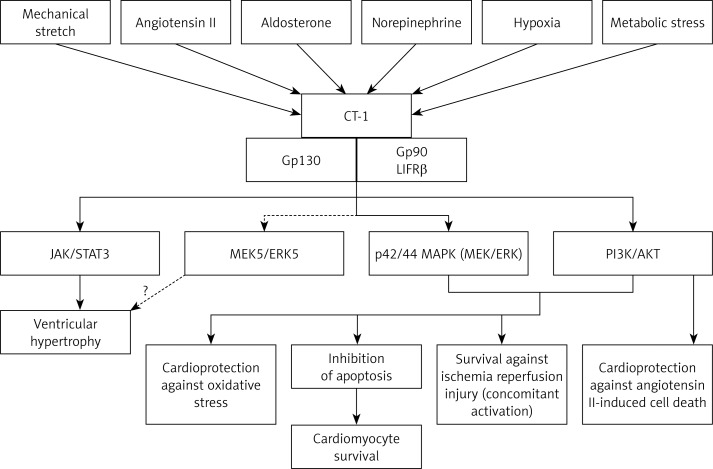
CT-1 is a survival promoter cytokine that is upregulated in cardiomyocytes and cardiac fibroblasts in response to mechanical (ventricular stretch), humoral (angiotensin II, aldosterone, catecholamines), metabolic (glucose, insulin) and hypoxic stress. In acute stress, CT-1 promotes cell survival. However, if stressful signals persist, chronic upregulation of CT-1 leads to cardiomyocyte hypertrophy and, finally, left ventricular dysfunction [4]. CT-1 exerts its biological activity through the leukemia inhibitory factor receptor β (LIFRβ)/glycoprotein 130 (gp130) heterodimer receptor that further activates multiple signaling pathways with different consequences (Figure 1): Janus kinase/signal transducer and activator of transcription 3 (JAK/STAT3) promotes ventricular hypertrophy while p42/44 mitogen-activated protein kinase (p42/44 MAPK) and the phosphoinositide 3-kinase/Akt (PI3K/Akt) pathways inhibit apoptosis and promote cardiomyocyte survival.
Figure 1.
Schematic representation of CT-1 signaling pathways and their different effects on the cardiac cell (see text for abbreviations)
It may be possible that myocyte hypertrophy is also mediated by the activation of mitogen-activated protein kinase kinase 5/extracellular-signal-regulated kinase 5 (MEK5/ERK5 pathway) [4, 18–20]. CT-1 is also expressed in vascular endothelial cells and has direct vascular effects, resulting in atherogenesis, vascular dysfunction, arterial stiffness, and increased blood pressure [21–23]. Overall, CT-1 is a potent profibrotic agent for the heart and vessels [4, 23].
Clinical studies
CT-1 is a key link between hypertension, left ventricular hypertrophy (LVH) and HF. In a meta-analysis performed by Song et al. [24] Cardiotrophin-1 levels were highest in hypertensive patients with LVH and HF compared to controls, followed by hypertensive patients with LVH without HF and hypertensive patients without LVH or HF. Moreover, CT-1 directly correlates with left ventricular mass index (LVMI) in hypertensive patients independently of systolic blood pressure: patients with CT-1 above the cut-off value of 39 fmol/ml are six times more likely to have LVH. Thus CT-1 could be applied in the screening and diagnosis of hypertensive heart disease, helping to select patients that would best benefit from echocardiography [25].
Regarding CT-1 and HF, Talwar et al. [26] were the first to report that plasma CT-1 levels correlate with the degree of systolic dysfunction in HF patients. Plasma CT-1 levels are also increased in patients with diastolic HF and positively correlate with left ventricular filling pressures [27]. Furthermore, CT-1 levels significantly predict overt HF in hypertensive patients: CT-1 concentration positively correlates with NT-proBNP and negatively correlates with left ventricular ejection fraction [28, 29]. The cut-off value for CT-1 plasma levels of 152 pg/ml has 77% sensitivity and 85% specificity for detecting the occurrence of overt HF in hypertensive patients [28]. Also, CT-1 is a predictor of mortality in chronic HF irrespective of etiology and independently of BNP [30]. Thresholds and potential indications for CT-1 use are presented in Table IV.
Table IV.
Novel potential biomarkers – indications and thresholds
| Biomarker | CV risk assessment/Diagnosis/Prognosis | Thresholds | Source | ||
|---|---|---|---|---|---|
| CT-1 | Diagnosis of HHD/LVH in hypertension | 39 fmol/ml 70% sensitivity 75% specificity |
LÓpez et al. [25] | ||
| Prognosis of overt HF in hypertension | 152.2 pg/ml 77% sensitivity 85% specificity |
Bielecka-Dabrowa et al. [28] | |||
| Prognosis of mortality in chronic HF (irrespective of etiology) | 658 fmol/ml 57% sensitivity 74% specificity |
Tsutamoto et al. [30] | |||
| Leptin | CHD risk assessment | N/A | Shanker et al. [51] Zeng et al. [41] |
||
| Adiponectin | CHD risk assessment | N/A | Shanker et al. [51] | ||
| Prognosis of CHD | N/A | Cavusoglu et al. [65] | |||
| Prognosis of mortality in chronic HF | 11.6 mg/l HR = 3.23 (1.11–9.69), p = 0.032 |
Kistorp et al. [64] | |||
| Resistin | Prediction of HF risk in non-CHD community dwelling-individuals | 1.2–10.9 ng/ml HR = 1 |
11–14.9 ng/ml HR = 2.87 (0.57–14.52) |
15–110 ng/ml HR = 5.2 (1.08–25.13) |
Frankel et al. [86] |
| Prognosis of multivessel CHD (major cardiac and cerebrovascular events) | 17.3 ng/ml OR = 13.5 (95% CI: 2.3–80.3) Area under ROC curve = 0.75 |
Kręcki et al. [69] | |||
| Diagnosis of AMI | Median (25th–75th percentile) levels at peak (> 12 h after pain onset): 5.74 ng/ml (438–7.51) Cut-off not available |
Lubos et al. [90] | |||
| Galectin-3 | Prognosis of patients with chronic heart failure (already FDA validated) | Low-risk < 17.8 ng/ml Intermediate risk: 17.8–25.9 ng/ml High-risk: > 25.9 ng/ml |
FDA 510 (k) Summary 2010 | ||
| Risk stratification/prognosis in acute HF (recommended by ACCF/AHA guidelines) | 17.8 ng/ml | Peacock et al. [110] | |||
| Prediction of incident HF | N/A | Jagodzinski et al. [104] | |||
ACCF/AHA – American College of Cardiology Foundation/American Heart Association, AMI – acute myocardial infarction, CHD – coronary heart disease, FDA – Food and Drug Administration, HF – heart failure, HHD – hypertensive heart disease, HR – hazard ratio, OR – odds ratio, ROC – receiver operating characteristic, N/A – not available, LVH – left ventricular hypertrophy.
CT-1 could also be used as a monitoring tool for therapeutic management in hypertensive heart disease patients: LVH and CT-1 levels had a tendency to decrease after losartan treatment rather than after treatment with atenolol [31].
Finally, although a promoter of ED, studies failed to show an association between CT-1 and coronary heart disease (CHD) [32].
Cardiotrophin-1 vs. standardly used biomarkers
With regards to predicting LVH onset in hypertensive patients, CT-1 performs much better than NT-proBNP, with 70% sensitivity and 75% specificity for CT-1 cut-off value vs. 40% sensitivity and 61% specificity for NT-proBNP cut-off value [25].
Even though NT-proBNP is the gold-standard biomarker for HF diagnosis, studies have shown that the association between CT-1 and NT-proBNP is superior to NT-proBNP alone in assessing the presence of HF in hypertensive patients [28, 29]. CT-1 is less specific but more sensitive than NT-proBNP for detecting stage C HF in hypertensive patients, thus improving NT-proBNP sensitivity for detecting HF [29]. The combination CT-1 and BNP is better than either marker alone in predicting mortality in chronic HF [30].
Also, CT-1 may compensate for some of the downsides of NT-proBNP: CT-1 is not influenced by kidney function, while NT-proBNP negatively correlates with estimated glomerular filtration rate (eGFR) [29]. Also, CT-1 correlates with echocardiographic parameters of LVH (LVMI), while NT-proBNP does not correlate with LVH in hypertensive patients with normal cardiac function. Thus, NT-proBNP is not useful in assessing hypertensive heart disease in patients with normal systolic function [25].
In summary, CT-1 is a promising biomarker for predicting maladaptive hypertensive heart disease in hypertensive patients. CT-1 could become a predictor of arterial hypertension prognosis in terms of occurrence of LVH in hypertensive patients with normal systolic function. Also, CT-1 could bring incremental power when used together with NT-proBNP for assessing HF in hypertension and for the prognosis of chronic HF. Eventually, circulating CT-1 determination could also be useful for establishing optimal individualized therapy and for monitoring disease evolution in these patients.
Adipo(cyto)kines
Adipocytokines (adipokines) are adipose-derived proteins (hormones and cytokines) produced by the adipose tissue that perform major functions concerning energy expenditure and metabolism, bone mass regulation, insulin sensitivity/resistance, inflammation and cardiovascular function [33, 34]. Recent research has focused on the role of adipocytokines in CV homeostasis.
Leptin
Leptin is a pleiotropic adipokine, a 167 amino-acid peptide secreted by adipocytes, and a member of the IL-6 family. It is the main anorexigenic hormone, regulating lipid and glucose metabolism, bone metabolism, immune function and the cardiovascular system [35, 36]. Leptin has recently emerged as a novel and potential risk factor for CVD, being one of the key elements that connects it with obesity.
Pathophysiology and experimental studies
Obese-associated hyperleptinemia has deleterious vascular effects [35]. By acting on the sympathetic nervous system (SNS), chronic leptin signaling impairs endothelium-dependent vasorelaxation and favors an increased blood pressure response to angiotensin II in murine models [37]. Hyperleptinemia also has proinflammatory [38], proatherogenic [36] and prothrombotic effects [39], thus promoting ED. The pathophysiology of deleterious actions of leptin is presented in Figure 2.
Figure 2.
Schematic representation of actions of leptin
ED – endothelial dysfunction, IL-6 – interleukin 6, IL-4 – interleukin 4, TNF-α – tumor necrosis factor α.
Clinical studies
In a case-control study performed by Karthick et al. [40], patients with non-thrombolysed AMI had significantly higher levels of leptin [40]. Moreover, in a meta-analysis performed by Zeng et al. [41], high leptin levels were significantly associated with an increase in CHD (including myocardial infarction) (OR = 1.90) and stroke risk (OR = 2.14) [41]. In support of these findings, hyperleptinemia positively and independently correlated with increased coagulation activity (high circulating levels of coagulation factor VIII, fibrin D-dimer, fibrinogen and von Willebrand factor) in a study performed by Wannamethee et al. [42]. However, the numerous discrepant results in the literature are not to be disregarded, and the following question arises: is the effect of hyperleptinemia on CVD exaggerated? The results were adjusted for age, blood pressure and body mass index (BMI) in all the studies included in the above meta-analysis, but there are other possible confounders that should be taken into consideration. One of them is inflammation, and CRP is an important link between obesity and CVD [43].
Hyperleptinemia is also an independent predictor of HF in older men without pre-existing CHD, after adjustment for BMI [44]. However, in overweight or obese men with pre-existing CHD, leptin seems to have no additional significant effect, as probably obesity-triggered increased cardiac workload is enough to increase HF risk [45]. Despite high levels in HF, leptin is not a reliable marker as it is highly dependent on body fat: normal/low leptin levels may be encountered in HF patients with cachexia [45].
Controversies
There are also studies that have reported paradoxical results regarding the relationship between leptin and CVD risk. In the NAMIS study, low leptin levels 7 days after the onset of an AMI were associated with poor outcome defined as higher risk for adverse cardiovascular events and poor survival [46]. Also, Scholze et al. [47] provided proof that low leptin levels are in fact an independent predictor of all-cause death in hemodialysis patients, with the most frequent cause being CVD [47].
Such contradictory findings are reminiscent of the concept of the “obesity paradox” or “reverse epidemiology”: although a high body mass is a conventional risk factor for CVD in apparently healthy individuals, there seems to be a reversal of the relationship between overnutrition and long-term outcome in high-risk patients (chronic kidney disease and/or with already established CHD or HF), where, paradoxically, overnutrition has a protective effect against mortality [48, 49]. The concept may apply to hyperleptinemia as well, as leptin is a reflection of body fat [33].
Leptin vs. standardly used biomarkers/other novel potential biomarkers
In a large prospective population-based study of 6502 participants with a mean follow-up of 11.4 years, leptin did not correlate with incident ischemic heart disease or stroke, after adjustment for sex, age, blood pressure, lipid profile, smoking, comorbidities such as hypertension and diabetes, BMI, insulin resistance parameters, eGFR and CRP. However, CRP remained an independent predictor of ischemic heart disease (fatal and non-fatal) after adjustment for all the above variables and also for leptin and adiponectin [43]. It seems that the association between leptin and CVD is mediated by inflammation/CRP; indeed, leptin and CRP levels seem to correlate and leptin is able to directly regulate CRP synthesis [50]. However, despite the power of leptin alone being limited, it may be useful in conjunction with other biomarkers: the association between leptin, adiponectin and traditional CV risk performed significantly better in predicting CHD when compared to a model that included only the conventional risk factors, with maximum reclassification in the intermediate risk group [51].
Overall, these data do not yet support the use of leptin as a biomarker in clinical practice for assessing the risk of CHD. Leptin levels should be interpreted with caution, according to the underlying condition.
Adiponectin
Adiponectin is a 244 amino acid adipokine, part of the collagen superfamily. The most abundant form in the plasma is the high molecular weight (HMW) adiponectin, the most stable and with the longest half-life [33, 52].
Pathophysiology and experimental studies
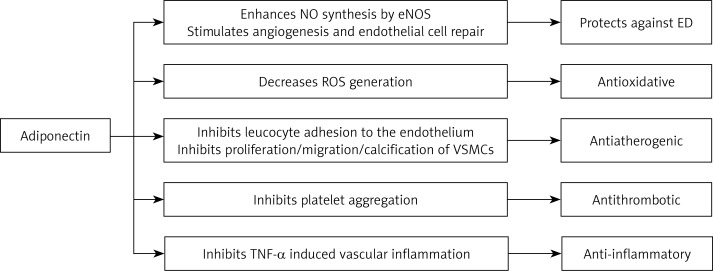
Adiponectin improves insulin sensitivity [53] and fatty acid oxidation [52] and inhibits vascular inflammation [54]. Adiponectin also protects against ED by increasing endothelial nitric oxide synthase (eNOS) activity and by promoting angiogenesis and endothelial cell repair [52, 55, 56] (Figure 3).
Figure 3.
Schematic representation of adiponectin actions
ED – endothelial dysfunction, TNF-α – tumor necrosis factor α, VSMCs – vascular smooth muscle cells, eNOS – endothelial nitric oxide synthase, NO – nitric oxide, ROS – reactive oxygen species.
In addition, it inhibits atherogenesis and also has antithrombotic effects [52, 57].
Clinical studies
In a longitudinal population-based cohort study, baseline adiponectin levels were negatively and independently associated with the development of CHD in a 10-year follow-up [58]. Moreover, Wang et al. [59] showed that adiponectin concentration inversely correlates with the severity of coronary artery disease assessed through the Gensini score. In their study, the HMW isoform of adiponectin showed a stronger correlation with the severity of coronary artery disease than total adiponectin did, pointing to the utility of HMW adiponectin as a potential biomarker for detecting the risk of CHD [59]. Also in type 2 diabetes mellitus (DM) patients, the C1q-adiponectin/total adiponectin ratio is a significant independent predictor of the atherosclerosis score with 72.2% sensitivity and a positive predictive value of 78.8%. The above relationship could be explained by the anti-atherogenic actions of adiponectin: binding of adiponectin to complement fraction C1q protects against the C1q-induced inflammatory process in systemic atherosclerosis [60].
Adiponectin may protect against hypertension onset: in a systematic review and meta-analysis performed by Kim et al. [61], adiponectin levels were lower in patients with hypertension than in controls. Also, the risk of developing hypertension decreased with the increase of adiponectin levels.
Controversies
High adiponectin levels are encountered in HF, where adiponectin increases with left ventricular dysfunction progression. Adiponectin is directly correlated with BNP and NT-proBNP and also with TNF-α: adiponectin may rise in congestive HF to mitigate the actions of TNF-α. Also, HF patients with higher adiponectin levels have a significantly higher mortality risk [62–64]. Thus, adiponectin could be considered in the future as a marker of HF severity and also a predictor of mortality.
Also, adiponectin in the upper quartile in CHD patients is associated with a higher risk of recurrent adverse events [51]. High adiponectin levels are an independent predictor of all-cause and cardiac mortality in patients undergoing coronary angiography for stable angina or ACS [65], in patients with carotid atherosclerosis undergoing carotid endarterectomy [66] and in elderly patients (≥ 65 years) [67]. Also, despite an increase 3 months after coronary artery bypass grafting (CABG) in severe CHD patients, adiponectin directly correlated with markers of ED [68]. It may be speculated that adiponectin increases as a compensatory mechanism to counteract ED that occurs after CABG. Other authors observed a decrease in adiponectin concentration 12 months after CABG together with a significant improvement of ischemic and HF symptoms [69]. BNP is a strong predictor of adiponectin levels and is associated with adiponectin release from the adipose tissue [70]. Also, high adiponectin levels may be a reflection of adiponectin resistance at the receptor level [69]. This may partially explain why adiponectin predicts negative outcomes in subjects with advanced CVD and also why adiponectin declines as improvement of cardiovascular function occurs after CABG.
Therefore, the interpretation of adiponectin concentration should be made according to underlying CVD severity, as it has different predictive implications (the “adiponectin paradox”) [67]: in disease-free subjects, high adiponectin is a marker of low risk for acute or chronic CHD, while in patients with established CHD/HF, high adiponectin levels seem to be independent predictors of future adverse events [71].
Adiponectin vs. standardly used biomarkers
In order to evaluate the prognostic power of adiponectin in patients with established CHD, Ang et al. [72] compared adiponectin to BNP with regards to predicting a future adverse outcome (all-cause death, readmission for ACS or congestive HF) in patients with ACS: an increase in adiponectin following admission for ACS better predicted adverse outcome than single determinations at baseline and 7 weeks, but this relationship was lost after adjustment for BNP. Therefore, BNP performs better than adiponectin for prognosis of adverse events in ACS [72].
As mentioned above, adiponectin is independently associated with NP and parallels the evolution of HF [62]. Adiponectin is a predictor of mortality in HF, independently of NP (Table IV) [64]. Subsequently, the hypothesis that adiponectin could be used in conjunction with NP to improve mortality risk assessment in HF is to be verified.
Resistin
Resistin is a proinflammatory cytokine, a member of the adipokines, predominantly produced by macrophages but also released from the adipose tissue [73]. Resistin is involved in the pathogenesis of insulin resistance (“resists” insulin actions) and is also thought to be a contributory factor in CVD development [73].
Pathophysiology and experimental studies
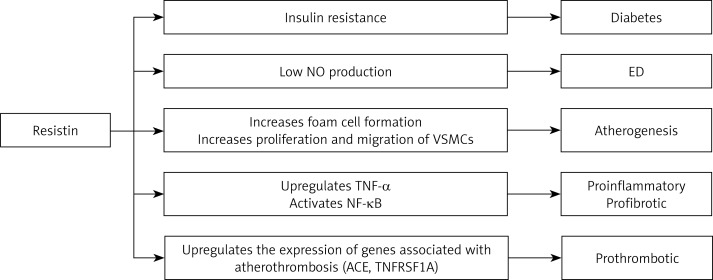
Firstly, high levels of resistin are associated with insulin resistance and diabetes: resistin upregulates the suppressor of cytokine signaling-3 (SOCS-3) pathway that leads to downregulation of insulin receptor expression and also inhibits phosphatidyl inositol 3-kinase activation that mediates insulin metabolic functions [53]. Secondly, resistin promotes ED, atherogenesis and atherothrombosis, having direct effects on the vessels: (1) resistin down-regulates eNOS gene expression, leading to low nitric oxide levels with reduced vasorelaxation function [73], (2) resistin increases foam cell formation [74] and also proliferation and migration of VSMCs [75, 76], (3) resistin upregulates the expression of genes associated with atherothrombosis [77]. Vascular thrombosis manifested as increased platelet reactivity and adherence to the endothelium and ED are the main triggers of CVD [78]. Finally, resistin also upregulates TNF-α expression and activates the NF-κB proinflammatory signaling pathway in the myocardium, leading to cardiac inflammation and fibrosis [79]. These findings led to the investigation of resistin as a possible marker and therapeutic target in CVD (Figure 4).
Figure 4.
Schematic representations of resistin actions
ED – endothelial dysfunction, NO – nitric oxide, NF-κB – nuclear factor κB, ACE – angiotensin converting enzyme, TNFRSF1At – tumor necrosis factor receptor superfamily member 1A, TNF-α – tumor necrosis factor α, VSMCs – vascular smooth muscle cells.
Clinical studies
In prospective studies, high resistin significantly increases the risk for all CVD, chronic CHD [80] and acute coronary syndrome, independently of other CV risk factors, including markers of inflammation (IL-6 and CRP) [81]. A resistin concentration equal to or above 17.3 ng/ml is associated with a 13-fold increase in the risk of major cardiac and cerebrovascular events in patients with multivessel coronary disease (Table IV) [82]. Resistin levels also rise 24 h after CABG as a marker of ischemia-reperfusion injury and are correlated with oxidative stress [83]. However, resistin levels do not seem to change after 12 months of medical treatment or CABG in these patients and thus may not be helpful for follow-up after therapeutic intervention [69]. Also, adding resistin to traditional CV risk factors significantly improves the risk assessment for ischemic stroke [84].
Nevertheless, resistin is a strong independent predictor of incident HF in community-dwelling older individuals and is also correlated with NYHA functional class in prospective studies. Whether resistin has a negative direct impact on the myocardium or it is merely a reflection of the vascular inflammatory process, it can be proposed as a biomarker for HF risk assessment and stratification [45, 85, 86].
Moreover, a meta-analysis performed by Fontana et al. [87] regarding the influence of resistin on mortality in high-risk patients (patients with CHD, myocardial infarction, ischemic stroke, type 2 DM, end-stage renal disease) showed that resistin significantly increased the risk for all-cause mortality, with a hazard ratio (HR) of 1.24 for 1 standard deviation increment in resistin levels. In the same meta-analysis, the effect of resistin on cardiovascular mortality was however neutral (HR = 1.05 for 1 standard deviation increase in resistin levels) [87].
Controversies
In the prospective study of Zhang et al. [88], resistin did not provide prognostic information beyond traditional risk factors regarding the development of HF in CHD patients. Therefore, resistin may be proposed as a biomarker for HF in non-CHD individuals.
Resistin vs. standardly used biomarkers
Although resistin significantly correlates with CRP and BNP levels, resistin is a predictor of HF in the community, independently of these biomarkers, and the predictive power is maintained after the exclusion of CHD individuals. The HRs for incident HF in the second and third tertile of resistin after adjusting for various cardiovascular risk factors, CRP and BNP are presented in Table IV [86].
Resistin levels correlate with troponin 24 h after CABG, reflecting myocardial injury [83]. Resistin levels are also significantly increased in AMI and unstable and stable angina patients, with resistin being significantly higher in AMI than in unstable/stable angina. Resistin correlates with myocardial injury enzymes, troponin I and CRP in AMI, but not in unstable/stable angina patients [89]. Resistin rises 3–6 h after chest pain onset and peaks 12 h after chest pain onset (Table IV) [90]. Thus, resistin could become a biomarker for AMI.
Overall, increasing clinical evidence supports the use of resistin as a cardiovascular marker for risk stratification and prognostic in CHD and as a predictor of HF in community-dwelling individuals. Clinical studies that evaluate the prognostic power of resistin in comparison to that of classical biomarkers are needed.
Galectin-3
Galectin-3 is a member of the lectin family, encoded by the LGALS3 gene located on chromosome 14. It is expressed predominantly by macrophages and plays an important role in the pathophysiology of immunity/inflammation related diseases (cancer, metabolic diseases). Galectin-3 is a marker of tissue fibrosis, including cardiovascular fibrosis, and also of atherosclerosis. Consequently, galectin-3 has emerged as a new biomarker for CV disease, especially HF but not only [91, 92].
Pathophysiology and experimental studies
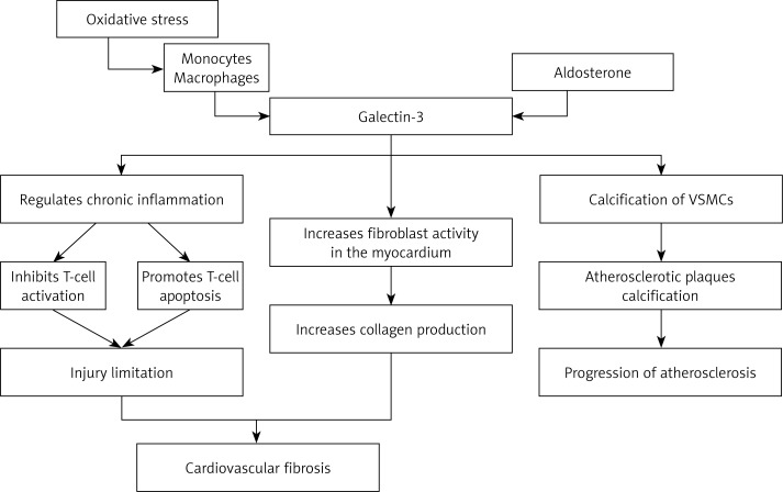
Galectin-3 is one of the key links between inflammation and fibrosis at the cardiovascular level [91, 92]. It regulates chronic inflammation within the vessels by promoting osteogenic differentiation of VSMCs, leading to macrocalcifications in the atherosclerotic plaques [92, 93]. Galectin-3 is a profibrotic agent by itself and also mediates aldosterone-induced cardiac, vascular and renal fibrosis [91, 94]. High galectin-3 levels in the myocardium stimulate excessive collagen production [95] and are correlated with abundant macrophages, increased fibroblast activity and increased accumulation of extracellular matrix [96]. Galectin-3 levels also show a later peak in non-infarcted, remodeling areas in rat models of myocardial infarction [97]. Thus, galectin-3 is a marker of cardiac fibrosis and remodeling (Figure 5).
Figure 5.
Schematic representation of galectin-3 actions
VSMCs – vascular smooth muscle cells.
Clinical studies
Chronic heart failure
In the above-mentioned context, it is not surprising that galectin-3 is a biomarker for chronic HF prognosis [91]. Although galectin-3 lost its predictive power for future adverse outcome in chronic HF after adjusting for NT-proBNP in some studies [98, 99], it remains a significant independent predictor of all-cause mortality [14], adverse CV events (including cardiac death) and left ventricular remodeling in patients with left ventricular systolic dysfunction in most trials [14, 100, 101]. It is also a significant independent predictor of all-cause death and hospitalization in patients with HF with preserved ejection fraction [102].
Acute heart failure
Galectin-3 is also a promising biomarker in acute heart failure (AHF). In a pooled analysis from 3 cohorts of patients admitted to hospital for AHF (AHF patients from COACH, PRIDE and UMD H-23258 trials), galectin-3 was a significant predictor of rehospitalization for HF at 30, 60, 90 and 120 days, independently of New York Heart Association (NYHA) functional class, renal function, left ventricular ejection fraction and BNP [103]. It was also a significant independent predictor of the composite outcome all-cause mortality and HF rehospitalization in PRIDE and UMD H-23258 cohorts [103].
Non-heart failure subjects
Galectin-3 may also be used as a biomarker for CV adverse outcome in non-HF individuals. Interestingly, galectin-3 significantly and independently predicted incident HF and all-cause mortality in the largest population-based cohort in which the relationship between galectin-3, HF and mortality risk was assessed (FINRISK 1997 cohort) [104].
Controversies
Renal function seems to be an important confounder when assessing the prognostic value of galectin-3: the association between high levels of galectin-3 and prognosis of HF was lost after adjusting for eGFR in some studies [105, 106]. Galectin-3 levels strongly correlate with eGFR (inverse relationship) in HF patients [14]. Therefore, the interpretation of galectin-3 should take into consideration renal function.
Guidelines
The use of the galectin-3 assay is currently approved by the FDA for the prognosis of chronic HF together with clinical assessment [91]. The 2013 American College of Cardiology Foundation/American Heart Association (ACCF/AHA) Guideline for management of HF also states that galectin-3 may be considered an additional biomarker for both chronic (class IIb recommendation, level of evidence B) and acute HF (class IIb recommendation, level of evidence A) improved risk stratification [8]. Whether galectin-3 may be useful in predicting CV risk in non-HF patients still needs to be established.
Galectin-3 vs. standardly used biomarkers
Galectin-3 is a prognostic marker for all-cause mortality and rehospitalization in chronic HF, independently of NP [8, 14, 100, 101]. Also, galectin-3 adds prognostic information to that provided by NT-proBNP in HF patients with NT-proBNP above the median (Table IV) [107].
In a recent head-to-head comparison of ST-2 and galectin-3 for risk stratification in chronic HF performed by Bayes-Genis et al. [108], ST-2 proved superior: ST-2 brought a significant increase in the predictive power of a risk model for HF composed of 11 classical risk factors and NT-proBNP, while the contribution of galectin-3 was negligible – hence the observation that galectin-3 may be more useful in early stages of HF and the need for trials that investigate the predictive utility of galectin-3 according to HF stages [108].
Despite NT-proBNP being more sensitive than galectin-3 in diagnosing AHF, galectin-3 was a better predictor for 60-day mortality and for the composite end-point death and recurrent HF within 60 days in AHF patients. The combination of galectin-3 and NT-proBNP best predicts death/recurrent HF in AHF [109].
Peacock [110] developed an algorithm for the emergency department recommending managing HF presentations (emergency department treatment and discharge, observation unit admission with 24-hour re-evaluation or immediate hospitalization) according to BNP and galectin-3 levels. The algorithm has not yet been validated. Galectin-3 in combination with NT-proBNP may be useful in short-term risk reclassification of patients with AHF and could help decision-making in order to better identify candidates for admission to intensive care units and candidates for discharge.
The multimarker approach
Cardiovascular disease have multiple pathophysiological backgrounds: HF is a result of inflammation, myocardial remodeling, cardiomyocyte injury and neurohormonal activation, while CHD is the result of vascular inflammation, atherosclerosis, thrombosis and ED. Therefore, a multibiomarker assessment could provide a global assessment of the disease, thus increasing the predictive and prognosis accuracy. Also, despite its limited utility when used alone, a biomarker may still be helpful when integrated into a biomarker panel [5]. In this regard, Ky et al. [111] investigated the utility of a panel consisting of 8 biomarkers reflecting different biological processes as a prognostic tool in chronic HF. Established biomarkers such as troponin I, BNP, ST-2, CRP, creatinine, and uric acid were included together with markers of oxidative stress and vascular growth and remodeling. The multimarker score significantly reclassified 24% of patients as higher risk when added to clinical risk scores.
The difficulty consists in identifying those multibiomarker panels that perform best for the minimum cost and are easy to assay. Also, the optimal biomarker panel should best guide therapeutic management [112]. As numerous potential biomarkers have been discovered, now the great challenge is to identify the optimal multibiomarker test.
Most of the above-mentioned molecules proved to be significant predictors of CVD and associated mortality, alone and in conjunction with traditional biomarkers. The combinations of biomarkers that are promising for further analysis in clinical trials are presented in Table V.
Table V.
Combinations of biomarkers that proved superior to single biomarker assessment and proposed multimarker associations to assess in future trials
| Proposed association of biomarkers | Type of information and indication | Advantages | Pitfalls/confounders that may influence the power of prediction/risk stratification | Source |
|---|---|---|---|---|
| CT-1 + NT-proBNP | Diagnosis of HF in hypertension | Increases sensitivity for diagnosing HF (78%) compared to NT-proBNP alone (72%) and slightly improves AUC (from 0.818 for NT-proBNP alone to 0.854) | Metabolic syndrome (also increased levels of CT-1) Chronic kidney disease (increases NT-proBNP) | Lopez et al. [29] |
| CT-1 + BNP | Prognosis of mortality in chronic HF | HR = 2.48 for BNP > 170 pg/ml + CT-1 > 658 fmol/ml vs. BNP > 170 pg/ml + CT-1 < 658 fmol/ml (p = 0.01) | Metabolic syndrome (also increased levels of CT-1) Chronic kidney disease (increases NT-proBNP) | Tsutamoto et al. [30] |
| Galectin-3 + NT-proBNP | Prognosis of mortality in chronic HF | HR = 1.84 for NT-proBNP > 932 pg/ml + galectin-3 > 16.2 ng/ml | Chronic kidney disease (both correlate with kidney function) | Anand et al. [107] (Val-HeFT trial) |
| Galectin-3 + NT-proBNP | Prognosis of death/recurrent HF in AHF | Galectin-3 > 9.42 ng/ml + NT-proBNP > 5 562 pg/ml was associated with significantly higher risk of mortality/recurrent heart failure than either marker alone | Chronic kidney disease (both correlate with kidney function) | Van Kimmenade et al. [109] |
| Potential multimarker assessments to consider in future clinical trials | ||||
| Chronic HF prediction and prognosis Resistin + BNP/NT-proBNP + CT-1 | ||||
| Chronic HF prognosis Adiponectin + BNP/NT-proBNP + CT-1 | ||||
| CHD risk stratification Resistin + BNP/NT-proBNP | ||||
| Diagnosis of ACS Resistin + myocardial enzymes + troponin | ||||
| CHD risk assessment Leptin + adiponectin + CRP | ||||
ACS – acute coronary syndrome, AHF – acute heart failure, AUC – area under the curve, CHD – coronary heart disease, HF – heart failure, HR – hazard ratio.
Conclusions
Combining various new risk markers could help improve risk-based intervention and achieve the optimal therapeutic approach, as traditional CV risk factors are in many cases not able to provide a clear assessment of risk strata. Nevertheless, CT-1 is a promising biomarker for assessing risk in hypertensive patients. It would allow the identification of patients at high risk for developing HF, and it could also be a useful tool for assessing treatment outcome. High leptin, low adiponectin and high resistin levels are biomarkers of vascular inflammation, dysfunction, atherosclerosis, and increased stiffness and could be used to predict CV disease and outcome, according to the underlying CV state. Galectin-3 is already recommended for establishing prognosis in both acute and chronic HF patients. Although clinical trials have also shown promising results for galectin-3 as an independent prognostic factor for assessing CV risk in non-HF patients, its best predictive value seems to be in high-risk patients.
Most studies are in favor of using the above-mentioned molecules as new biomarkers for CVD and mortality. The practical use of a multi-biomarker evaluation in conjunction with risk score models already available could bring valuable information regarding disease or risk prognosis, especially as these new biomarkers promise to bring a significant increase in the predictive power of already established biomarkers (e.g. BNP) and/or clinical factor algorithms.
Acknowledgments
This work received financial support through the “CERO - CAREER PROFILE: Romanian researchers”, contract no. POSDRU/159/1.5/S/135760, a project co-financed by the European Social Fund Operational Programme Human Resources Development 2007-2013.
Simona Hogas, Stefana C. Bilha – These authors contributed equally to this work.
Conflict of interest
The authors declare no conflict of interest.
References
- 1.Dorobantu M, Tautu OF, Darabont F, et al. Objectives and methodology of Romanian SEPHAR II Survey. Project for comparing the prevalence and control of cardiovascular risk factors in two East-European countries: Romania and Poland. Arch Med Sci. 2015;11:715–23. doi: 10.5114/aoms.2015.53290. [DOI] [PMC free article] [PubMed] [Google Scholar]
- 2.Mendis S, Puska P, Norrving B. Global atlas on cardiovascular disease prevention and control. Geneva: World Health Organization; 2011. Available at http://www.who.int/cardiovascular_diseases/publications/atlas_cvd/en/ [Google Scholar]
- 3.De Backer GG. New risk markers for cardiovascular prevention. Curr Atheroscler Rep. 2014;16:427. doi: 10.1007/s11883-014-0427-z. [DOI] [PubMed] [Google Scholar]
- 4.Gonzalez A, Lopez B, Ravassa S, et al. Cardiotrophin-1 in hypertensive heart disease. Endocrine. 2012;42:9–17. doi: 10.1007/s12020-012-9649-4. [DOI] [PubMed] [Google Scholar]
- 5.Sun RR, Lu L, Liu M, et al. Biomarkers and heart disease. Eur Rev Med Pharmacol Sci. 2014;18:2927–35. [PubMed] [Google Scholar]
- 6.Latini R, Masson S. NT-PROBNP: a guide to improve the management of patients with heart failure. JIFCC. 2013;24:3–4. [PMC free article] [PubMed] [Google Scholar]
- 7.Battistoni A, Rubattu S, Volpe M. Circulating biomarkers with preventive, diagnostic and prognostic implications in cardiovascular diseases. Int J Cardiol. 2012;157:160–8. doi: 10.1016/j.ijcard.2011.06.066. [DOI] [PubMed] [Google Scholar]
- 8.Yancy CW, Jessup M, Bozkurt B, et al. 2013 ACCF/AHA guideline for the management of heart failure: a report of the American College of Cardiology Foundation/American Heart Association Task Force on Practice Guidelines. J Am Coll Cardiol. 2013;62:e147–239. doi: 10.1016/j.jacc.2013.05.019. [DOI] [PubMed] [Google Scholar]
- 9.Raizada A, Bhandari S, Khan MA, et al. Brain type natriuretic peptide (BNP) – a marker of new millenium in diagnosis of congestive heart failure. Ind J Clin Biochem. 2007;22:4–9. doi: 10.1007/BF02912873. [DOI] [PMC free article] [PubMed] [Google Scholar]
- 10.del Val Martin D, Sanmartin Fernandez M, Zamorano Gomez JL. Biomarkers in acute coronary syndrome. IJC Metabolic Endocrine. 2015;8:20–3. [Google Scholar]
- 11.US Food and Drug Administration. Review criteria for assessment of C-reactive protein (CRP), high sensitivity C-reactive protein, and cardiac C-reactive protein (cCRP) assays. 2005. http://www.fda.gov/downloads/MedicalDevices/DeviceRegulationandGuidance/GuidanceDocuments/ucm071017.pdf.
- 12.Ridker PM. High-sensitivity C-reactive protein: potential adjunct for global risk assessment in the primary prevention of cardiovascular disease. Circulation. 2001;103:1813–8. doi: 10.1161/01.cir.103.13.1813. [DOI] [PubMed] [Google Scholar]
- 13.French D, Wu AHB. Cardiac markers. In: Wild D, editor. The immunoassay handbook: theory and applications of ligand binding, ELISA and related techniques. Oxford: Elsevier; 2013. pp. 828–9. [Google Scholar]
- 14.Tang WHW, Shrestha K, Shao Z, et al. Usefulness of plasma galectin-3 levels in systolic heart failure to predict renal insuficiency and survival. Am J Cardiol. 2011;108:385–90. doi: 10.1016/j.amjcard.2011.03.056. [DOI] [PMC free article] [PubMed] [Google Scholar]
- 15.Kehl DW, Iqbal N, Fard A, et al. Biomarkers in acute myocardial injury. Transl Res. 2012;159:252–64. doi: 10.1016/j.trsl.2011.11.002. [DOI] [PubMed] [Google Scholar]
- 16.Cabaniss CD. Creatine kinase. In: Hall WD, Hurst JW, Walker HK, editors. Clinical methods: the history, physical, and laboratory examinations. 3rd ed. Boston: Butterworths; 1990. pp. 161–3. [PubMed] [Google Scholar]
- 17.Pennica D, King KL, Shaw KJ, et al. Expression cloning of cardiotrophin 1, a cytokine that induces cardiac myocyte hypertrophy. Proc Natl Acad Sci USA. 1995;92:1142–6. doi: 10.1073/pnas.92.4.1142. [DOI] [PMC free article] [PubMed] [Google Scholar]
- 18.Sheng Z, Knowlton K, Chen J, Hoshijima M, Brown JH, Chien KR. Cardiotrophin 1 (CT-1) inhibition of cardiac myocyte apoptosis via a mitogen-activated protein kinase-dependent pathway. Divergence from downstream CT-1 signals for myocardial cell hypertrophy. J Biol Chem. 1997;272:5783–91. doi: 10.1074/jbc.272.9.5783. [DOI] [PubMed] [Google Scholar]
- 19.Calabro P, Limongelli G, Riegler L, et al. Novel insights into the role of cardiotrophin-1 in cardiovascular diseases. J Mol Cell Cardiol. 2009;46:142–8. doi: 10.1016/j.yjmcc.2008.11.002. [DOI] [PubMed] [Google Scholar]
- 20.Takahashi N, Saito Y, Kuwahara K, et al. Hypertrophic responses to cardiotrophin-1 are not mediated by STAT3, but via a MEK5-ERK5 pathway in cultured cardiomyocytes. J Mol Cell Cardiol. 2005;38:185–92. doi: 10.1016/j.yjmcc.2004.10.016. [DOI] [PubMed] [Google Scholar]
- 21.Konii H, Sato K, Kikuchi S, et al. Stimulatory effects of cardiotrophin-1 on atherosclerosis. Hypertension. 2013;62:942–50. doi: 10.1161/HYPERTENSIONAHA.113.01653. [DOI] [PubMed] [Google Scholar]
- 22.Ichiki T, Jougasaki M, Setoguchi M, et al. Cardiotrophin-1 stimulates intercellular adhesion molecule-1 and monocyte chemoattractant protein-1 in human aortic endothelial cells. Am J Physiol Heart Circ Physiol. 2008;294:H750–63. doi: 10.1152/ajpheart.00161.2007. [DOI] [PubMed] [Google Scholar]
- 23.Lopez Andres N, Calvier L, Labat C, et al. Absence of cardiotrophin 1 is associated with decreased age-dependent arterial stiffness and increased longevity in mice. Hypertension. 2013;61:120–9. doi: 10.1161/HYPERTENSIONAHA.112.201699. [DOI] [PubMed] [Google Scholar]
- 24.Song K, Wang S, Huang B, Luciano A, Srivastava R, Mani A. Plasma cardiotrophin-1 levels are associated with hypertensive heart disease: a meta-analysis. J Clin Hypertens (Greenwich) 2014;16:686–92. doi: 10.1111/jch.12376. [DOI] [PMC free article] [PubMed] [Google Scholar]
- 25.LÓpez B, GonzÁlez A, Lasarte JJ, et al. Is plasma cardiotrophin-1 a marker of hypertensive heart disease? J Hypertens. 2005;23:625–32. doi: 10.1097/01.hjh.0000160221.09468.d3. [DOI] [PubMed] [Google Scholar]
- 26.Talwar S, Squire IB, Downie PF, O’Brien RJ, Davies JE, Ng LL. Elevated circulating cardiotrophin-1 in heart failure: relationship with parameters of left ventricular systolic dysfunction. Clin Sci. 2000;99:83–8. [PubMed] [Google Scholar]
- 27.Celik A, Sahin S, Koc F, et al. Cardiotrophin-1 plasma levels are increased in patients with diastolic heart failure. Med Sci Monit. 2012;18:CR25–31. doi: 10.12659/MSM.882197. [DOI] [PMC free article] [PubMed] [Google Scholar]
- 28.Bielecka-Dabrowa A, Gluba-Brzozka A, Michalska-Kasiczak M, Misztal M, Rysz J, Banach M. The multi-biomarker approach for heart failure in patients with hypertension. Int J Mol Sci. 2015;16:10715–33. doi: 10.3390/ijms160510715. [DOI] [PMC free article] [PubMed] [Google Scholar]
- 29.Lopez B, Gonzalez A, Querejeta R, Barba J, Diez J. Association of plasma cardiotrophin-1 with stage C heart failure in hypertensive patients: potential diagnostic implications. J Hypertens. 2009;27:418–24. doi: 10.1097/HJH.0b013e32831ac981. [DOI] [PubMed] [Google Scholar]
- 30.Tsutamoto T, Asai S, Tanaka T, et al. Plasma level of cardiotrophin-1 as a prognostic predictor in patients with chronic heart failure. Eur J Heart Fail. 2007;9:1032–7. doi: 10.1016/j.ejheart.2007.07.015. [DOI] [PubMed] [Google Scholar]
- 31.Gonzalez A, Lopez B, Martin-Raymondi D, et al. Usefulness of plasma cardiotrophin-1 in assessment of left ventricular hypertrophy regression in hypertensive patients. J Hypertens. 2005;23:2297–304. doi: 10.1097/01.hjh.0000184406.12634.f9. [DOI] [PubMed] [Google Scholar]
- 32.Vespasiani-Gentilucci U, De Vincentis A, Argemi J, et al. Cardiotrophin-1 is not associated with carotid or coronary disease and is inversely associated with obesity in patients undergoing coronary angiography. Arch Med Sci. 2013;9:635–9. doi: 10.5114/aoms.2013.37272. [DOI] [PMC free article] [PubMed] [Google Scholar]
- 33.Mattu HS, Randeva HS. Role of adipokines in cardiovascular disease. J Endocrinol. 2013;216:T17–36. doi: 10.1530/JOE-12-0232. [DOI] [PubMed] [Google Scholar]
- 34.Mohiti-Ardekani J, Soleymani-Salehabadi H, Owlia MB, Mohiti A. Relationships between serum adipocyte hormones (adiponectin, leptin, resistin), bone mineral density and bone metabolic markers in osteoporosis patients. J Bone Miner Metab. 2014;32:400–4. doi: 10.1007/s00774-013-0511-4. [DOI] [PubMed] [Google Scholar]
- 35.Mantzoros C, Magkos F, Brinkoetter M, et al. Leptin in human physiology and pathophysiology. Am J Physiol Endocrinol Metab. 2011;301:E567–84. doi: 10.1152/ajpendo.00315.2011. [DOI] [PMC free article] [PubMed] [Google Scholar]
- 36.Patel S, Reams GP, Spear RM, Freeman RH, Villarreal D. Leptin: linking obesity, the metabolic syndrome, and cardiovascular disease. Curr Hypertens Rep. 2008;10:131–7. doi: 10.1007/s11906-008-0025-y. [DOI] [PubMed] [Google Scholar]
- 37.Wang J, Wang H, Luo W, et al. Leptin-induced endothelial dysfunction is mediated by sympathetic nervous system activity. J Am Heart Assoc. 2013;2:e000299. doi: 10.1161/JAHA.113.000299. [DOI] [PMC free article] [PubMed] [Google Scholar]
- 38.Singh P, Hoffmann M, Wolk R, Shamsuzzaman ASM, Somers VK. Leptin induces C-reactive protein expression in vascular endothelial cells. Arterioscler Thromb Vasc Biol. 2007;27:e302–7. doi: 10.1161/ATVBAHA.107.148353. [DOI] [PubMed] [Google Scholar]
- 39.Rafaila S, Ritisa K, Schaeferb K, et al. Leptin induces the expression of functional tissue factor in human neutrophils and peripheral blood mononuclear cells through JAK2-dependent mechanisms and TNFalpha involvement. Thromb Res. 2008;122:366–75. doi: 10.1016/j.thromres.2007.12.018. [DOI] [PubMed] [Google Scholar]
- 40.Karthick R, Nalini D, Manohar G, Malathi R. Obesity, inflammation and acute myocardial infarction – expression of leptin, IL-6 and high-sensitivity-CRP in Chennai based population. Thromb J. 2012;10:13. doi: 10.1186/1477-9560-10-13. [DOI] [PMC free article] [PubMed] [Google Scholar]
- 41.Zeng R, Xu CH, Xu YN, Wang YL, Wang M. Association of leptin levels with pathogenetic risk of coronary heart disease and stroke: a meta-analysis. Arq Bras Endocrinol Metab. 2014;58:817–23. doi: 10.1590/0004-2730000003390. [DOI] [PubMed] [Google Scholar]
- 42.Wannamethee SG, Tchernova J, Whincup P, et al. Plasma leptin: associations with metabolic, inflammatory and haemostatic risk factors for cardiovascular disease. Atherosclerosis. 2007;191:418–26. doi: 10.1016/j.atherosclerosis.2006.04.012. [DOI] [PubMed] [Google Scholar]
- 43.Seven E, Husemoen LL, Sehested TS, et al. Adipocytokines, C-reactive protein, and cardiovascular disease: a population-based prospective study. PLoS One. 2015;10:e0128987. doi: 10.1371/journal.pone.0128987. [DOI] [PMC free article] [PubMed] [Google Scholar]
- 44.Wannamethee SG, Shaper AG, Whincup PH, Lennon L, Sattar N. Obesity and risk of incident heart failure in older men with and without pre-existing coronary heart disease. J Am Coll Cardiol. 2011;58:1870–7. doi: 10.1016/j.jacc.2011.06.057. [DOI] [PubMed] [Google Scholar]
- 45.Yanavitski M, Givertz MM. Novel biomarkers in acute heart failure. Curr Heart Fail Rep. 2011;8:206–11. doi: 10.1007/s11897-011-0065-5. [DOI] [PubMed] [Google Scholar]
- 46.Morita Y, Maeda K, Kondo T, et al. Impact of adiponectin and leptin on long-term adverse events in Japanese patients with acute myocardial infarction. Results from the Nagoya Acute Myocardial Infarction Study (NAMIS) Circ J. 2013;77:2778–85. doi: 10.1253/circj.cj-13-0251. [DOI] [PubMed] [Google Scholar]
- 47.Scholze A, Rattensperger D, Zidek W, Tepel M. Low serum leptin predicts mortality in patients with chronic kidney disease stage 5. Obesity. 2007;15:1617–22. doi: 10.1038/oby.2007.191. [DOI] [PubMed] [Google Scholar]
- 48.Chrysant SG, Chrysant GS. New insights into the true nature of the obesity paradox and the lower cardiovascular risk. J Am Soc Hypertens. 2013;7:85–94. doi: 10.1016/j.jash.2012.11.008. [DOI] [PubMed] [Google Scholar]
- 49.Kalantar-Zadeh K, Block G, Humphreys MH, Kopple JD. Reverse epidemiology of cardiovascular risk factors in maintenance dialysis patients. Kidney Int. 2003;63:793–808. doi: 10.1046/j.1523-1755.2003.00803.x. [DOI] [PubMed] [Google Scholar]
- 50.Hribal ML, Fiorentino TV, Sesti G. Role of C reactive protein (CRP) in leptin resistance. Curr Pharm Des. 2014;20:609–15. doi: 10.2174/13816128113199990016. [DOI] [PMC free article] [PubMed] [Google Scholar]
- 51.Shanker J, Rao VS, Ravindran V, et al. Relationship of adiponectin and leptin to coronary artery disease, classical cardiovascular risk factors and atherothrombotic biomarkers in the IARS cohort. Thromb Haemost. 2012;108:769–80. doi: 10.1160/TH12-04-0263. [DOI] [PubMed] [Google Scholar]
- 52.Ebrahimi-Mamaeghani M, Mohammadi S, Arefhosseini SR, Fallah P, Bazi Z. Adiponectin as a potential biomarker of vascular disease. Vasc Health Risk Manag. 2015;11:55–70. doi: 10.2147/VHRM.S48753. [DOI] [PMC free article] [PubMed] [Google Scholar]
- 53.Papaetis GS, Papakyriakou P, Panagiotou TN. Central obesity, type 2 diabetes and insulin: exploring a pathway full of thorns. Arch Med Sci. 2015;11:463–82. doi: 10.5114/aoms.2015.52350. [DOI] [PMC free article] [PubMed] [Google Scholar]
- 54.Wang Y, Wang X, Lau WB, et al. Adiponectin inhibits tumor necrosis factor-alpha-induced vascular inflammatory response via caveolin-mediated ceramidase recruitment and activation. Circ Res. 2014;114:792–805. doi: 10.1161/CIRCRESAHA.114.302439. [DOI] [PMC free article] [PubMed] [Google Scholar]
- 55.Margaritis M, Antonopoulos AS, Digby J, et al. Interactions between vascular wall and perivascular adipose tissue reveal novel roles for adiponectin in the regulation of endothelial nitric oxide synthase function in human vessels. Circulation. 2013;127:2209–21. doi: 10.1161/CIRCULATIONAHA.112.001133. [DOI] [PubMed] [Google Scholar]
- 56.Adya R, Tan BK, Chen J, Randeva HS. Protective actions of globular and full-length adiponectin on human endothelial cells: novel insights into adiponectin-induced angiogenesis. J Vasc Res. 2012;49:534–43. doi: 10.1159/000338279. [DOI] [PubMed] [Google Scholar]
- 57.Kato H, Kashiwagi H, Shiraga M, et al. Adiponectin acts as an endogenous antithrombotic factor. Arterioscler Thromb Vasc Biol. 2006;26:224–30. doi: 10.1161/01.ATV.0000194076.84568.81. [DOI] [PubMed] [Google Scholar]
- 58.Frystyk J, Berne C, Berglund L, Jensevik K, Flyvbjerg A, Zethelius B. Serum adiponectin is a predictor of coronary heart disease: a population-based 10-year follow-up study in elderly men. J Clin Endocrinol Metab. 2007;92:571–6. doi: 10.1210/jc.2006-1067. [DOI] [PubMed] [Google Scholar]
- 59.Wang Y, Zheng A, Yan Y, et al. Association between HMW adiponectin, HMW-total adiponectin ratio and early-onset coronary artery disease in Chinese population. Atherosclerosis. 2014;235:392–7. doi: 10.1016/j.atherosclerosis.2014.05.910. [DOI] [PubMed] [Google Scholar]
- 60.Hirata A, Kishida K, Kobayashi H, Nakatsuji H, Funahashi T, Shimomura I. Correlation between serum C1q-adiponectin/total adiponectin ratio and polyvascular lesions detected by vascular ultrasonography in Japanese type 2 diabetics. Metabolism. 2013;62:376–85. doi: 10.1016/j.metabol.2012.08.009. [DOI] [PubMed] [Google Scholar]
- 61.Kim DH, Kim C, Ding EL, Townsend MK, Lipsitz LA. Adiponectin levels and the risk of hypertension: a systematic review and meta-analysis. Hypertension. 2013;62:27–32. doi: 10.1161/HYPERTENSIONAHA.113.01453. [DOI] [PMC free article] [PubMed] [Google Scholar]
- 62.Nakamura T, Funayama H, Kubo N, et al. Association of hyperadiponectinemia with severity of ventricular dysfunction in congestive heart failure. Circ J. 2006;70:1557–62. doi: 10.1253/circj.70.1557. [DOI] [PubMed] [Google Scholar]
- 63.Sokhanvar S, Sheykhi M, Mazlomzade S, Taran L, Golmohammadi Z. The relationship between serum adiponectin and prognosis in patients with heart failure. Bratisl Lek Listy. 2013;114:455–9. doi: 10.4149/bll_2013_095. [DOI] [PubMed] [Google Scholar]
- 64.Kistorp C, Faber J, Galatius S, et al. Plasma adiponectin, body mass index, and mortality in patients with chronic heart failure. Circulation. 2005;112:1756–62. doi: 10.1161/CIRCULATIONAHA.104.530972. [DOI] [PubMed] [Google Scholar]
- 65.Cavusoglu E, Ruwende C, Chopra V, et al. Adiponectin is an independent predictor of all-cause mortality, cardiac mortality, and myocardial infarction in patients presenting with chest pain. Eur Heart J. 2006;27:2300–9. doi: 10.1093/eurheartj/ehl153. [DOI] [PubMed] [Google Scholar]
- 66.Persson J, Folkersen L, Ekstrand J, et al. High plasma adiponectin concentrations is associated with all-cause mortality in patients with carotid atherosclerosis. Atherosclerosis. 2012;225:491–6. doi: 10.1016/j.atherosclerosis.2012.09.036. [DOI] [PubMed] [Google Scholar]
- 67.Choi SH, Ku EJ, Hong ES, et al. High serum adiponectin concentration and low body mass index are significantly associated with increased all-cause and cardiovascular mortality in an elderly cohort, “adiponectin paradox”: The Korean Longitudinal Study on Health and Aging (KLoSHA) Int J Cardiol. 2015;183:91–7. doi: 10.1016/j.ijcard.2015.01.057. [DOI] [PubMed] [Google Scholar]
- 68.Lisowska A, Lisowski P, Knapp M, et al. Serum adiponectin and markers of endothelial dysfunction in stable angina pectoris patients undergoing coronary artery bypass grafting (CABG) Adv Med Sci. 2014;59:245–9. doi: 10.1016/j.advms.2014.05.001. [DOI] [PubMed] [Google Scholar]
- 69.Kręcki R, Krzemińska-Pakuła M, Peruga JZ, et al. Influence of treatment strategy on serum adiponectin, resistin and angiogenin concentrations in patients with stable multivessel coronary artery disease after one-year follow-up. Kardiol Pol. 2010;68:1313–20. [PubMed] [Google Scholar]
- 70.Antonopoulos AS, Margaritis M, Coutinho P, et al. Reciprocal effects of systemic inflammation and brain natriuretic peptide on adiponectin biosynthesis in adipose tissue of patients with ischemic heart disease. Arterioscler Thromb Vasc Biol. 2014;34:2151–9. doi: 10.1161/ATVBAHA.114.303828. [DOI] [PubMed] [Google Scholar]
- 71.Wilson SR, Sabatine MS, Wiviott SD, et al. Assessment of adiponectin and the risk of recurrent cardiovascular events in patients presenting with an acute coronary syndrome: observations from the Pravastatin Or atorVastatin Evaluation and Infection Trial-Thrombolysis in Myocardial Infarction 22. Am Heart J. 2011;161:1147–55. doi: 10.1016/j.ahj.2011.02.014. [DOI] [PubMed] [Google Scholar]
- 72.Ang DS, Welsh P, Watt P, Nelson SM, Struthers A, Sattar N. Serial changes in adiponectin and BNP in ACS patients: paradoxical associations with each other and with prognosis. Clin Sci (Lond) 2009;117:41–8. doi: 10.1042/CS20080506. [DOI] [PubMed] [Google Scholar]
- 73.Jamaluddin MS, Weakly SM, Yao Q, Chen C. Resistin: functional roles and therapeutic considerations for cardiovascular disease. Br J Pharmacol. 2012;165:622–32. doi: 10.1111/j.1476-5381.2011.01369.x. [DOI] [PMC free article] [PubMed] [Google Scholar]
- 74.Lee TS, Lin CY, Tsai JY, et al. Resistin increases lipid accumulation by affecting class A scavenger receptor, CD36 and ATP-binding cassette transporter-A1 in macrophages. Life Sci. 2009;84:97–104. doi: 10.1016/j.lfs.2008.11.004. [DOI] [PubMed] [Google Scholar]
- 75.Hirai H, Satoh H, Kudoh A, Watanabe T. Interaction between resistin and adiponectin in the proliferation of rat vascular smooth muscle cells. Mol Cell Endocrinol. 2013;366:108–16. doi: 10.1016/j.mce.2012.12.009. [DOI] [PubMed] [Google Scholar]
- 76.Ding Q, Chai H, Mahmood N, Tsao J, Mochly-Rosen D, Zhou W. Matrix metalloproteinases modulated by protein kinase Cepsilon mediate resistin-induced migration of human coronary artery smooth muscle cells. J Vasc Surg. 2011;53:1044–51. doi: 10.1016/j.jvs.2010.10.117. [DOI] [PMC free article] [PubMed] [Google Scholar]
- 77.Fang WQ, Zhang Q, Peng YB, et al. Resistin level is positively correlated with thrombotic complications in Southern Chinese metabolic syndrome patients. J Endocrinol Invest. 2011;34:e36–42. doi: 10.1007/BF03347059. [DOI] [PubMed] [Google Scholar]
- 78.Dudzinska D, Boncler M, Watala C. The cardioprotective power of leaves. Arch Med Sci. 2015;11:819–39. doi: 10.5114/aoms.2015.53303. [DOI] [PMC free article] [PubMed] [Google Scholar]
- 79.Chemaly ER, Hadri L, Zhang S, et al. Long-term in vivo resistin overexpression induces myocardial dysfunction and remodeling in rats. J Mol Cell Cardiol. 2011;51:144–55. doi: 10.1016/j.yjmcc.2011.04.006. [DOI] [PMC free article] [PubMed] [Google Scholar]
- 80.Muse ED, Feldman DI, Blaha MJ, et al. The association of resistin with cardiovascular disease in the MultiEthnic Study of Atherosclerosis. Atherosclerosis. 2015;239:101–8. doi: 10.1016/j.atherosclerosis.2014.12.044. [DOI] [PMC free article] [PubMed] [Google Scholar]
- 81.Weikert C, Westphal S, Berger K, et al. Plasma resistin levels and risk of myocardial infarction and ischemic stroke. J Clin Endocrinol Metab. 2008;93:2647–53. doi: 10.1210/jc.2007-2735. [DOI] [PubMed] [Google Scholar]
- 82.Krecki R, Krzeminska-Pakula M, Peruga JZ, et al. Elevated resistin opposed to adiponectin or angiogenin plasma levels as a strong, independent predictive factor for the occurrence of major adverse cardiac and cerebrovascular events in patients with stable multivessel coronary artery disease over 1-year. Med Sci Monit. 2011;17:CR26–32. doi: 10.12659/MSM.881325. [DOI] [PMC free article] [PubMed] [Google Scholar]
- 83.Laurikka A, Vuolteenaho K, Toikkanen V, et al. Adipocytokine resistin correlates with oxidative stress and myocardial injury in patients undergoing cardiac surgery. Eur J Cardiothorac Surg. 2014;46:729–36. doi: 10.1093/ejcts/ezt634. [DOI] [PubMed] [Google Scholar]
- 84.Prugger C, Luc G, Haas B, et al. Multiple biomarkers for the prediction of ischemic stroke: the PRIME study. Arterioscler Thromb Vasc Biol. 2013;33:659–66. doi: 10.1161/ATVBAHA.112.300109. [DOI] [PubMed] [Google Scholar]
- 85.Butler J, Kalogeropoulos A, Georgiopoulou V, et al. Serum resistin concentrations and risk of new onset heart failure in older persons: the Health, Aging, and Body Composition (Health ABC) study. Arterioscler Thromb Vasc Biol. 2009;29:1144–9. doi: 10.1161/ATVBAHA.109.186783. [DOI] [PMC free article] [PubMed] [Google Scholar]
- 86.Frankel DS, Vasan RS, D’Agostino RB, et al. Resistin, adiponectin, and risk of heart failure: the Framingham Offspring study. J Am Coll Cardiol. 2009;53:754–62. doi: 10.1016/j.jacc.2008.07.073. [DOI] [PMC free article] [PubMed] [Google Scholar]
- 87.Fontana A, Spadaro S, Copetti M, et al. Association between resistin levels and all-cause and cardiovascular mortality: a new study and a systematic review and meta-analysis. PLoS One. 2015;10:e0120419. doi: 10.1371/journal.pone.0120419. [DOI] [PMC free article] [PubMed] [Google Scholar]
- 88.Zhang MH, Na B, Schiller NB, Whooley MA. Association of resistin with heart failure and mortality in patients with stable coronary heart disease: data from the heart and soul study. J Card Fail. 2011;17:24–30. doi: 10.1016/j.cardfail.2010.08.007. [DOI] [PubMed] [Google Scholar]
- 89.Qiao XZ, Yang YM, Xu ZR, Yang LA. Relationship between resistin level in serum and acute coronary syndrome or stable angina pectoris. J Zhejiang Univ Sci B. 2007;8:875–80. doi: 10.1631/jzus.2007.B0875. [DOI] [PMC free article] [PubMed] [Google Scholar]
- 90.Lubos E, Messow CM, Schnabel R, et al. Resistin, acute coronary syndrome and prognosis results from the AtheroGene study. Atherosclerosis. 2007;193:121–8. doi: 10.1016/j.atherosclerosis.2006.05.039. [DOI] [PubMed] [Google Scholar]
- 91.Filipe MD, Meijers WC, van der Velde AR, Boer RA. Galectin-3 and heart failure: prognosis, prediction and clinical utility. Clin Chim Acta. 2015;443:48–56. doi: 10.1016/j.cca.2014.10.009. [DOI] [PubMed] [Google Scholar]
- 92.Pugliese G, Iacobini C, Pesce CM, Menini S. Galectin-3: an emerging all-out player in metabolic disorders and their complications. Glycobiology. 2015;25:136–50. doi: 10.1093/glycob/cwu111. [DOI] [PubMed] [Google Scholar]
- 93.Menini S, Iacobini C, Ricci C, Blasetti Fantauzzi C, Salvi L. The galectin-3/RAGE dyad modulates vascular osteogenesis in atherosclerosis. Cardiovasc Res. 2013;100:472–80. doi: 10.1093/cvr/cvt206. [DOI] [PubMed] [Google Scholar]
- 94.Calvier L, Miana M, Reboul P, et al. Galectin-3 mediates aldosterone-induced vascular fibrosis. Arterioscler Thromb Vasc Biol. 2013;33:67–75. doi: 10.1161/ATVBAHA.112.300569. [DOI] [PubMed] [Google Scholar]
- 95.Yu L, Ruifrok WP, Meissner M, et al. Genetic and pharmacological inhibition of galectin-3 prevents cardiac remodeling by interfering with myocardial fibrogenesis. Circ Heart Fail. 2013;6:107–17. doi: 10.1161/CIRCHEARTFAILURE.112.971168. [DOI] [PubMed] [Google Scholar]
- 96.Sharma UC, Pokharel S, van Brakel TJ, et al. Galectin-3 marks activated macrophages in failure-prone hypertrophied hearts and contributes to cardiac dysfunction. Circulation. 2004;110:3121–8. doi: 10.1161/01.CIR.0000147181.65298.4D. [DOI] [PubMed] [Google Scholar]
- 97.Sanchez-Mas, Lax A, Asensio-Lopez MC, et al. Galectin-3 expression in cardiac remodeling after myocardial infarction. Int J Cardiol. 2014;127:e98–e101. doi: 10.1016/j.ijcard.2013.12.129. [DOI] [PubMed] [Google Scholar]
- 98.Felker GM, Fiuzat M, Shaw LK, et al. Galectin-3 in ambulatory patients with heart failure: results from the HF-ACTION study. Circ Heart Fail. 2012;5:72–8. doi: 10.1161/CIRCHEARTFAILURE.111.963637. [DOI] [PMC free article] [PubMed] [Google Scholar]
- 99.Gullestad L, Ueland T, Kjekshus J, et al. The predictive value of galectin-3 for mortality and cardiovascular events in the Controlled Rosuvastatin Multinational Trial in Heart Failure (CORONA) Am Heart J. 2012;164:878–83. doi: 10.1016/j.ahj.2012.08.021. [DOI] [PubMed] [Google Scholar]
- 100.Motiwala SR, Szymonifka J, Belcher A, et al. Serial measurement of galectin-3 in patients with chronic heart failure: results from the ProBNP Outpatient Tailored Chronic Heart Failure Therapy (PROTECT) study. Eur J Heart Fail. 2013;15:1157–63. doi: 10.1093/eurjhf/hft075. [DOI] [PubMed] [Google Scholar]
- 101.Lok DJ, Lok SI, Bruggink-Andre de la Porte PW, et al. Galectin-3 is an independent marker for ventricular remodeling and mortality in patients with chronic heart failure. Clin Res Cardiol. 2013;102:103–10. doi: 10.1007/s00392-012-0500-y. [DOI] [PubMed] [Google Scholar]
- 102.Edelmann F, Holzendorf V, Wachter R, et al. Galectin-3 in patients with heart failure with preserved ejection fraction: results from the Aldo-DHF trial. Eur J Heart Fail. 2015;17:214–23. doi: 10.1002/ejhf.203. [DOI] [PubMed] [Google Scholar]
- 103.Meijers WC, Januzzi JL, deFilippi C, et al. Elevated plasma galectin-3 is associated with near-term rehospitalization in heart failure: a pooled analysis of 3 clinical trials. Am Heart J. 2014;167:853–60. doi: 10.1016/j.ahj.2014.02.011. [DOI] [PubMed] [Google Scholar]
- 104.Jagodzinski A, Havulinna AS, Appelbaum S, et al. Predictive value of galectin-3 for incident cardiovascular disease and heart failure in the population-based FINRISK 1997 cohort. Int J Cardiol. 2015;192:33–9. doi: 10.1016/j.ijcard.2015.05.040. [DOI] [PubMed] [Google Scholar]
- 105.Lopez-Andres N, Rossignol P, Iraqi W, et al. Association of galectin-3 and fibrosis markers with long-term cardiovascular outcomes in patients with heart failure, left ventricular dysfunction and dyssynchrony: insights from the CARE-HF (Cardiac Resynchronization in Heart Failure) trial. Eur J Heart Fail. 2012;14:74–81. doi: 10.1093/eurjhf/hfr151. [DOI] [PubMed] [Google Scholar]
- 106.Zamora E, Lupon J, de Antonio M, et al. Renal function largely influences galectin-3 prognostic value in heart failure. Int J Cardiol. 2014;177:171–7. doi: 10.1016/j.ijcard.2014.09.011. [DOI] [PubMed] [Google Scholar]
- 107.Anand IS, Rector TS, Kuskowski M, Adourian A, Muntendam P, Cohn JN. Baseline and serial measurements of galectin-3 in patients with heart failure: relationship to prognosis and effect of treatment with valsartan in the Val-HeFT. Eur J Heart Fail. 2013;15:511–8. doi: 10.1093/eurjhf/hfs205. [DOI] [PubMed] [Google Scholar]
- 108.Bayes-Genis A, de Antonio M, Vila J, et al. Head-to-head comparison of 2 myocardial fibrosis biomarkers for long-term heart failure risk stratification: ST2 versus galectin-3. J Am Coll Cardiol. 2014;63:158–66. doi: 10.1016/j.jacc.2013.07.087. [DOI] [PubMed] [Google Scholar]
- 109.van Kimmenade RR, Januzzi JL, Jr, Ellinor PT, et al. Utility of amino-terminal pro-brain natriuretic peptide, galectin-3, and apelin for the evaluation of patients with acute heart failure. J Am Coll Cardiol. 2006;48:1217–24. doi: 10.1016/j.jacc.2006.03.061. [DOI] [PubMed] [Google Scholar]
- 110.Peacock WF. How galectin-3 changes acute heart failure decision making in the emergency department. Clin Chem Lab Med. 2014;52:1409–12. doi: 10.1515/cclm-2014-0221. [DOI] [PubMed] [Google Scholar]
- 111.Ky B, French B, Levy WC, et al. Multiple biomarkers for risk prediction in chronic heart failure. Circ Heart Fail. 2012;5:183–90. doi: 10.1161/CIRCHEARTFAILURE.111.965020. [DOI] [PMC free article] [PubMed] [Google Scholar]
- 112.Jaffe AS, Landt Y, Parvin CA, Abendschein DR, Geltman EM, Ladenson JH. Comparative sensitivity of cardiac troponin I and lactate dehydrogenase isoenzymes for diagnosing acute myocardial infarction. Clin Chem. 1996;42:1770–6. [PubMed] [Google Scholar]


