

Abstract
Purpose
To compare the prevalence of isolated lateral and medial meniscal tears in different aged populations.
Methods
A five-year retrospective review for meniscal procedures performed on a total of 782 patients. Each chart was reviewed to document the prevalence of medial or lateral meniscal injuries. Inclusion criteria were patients found to have documented evidence of meniscal tear, either lateral or medial, without any concomitant injuries and/or any other procedures performed. Patients excluded from the study were those with concomitant pathologies, such as chondromalacia, malalignment or ligamentous injuries. Patients were classified by age into three groups: < 20 years, 20-30 years and > 30 years old.
Results
68.7% of patients had medial meniscal tears, (average age 37.6 years), 17.1% of these were isolated medial meniscus injuries (average 31.9 years). 31.3% had lateral meniscal injuries (average 27.7 years). Of these, 18.8 % had isolated lateral meniscal injuries (average 22.8 years). All remaining patients had additional diagnoses/procedures. Isolated medial meniscal injuries were more common in older patients as 48 of the 92 isolated medial tears (52.2%) were found in patients > 30 years of age (p <0.001).
Isolated lateral meniscal injuries, on the other hand, were more common in younger patients. 29 of the 46 isolated lateral tears (63%) occurred in patients under 20 years (p = 0.002). Only seven (15.2%) isolated lateral tears were shown in patients older than 30 years.
Conclusion
Isolated lateral meniscal tears are more common in patients < 20 years, and decrease with age, while the prevalence of medial meniscal tears increase with age.
Keywords: meniscus, meniscal tears, meniscal lesions, medial, lateral, isolated meniscus tear
Introduction
Meniscal lesions are the most common intra-articular knee injury with arthroscopic partial meniscectomy the most frequent surgical procedures performed by orthopedists1-4. The mean annual prevalence of meniscal lesions has been reported to be 66 per 100,000 inhabitants, 61 of which result in meniscectomy5,6, with a male to female prevalence ratio between 2.5:1 and 4:1, and overall prevalence peaking at 20-29 years of age for both sexes5,7,8. Meniscal lesions occur in all age groups, with the main etiological and pathophysiological factors varying and being highly dependent upon the patient’s age4,9,10.
The prevalence of lateral meniscal tears are thought to be higher in younger populations, with medial tears occurring in lower rates in younger people and increasing with age. To our knowledge, no study has evaluated the prevalence of isolated meniscal tears with respect to age. It was hypothesized that isolated medial meniscal tears are more common with increasing age, while lateral meniscal tears are more common in the younger population. This information will guide clinicians when developing differential diagnoses for patients with lateral and/or medial knee pain in different age groups.
Methods
After obtaining institutional review board approval, a chart review was performed to identify patients who underwent arthroscopic meniscal procedures from July 1, 2007 – July 1, 2012 at a single institution, resulting in 782 patients who underwent an arthroscopic meniscal procedure. Each operative note was reviewed thoroughly to identify concomitant injuries such as cruciate or collateral ligament injuries, chrondromalacia, and any other pathology. We also documented procedural codes, such as microfracture, chondroplasty, OATS, HTO etc. Inclusion criteria were patients found to have documented evidence of meniscal tear, either lateral or medial, without any concomitant injuries and/or any other procedures performed. Patients excluded from the study were those with concomitant pathologies, such as chondromalacia, malalignment or ligamentous injuries.
Demographic data, structures injured, presence and grade of chondromalacia, and treatments were recorded. We reviewed each patient’s chart for any other orthopedic injuries to the same joint.
Patient database was reviewed to document the prevalence of isolated medial or lateral meniscal injuries. Patients were further classified by age into three groups: less than 20 years, 20-30 years, and greater than 30 years old. The Chi-square test was performed by department statistician to evaluate for statistical difference in prevalence of isolated meniscal tears among these age groups. A value of p < 0.05 was considered significant.
Results
782 total patients underwent meniscal procedures during the five-year period at our institution. 537 of these patients (68.7%) had medial meniscal tears at an average age of 37.6 years. Furthermore, 92 of these 537 (17.1%) were isolated medial meniscus injuries with an average age of 31.9 years (range 12-62). The remaining 445 patients had additional diagnoses and procedures performed (ligament surgery, chondroplasty).
245 of the 782 patients had lateral meniscal injuries at an average age of 27.7 years. Of these 245, 46 (18.8%) had isolated lateral meniscal injuries, average age 22.8 years (range 11-55). The remaining 199 patients had additional diagnoses/procedures.
When analyzed by age group, isolated medial meniscal injuries were more common in older patients. 48 of the 92 isolated medial tears (52.2%) were found in patients older than 30 years of age (p < 0.001) while only 32 (34.8%) and 12 (13.0%) of these injuries were found in patients younger than 20 years old and age 20-30, respectively (Table 1).
Table I.
Table 1. Isolated medial and lateral meniscal injuries by age group. Isolated medial meniscal injuries were more common in older patients (p < 0.001), while isolated lateral meniscal injuries were more common in younger patients (p = 0.002).
| < 20 years old | 20-30 years old | > 30 years old | |
|---|---|---|---|
| Medial | 32/92 (34.8%) | 12/92 (13%) | 48/92 (52.2%) |
| Lateral | 29/46 (63%) | 10/46 (21.7%) | 7/46 (15.2%) |
| p - value | 0.002 | 0.19 | < 0.001 |
Isolated lateral meniscal injuries, on the other hand, were more common in younger patients. 29 of the 46 isolated lateral tears (63%) occurred in patients under 20 years (p = 0.002). Only seven (15.2%) isolated lateral tears were shown in patients older than 30 years (Figure 1). Ten isolated lateral tears (21.7%) were found in our 20-30 age group (p = 0.19).
Figure 1.
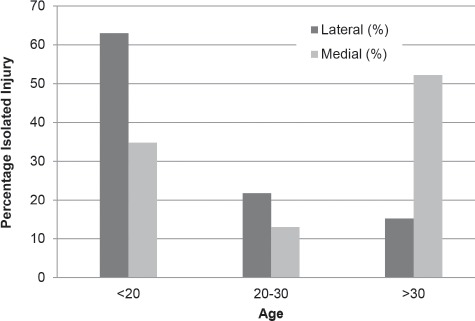
Percentage of Isolated Injuries vs Age. Isolated lateral meniscus injuries were more common in patients under 20 years of age. Isolated medial meniscus injuries were more common in patients greater than 30 years old.
Discussion
The purpose of this study was to determine the prevalence of isolated meniscal injuries as it relates to patient age. Anecdotally, orthopedic surgeons have assumed that medial meniscal tears occur less frequently in younger patients, and more commonly in older patients, while lateral tears occur more often in a younger population. However, no prior studies were found that sought to prove this hypothesis. This study ultimately confirms this to be true. This information is valuable for providers in the clinical setting when developing differential diagnoses while evaluating patients of different ages with lateral and/or medial sided knee symptoms.
The medial meniscus covers approximately 50% of the medial tibial plateau and is more tightly adherent to the joint capsule than the lateral meniscus11. The lateral meniscus is smaller than the medial meniscus and covers approximately 70% of the lateral surface of the tibial plateau11. The lateral meniscus is loosely attached to the joint capsule, particularly in the posterolateral corner where the posterior third attaches to the capsule via the popliteomeniscal fascicles. Medial and lateral menisci have distinctly different dimensions: lateral menisci are approximately 32.4-35.7 mm in length and 26.6-29.3 mm wide, while medial menisci are 40.5-45.5 mm long and 27 mm wide12,13. Although both menisci are roughly wedgeshaped and semilunar, lateral menisci display greater variety in size, shape, thickness, and mobility than medial menisci14,15. Lateral menisci also cover a larger portion of the tibial plateau (75-93% laterally) in comparison to medial menisci (51%-74% medially)2.
The propensity of lateral meniscal tears to occur in isolation in younger persons is likely due to its anatomic and biomechanical differences compared to the medial meniscus. The lateral meniscus is more mobile, has less staunch capsular attachments and covers a greater surface area of the lateral plateau than the medial meniscus. These characteristics make isolated tears of the lateral meniscus more likely in a traumatic setting in younger patients.
This study is not without limitations. In addition to the inherent weaknesses of a retrospective review, a further weakness of this study is that only meniscus injuries that underwent surgical intervention were included. Clinical outcomes and improvement post-meniscectomy is controversial in patients older than 50 years of age4. This has led many surgeons to refrain from arthroscopic treatment for meniscal tears in older patients. This likely leads to a gross underestimation of the prevalence of meniscal tears in the older population. However, this may not affect the results as it was found that microscopic degeneration was an almost invariable finding in patients over the age of forty and was as common in lateral as in medial menisci10. Furthermore, characteristics or tear patterns of the isolated tears, as well as treatment therein were not delineated, which could be interpreted as another weakness, or rather, may have provided further insight. However, the purpose of this study was to report on prevalence of medial and lateral meniscal tears. It was felt that, overall, this study accomplished the primary goal, supporting the proposed hypothesis that isolated medial meniscal tears are more common with increasing age, while lateral meniscal tears are more common in the younger population.
Conclusion
This study showed that isolated lateral meniscal tears occur more often in younger people. The prevalence of isolated lateral meniscal tears was more common in our defined young patient population of under 20 years of age, and in decreasing prevalence in our older patient groups (20-30, > 30 years of age). Additionally, the prevalence of isolated medial meniscal tears increases with age. This information is valuable in guiding clinicians when developing differential diagnoses upon evaluating patients of different ages with lateral and/or medial sided knee symptoms.
Acknowledgements
Department statistician Yubo Gao, PhD.
References
- 1.Englund M, Roos EM, Roos HP, Lohmander LS. Patient-relevant outcomes fourteen years after meniscectomy: Influence of type of meniscal tear and size of resection. Rheumatology (Oxford) 2001;40(6):631–9. doi: 10.1093/rheumatology/40.6.631. [PMID: 11426019] [DOI: 10.1093/ rheumatology/40.6.631] [DOI] [PubMed] [Google Scholar]
- 2.Garrett WE, Jr., Swiontkowski MF, Weinstein JN, Callaghan J, Rosier RN, Berry DJ, Harrast J, Derosa GP. American Board of Orthopaedic Surgery Practice of the Orthopaedic Surgeon: Part-II, certification examination case mix. J Bone Joint Surg Am. 2006;88(3):660–7. doi: 10.2106/JBJS.E.01208. [PMID: 16510834] [DOI: 10.2106/JBJS.E.01208] [DOI] [PubMed] [Google Scholar]
- 3.Morgan CD, Wojtys EM, Casscells CD, Casscells SW. Arthroscopic meniscal repair evaluated by second-look arthroscopy. Am J Sports Med. 1991;19(6):632–7. doi: 10.1177/036354659101900614. discussion 7-8. [PMID: 1781503] [DOI: 10.1177/036354659101900614] [DOI] [PubMed] [Google Scholar]
- 4.Salata MJ, Gibbs AE, Sekiya JK. A systematic review of clinical outcomes in patients undergoing meniscectomy. Am J Sports Med. 2010;38(9):1907–16. doi: 10.1177/0363546510370196. [PMID: 20587698] [DOI: 10.1177/0363546510370196. [DOI] [PubMed] [Google Scholar]
- 5.Baker BE, Peckham AC, Pupparo F, Sanborn JC. Review of meniscal injury and associated sports. Am J Sports Med. 1985;13(1):1–4. doi: 10.1177/036354658501300101. [PMID: 3838420] [DOI: 10.1177/036354658501300101. [DOI] [PubMed] [Google Scholar]
- 6.Hede A, Jensen DB, Blyme P, Sonne-Holm S. Epidemiology of meniscal lesions in the knee. 1,215 open operations in Copenhagen 1982-84. Acta Orthop Scand. 1990;61(5):435–7. doi: 10.3109/17453679008993557. [PMID: 2239168] [DOI: 10.3109/17453679008993557. [DOI] [PubMed] [Google Scholar]
- 7.Steinbruck K. Epidemiology of sports injuries. 25-year analysis of sports orthopedic-traumatologic ambulatory care. Sportverletz Sportschaden. 1999;13(2):38–52. doi: 10.1055/s-2007-993313. [PMID: 10478388] [DOI: 10.1055/s-2007-993313. [DOI] [PubMed] [Google Scholar]
- 8.Steinbruck K. Epidemiology of sports injuries. A 15-year analysis of sports orthopedic ambulatory care. Sportverletz Sportschaden. 1987;1(1):2–12. doi: 10.1055/s-2007-993688. [PMID: 3509867] [DOI: 10.1055/s-2007-993688. [DOI] [PubMed] [Google Scholar]
- 9.Makris EA, Hadidi P, Athanasiou KA. The knee meniscus: Structure-function, pathophysiology, current repair techniques, and prospects for regeneration. Biomaterials. 2011;32(30):7411–31. doi: 10.1016/j.biomaterials.2011.06.037. [PMID: 21764438] DOI: 10.1016/j.biomaterials.2011.06.037. [DOI] [PMC free article] [PubMed] [Google Scholar]
- 10.Noble J, Hamblen DL. The pathology of the degenerate meniscus lesion. J Bone Joint Surg Br. 1975;57(2):180–6. PMID: 1173585. [PubMed] [Google Scholar]
- 11.Rath E, Richmond JC. The menisci: Basic science and advances in treatment. Br J Sports Med. 2000;34(4):252–7. doi: 10.1136/bjsm.34.4.252. [PMID: 10953895] [DOI: 10.1136/ bjsm.34.4.252. [DOI] [PMC free article] [PubMed] [Google Scholar]
- 12.McDermott ID, Sharifi F, Bull AMJ, Gupte CM, Thomas RW, Amis AA. An anatomical study of meniscal allograft sizing. Knee Surg Sports Traumatol Arthrosc. 2004;12(2):130–5. doi: 10.1007/s00167-003-0366-7. [PMID: 12756521] DOI: 10.1007/s00167-003-0366-7. [DOI] [PubMed] [Google Scholar]
- 13.Shaffer B, Kennedy S, Klimkiewicz J, Yao L. Preoperative sizing of meniscal allografts in meniscus transplantation. Am J Sports Med. 2000;28(4):524–33. doi: 10.1177/03635465000280041301. PMID: 10921644. [DOI] [PubMed] [Google Scholar]
- 14.Clark CR, Ogden JA. Development of the menisci of the human knee joint. Morphological changes and their potential role in childhood meniscal injury. J Bone Joint Surg Am. 1983;65(4):538–47. PMID: 6833331. [PubMed] [Google Scholar]
- 15.Greis PE, Bardana DD, Holmstrom MC, Burks RT. Meniscal injury: I. Basic science and evaluation. J Am Acad Orthop Surg. 2002;10(3):168–76. doi: 10.5435/00124635-200205000-00003. PMID: 12041938. [DOI] [PubMed] [Google Scholar]