Abstract
A 55-year-old nurse was referred with a 5-month history of right eye corneal abscess. The initial injury occurred when doing lawn work. The infection worsened despite multiple antibiotic, antiviral, and steroid treatments. Visual acuity was limited to hand motion. On examination, there was keratitis, ocular hypertension, and a secondary cataract. Corneal scrapings grew a filamentous fungus, identified as Metarhizium anisopliae (MA). Despite intensive antifungal treatment with topical, intravitreous, and systemic voriconazole, purulent corneal melting and scleritis with endophthalmitis rapidly appeared. An emergency surgical procedure including sclerocorneal transplantation, cataract surgery, a pars plana vitrectomy using temporary keratoprosthesis, and scleral crosslinking was necessary. One year after the surgery, there was no recurrence of infection. Functional outcome remained very poor. This is the first case of sclerokeratitis and endophthalmitis caused by MA ever reported. The infection was successfully treated with an aggressive combination of medical and surgical treatments.
Keywords: Endophthalmitis, fungal, keratitis, Metarhizium anisopliae, scleritis
Fungal keratitis represents one of the most difficult manifestations of microbial keratitis regarding diagnosis and treatment. Risk factors include compromised corneas, topical steroids, contact lens wear, and surgical and external corneal trauma. More than 70 genera of filamentous fungi and yeast have been identified in fungal keratitis.[1] Progression of fungal keratitis to exogenous endophthalmitis, however, is relatively rare.[2]
Metarhizium anisopliae (MA) is an entomopathogenic ubiquitous fungus found in soil. It can act as a parasite in insects. In humans, it is an uncommon cause of fungal keratitis or sclerokeratitis, with only six cases previously described.[3]
We report a case of MA sclerokeratitis and subsequent endophthalmitis that was successfully treated with an aggressive combination of medical and surgical treatments.
Case Report
A 55-year-old female nurse from the Lorraine region in northeastern France was referred with a 5-month history of infectious keratitis and ocular hypertension in the right eye (OD). Despite many topical (ciprofloxacin, rifamycin, dexamethasone, and hexamidine) and systemic (valacyclovir) treatments, there was no significant improvement. Corneal scraping initially performed was negative. The patient did not wear contact lenses. She mentioned a vegetal foreign body in her OD after mowing the lawn before the beginning of the infection. Medical history was unremarkable.
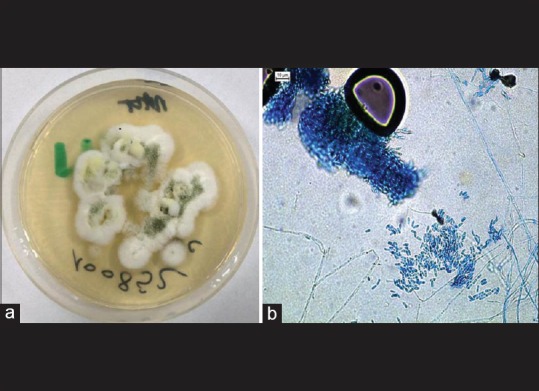
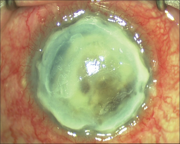
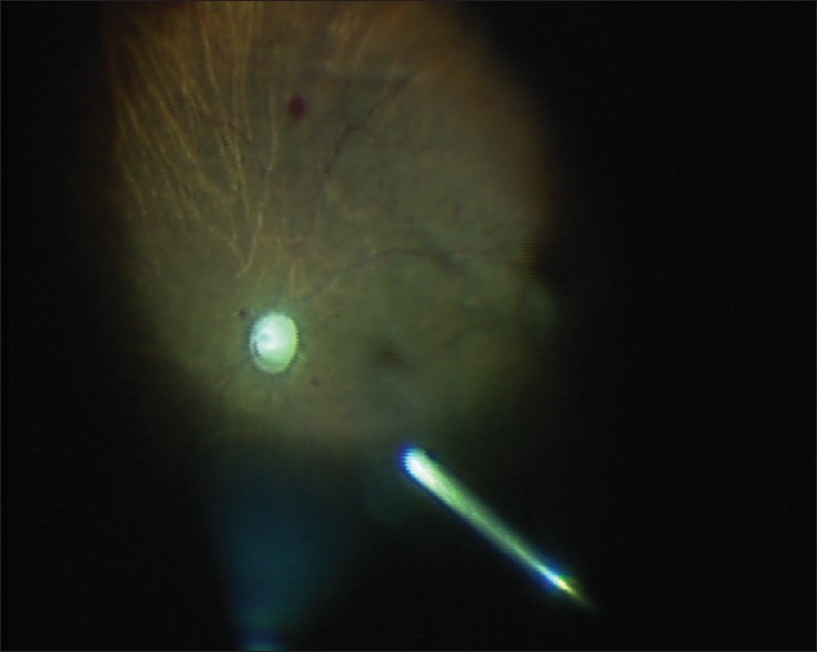
She presented with significant ocular pain and redness in the OD. Visual acuity was limited to hand motions OD and 20/20 left eye. Slit-lamp examination revealed a large central epithelial defect with an underlying deep white 6 mm × 7 mm central suppurative stromal infiltrate, keratic precipitates, and posterior synechiae. Intraocular pressure was 40 mmHg. Corneal scraping of the infiltrate was performed. No bacteria were isolated. Polymerase chain reaction for herpes viruses and Acanthamoeba was negative. After 8 days, two colonies of a fungus appeared on 2% malt extract medium at 27°C [Fig. 1]. The fungus was later identified by double-strand sequencing of the internal transcribed spacer (ITS1 and ITS4 primers) of the ribosomal DNA as MA. Susceptibility testing showed resistance to amphotericin B, itraconazole, and posaconazole, but susceptibility to voriconazole and caspofungin. Topical voriconazole (every hour) and oral voriconazole (200 mg b.i.d) treatment was started. As B-scan ultrasonography showed mild vitritis, two intravitreal injections of voriconazole (100 μg/0.1 ml) were administered 6 days apart. Despite these treatments, the abscess worsened, and purulent corneal melting involving sclera, with massive inflammation in a shallow anterior chamber, and a secondary cataract appeared [Fig. 2]. Visual acuity in the OD was reduced to light perception. A diagnosis of MA endophthalmitis and sclerokeratitis was made. To avoid evisceration, an emergency surgical procedure was performed. The procedure started with an 8-mm trephination of the central cornea. Button and fibrin deposits were then removed from the anterior chamber and iridocorneal angle. Open-sky phacoemulsification and intraocular lens implantation were performed. A temporary Eckardt's keratoprosthesis was then sutured to the recipient cornea, allowing a 25-gauge pars plana vitrectomy to be performed [Fig. 3]. Retinal hemorrhaging and optic nerve atrophy were observed intraoperatively.
Figure 1.

(a) Metarhizium anisopliae: Culture grown on 2% malt extract medium at 27°C for 2 weeks. (b) Metarhizium anisopliae: Conidiophores with verticillate branching and cylindrical conidia. Lactophenol cotton blue stain, ×400
Figure 2.

Preoperative slit-lamp photography of the right eye showing conjunctival hyperemia, corneal melting involving sclera with massive inflammation in a shallow anterior chamber
Figure 3.

Pars plana vitrectomy was performed through the optic of Eckardt's keratoprosthesis. Retinal hemorrhaging and optic nerve atrophy were observed intraoperatively
After keratoprosthesis removal, a conjunctival peritomy was performed, and corneal trephination was manually enlarged to a diameter of 13 mm to remove the infected peripheral cornea and adjacent scleral tissues. A 14 mm sclerocorneal graft was manually prepared and sutured with 24 interrupted stitches of 10/0 nylon. At the end of the procedure, a scleral crosslinking was performed (10 mW/cm2 ultraviolet A [UVA] lamp and riboflavin, Horus, Saint-Laurent du Var, France) on the donor–recipient junction. Procedure time was 2 h and 45 min. Pathologic examination of the excised cornea showed the presence of septate hyphae. Postoperative medications included topical and oral voriconazole, oral acetazolamide, and topical cyclosporine 2% t.i.d. Topical dexamethasone was started 2 months after surgery. Antifungal treatment was discontinued after 4 months due to elevated liver enzymes.
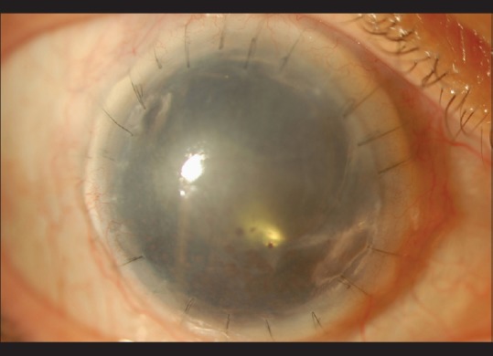
One year after the surgery, there was no recurrence of the infection [Fig. 4]. However, the sclerocorneal graft was rejected after 6 months, and persistent ocular hypertension required diode laser cyclophotocoagulation. Visual acuity was reduced to light perception.
Figure 4.

Slit-lamp photograph of the right eye 1 year after surgery revealing graft edema, mydriasis, but no sign of inflammation or infection
Discussion
MA is a fungus first described under the name Entomophthora anisoplia as a pathogen of the wheat cockchafer. It is an uncommon cause of cutaneous necrosis, chronic sinusitis, disseminated infection, and there were six published cases of keratitis or sclerokeratitis worldwide (Colombia, the USA, Australia, Japan, and France).[3,4,5,6] Three cases of keratitis were cured by topical natamycin eye drops alone or in combination with silver sulfadiazine 1% soluble cream,[4] bacitracin, and ciprofloxacin,[5] while the three other patients had sclerokeratitis. They were initially treated medically, but then underwent a therapeutic corneal graft.[3,6] Visual prognosis was poor for all cases, and we confirm that functional outcome is poor with this first case of sclerokeratitis and subsequent endophthalmitis. This may be due to retinal damage caused by inflammation and infection or might also be attributed to prolonged secondary glaucoma and subsequent optic neuropathy.
The main triggering incident found in our patient was the initial corneal trauma after lawn work. Apart from the history of trauma with vegetative matter, excessive usage of antibiotics and antivirals in combination with topical steroids might well have contributed to the destruction of local microflora, reduction in local immunity, and to epithelial toxicity, all of which might have predisposed to fungal infection.
The patient was treated with topical, intravitreal, and systemic voriconazole, the only molecule that was shown to be efficient on the isolate. Natamycin was not tested in our case. As the corneal infection progressed to the sclera and posterior segment, an emergency surgical intervention was decided. The procedure combined five steps: sclerocorneal allograft, cataract extraction, intraocular lens implantation, pars plana vitrectomy through temporary keratoprosthesis, and scleral crosslinking. As in previous cases, surgery was a decisive step in the management of the infection. It is now admitted that therapeutic keratoplasty or sclerokeratoplasty has a definitive role in the management of progressive microbial keratitis refractory to medical therapy.[2] The primary aim of the procedure is to eliminate infected tissues, especially when infection progresses to the peripheral cornea, limbus, and anterior sclera. The diameter of the trepanation was chosen to leave a 1-mm infection-free scleral margin. Riboflavin/UVA corneal collagen crosslinking has already been shown experimentally and clinically to be an interesting adjuvant treatment of advanced nonresponsive microbial keratitis.[7] Recently, collagen crosslinking of the sclera has been shown to increase the biomechanical strength of rabbit sclera without side effects on the retina or retinal pigment epithelium.[8] This new crosslinking method is considered to be a possible sclera-based treatment for progressive myopia. We suggest that scleral crosslinking could also be useful in the management of infectious scleritis. Experimental investigations are needed to confirm the anti-infectious and anti-inflammatory properties of the procedure in the scleral tissue. Finally, we were able to use Eckardt's temporary keratoprosthesis, a reusable device, which provided a clear view and watertight eye after corneal trephination. Its large optical diameter allowed the visualization of the peripheral fundus.
Conclusion
This is the first case of sclerokeratitis and endophthalmitis caused by MA ever reported. The patient's infection was successfully treated by an aggressive combination of medical and surgical treatments.
Financial support and sponsorship
Nil.
Conflicts of interest
There are no conflicts of interest.
References
- 1.Thomas PA. Fungal infections of the cornea. Eye (Lond) 2003;17:852–62. doi: 10.1038/sj.eye.6700557. [DOI] [PubMed] [Google Scholar]
- 2.Wang MX, Shen DJ, Liu JC, Pflugfelder SC, Alfonso EC, Forster RK. Recurrent fungal keratitis and endophthalmitis. Cornea. 2000;19:558–60. doi: 10.1097/00003226-200007000-00031. [DOI] [PubMed] [Google Scholar]
- 3.Eguchi H, Toibana T, Hotta F, Miyamoto T, Mitamura Y, Yaguchi T. Severe fungal sclerokeratitis caused by Metarhizium anisopliae: A case report and literature review. Mycoses. 2015;58:88–92. doi: 10.1111/myc.12279. [DOI] [PubMed] [Google Scholar]
- 4.De García MC, Arboleda ML, Barraquer F, Grose E. Fungal keratitis caused by Metarhizium anisopliae var. anisopliae. J Med Vet Mycol. 1997;35:361–3. [PubMed] [Google Scholar]
- 5.Jani BR, Rinaldi MG, Reinhart WJ. An unusual case of fungal keratitis: Metarhizium anisopliae. Cornea. 2001;20:765–8. doi: 10.1097/00003226-200110000-00020. [DOI] [PubMed] [Google Scholar]
- 6.Dorin J, Debourgogne A, Zaïdi M, Bazard MC, Machouart M. First unusual case of keratitis in Europe due to the rare fungus Metarhizium anisopliae. Int J Med Microbiol. 2015;305:408–12. doi: 10.1016/j.ijmm.2015.03.004. [DOI] [PubMed] [Google Scholar]
- 7.Vajpayee RB, Shafi SN, Maharana PK, Sharma N, Jhanji V. Evaluation of corneal collagen cross-linking as an additional therapy in mycotic keratitis. Clin Exp Ophthalmol. 2015;43:103–7. doi: 10.1111/ceo.12399. [DOI] [PubMed] [Google Scholar]
- 8.Wollensak G, Iomdina E. Long-term biomechanical properties of rabbit sclera after collagen crosslinking using riboflavin and ultraviolet A (UVA) Acta Ophthalmol. 2009;87:193–8. doi: 10.1111/j.1755-3768.2008.01229.x. [DOI] [PubMed] [Google Scholar]


