

Abstract
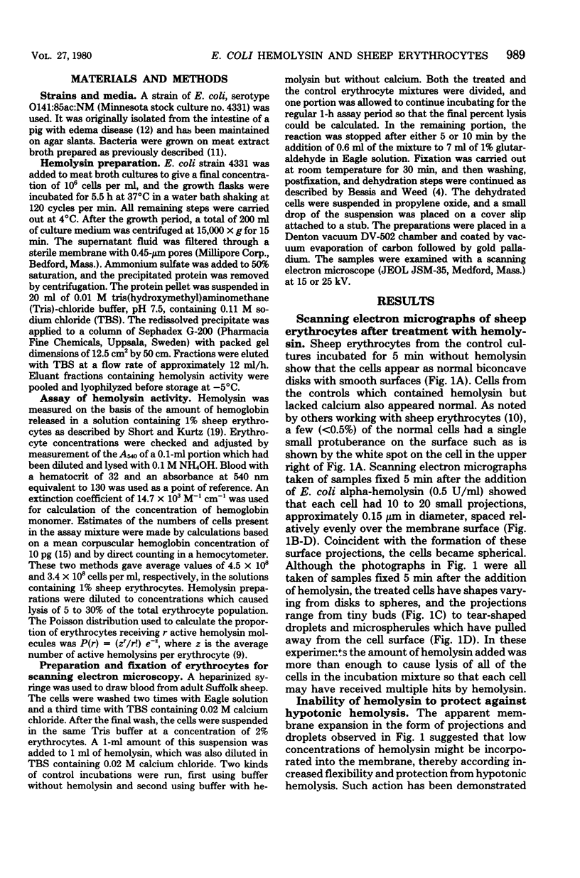
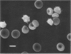
Scanning electron micrographs of sheep erythrocytes showed that attachment of the alpha hemolysin produced by Escherichia coli resulted in the formation of spherocytes, with 10 to 20 small projections spaced relatively evenly over the surface of the erythrocyte membrane. This shape change was induced within 5 min after treatment. If the hemolysin concentration was reduced to a level which would lyse only a fraction of the total erythrocytes, the affected cells were easily identified against a background of normal, unaffected cells. Unlike sodium lauryl sulfate and other amphipathic agents which enter cell membranes and increase their flexibility, low concentrations of hemolysin did not provide protection against hypotonic hemolysis. These findings indicate that the surface projections were not the result of membrane expansion caused by incorporation of hemolysin into the outer portion of the lipid bilayer. The ability of a given amount of hemolysin to release a constant amount of hemoglobin in the presence of increasing concentrations of red cells confirmed that a single hit is sufficient for lysis. These results suggest that a single hemolysin molecule can bind to a sheep erythrocyte and trigger internal reactions which result in the derangement of membrane integrity at multiple sites on the surface. Confirmation of one-hit kinetics indicates that measurement of E. coli hemolysin activity should be carried out at low ratios of hemolysin to erythrocyte to decrease the possibility of multiple hits on a single cell.
Full text
PDF


Images in this article

Selected References
These references are in PubMed. This may not be the complete list of references from this article.
- Allan D., Michell R. H. Accumulation of 1,2-diacylglycerol in the plasma membrane may lead to echinocyte transformation of erythrocytes. Nature. 1975 Nov 27;258(5533):348–349. doi: 10.1038/258348a0. [DOI] [PubMed] [Google Scholar]
- Allan D., Watts R., Michell R. H. Production of 1,2-diacylglycerol and phosphatidate in human erythrocytes treated with calcium ions and ionophore A23187. Biochem J. 1976 May 15;156(2):225–232. doi: 10.1042/bj1560225. [DOI] [PMC free article] [PubMed] [Google Scholar]
- Bessis M. Red cell shapes. An illustrated classification and its rationale. Nouv Rev Fr Hematol. 1972 Nov-Dec;12(6):721–745. [PubMed] [Google Scholar]
- Birchmeier W., Singer S. J. On the mechanism of ATP-induced shape changes in human erythrocyte membranes. II. The role of ATP. J Cell Biol. 1977 Jun;73(3):647–659. doi: 10.1083/jcb.73.3.647. [DOI] [PMC free article] [PubMed] [Google Scholar]
- Collier R. J., Kandel J. Structure and activity of diphtheria toxin. I. Thiol-dependent dissociation of a fraction of toxin into enzymically active and inactive fragments. J Biol Chem. 1971 Mar 10;246(5):1496–1503. [PubMed] [Google Scholar]
- Eaton J. W., Berger E., Nelson D., White J. G., Rundquist O. Intracellular calcium: lack of effect on ovine red cells. Proc Soc Exp Biol Med. 1978 Mar;157(3):506–510. doi: 10.3181/00379727-157-40086. [DOI] [PubMed] [Google Scholar]
- Inoue K., Akiyama Y., Kinoshita T., Higashi Y., Amano T. Evidence for a one-hit theory in the immune bactericidal reaction and demonstration of a multi-hit response for hemolysis by streptolysin O and Clostridium perfringens theta-toxin. Infect Immun. 1976 Feb;13(2):337–344. doi: 10.1128/iai.13.2.337-344.1976. [DOI] [PMC free article] [PubMed] [Google Scholar]
- Jain N. C., Kono C. S. Scanning electron microscopy of erythrocytes of dog, cat, cow, horse, sheep and goat. Res Vet Sci. 1972 Sep;13(5):489–491. [PubMed] [Google Scholar]
- Jorgensen S. E., Short E. C., Jr, kurtz H. J., Mussen H. K., Wu G. K. Studies on the origin of the alpha-haemolysin produced by Escherichia coli. J Med Microbiol. 1976 May;9(2):173–189. doi: 10.1099/00222615-9-2-173. [DOI] [PubMed] [Google Scholar]
- NAKAO M., NAKAO T., YAMAZOE S., YOSHIKAWA H. Adenosine triphosphate and shape of erythrocytes. J Biochem. 1961 Jun;49:487–492. doi: 10.1093/oxfordjournals.jbchem.a127333. [DOI] [PubMed] [Google Scholar]
- Rennie R. P., Arbuthnott J. P. Partial characterisation of Escherichia coli haemolysin. J Med Microbiol. 1974 May;7(2):179–188. doi: 10.1099/00222615-7-2-179. [DOI] [PubMed] [Google Scholar]
- SMITH H. W. The haemolysins of Escherichia coli. J Pathol Bacteriol. 1963 Jan;85:197–211. doi: 10.1002/path.1700850119. [DOI] [PubMed] [Google Scholar]
- Sheetz M. P., Singer S. J. Equilibrium and kinetic effects of drugs on the shapes of human erythrocytes. J Cell Biol. 1976 Jul;70(1):247–251. doi: 10.1083/jcb.70.1.247. [DOI] [PMC free article] [PubMed] [Google Scholar]
- Sheetz M. P., Singer S. J. On the mechanism of ATP-induced shape changes in human erythrocyte membranes. I. The role of the spectrin complex. J Cell Biol. 1977 Jun;73(3):638–646. doi: 10.1083/jcb.73.3.638. [DOI] [PMC free article] [PubMed] [Google Scholar]
- Short E. C., Kurtz H. J. Properties of the Hemolytic Activities of Escherichia coli. Infect Immun. 1971 May;3(5):678–687. doi: 10.1128/iai.3.5.678-687.1971. [DOI] [PMC free article] [PubMed] [Google Scholar]
- Snyder I. S., Koch N. A. Production and characteristics of hemolysins of Escherichia coli. J Bacteriol. 1966 Feb;91(2):763–767. doi: 10.1128/jb.91.2.763-767.1966. [DOI] [PMC free article] [PubMed] [Google Scholar]
- Weed R. I., Chailley B. Calcium-pH interactions in the production of shape change in erythrocytes. Nouv Rev Fr Hematol. 1972 Nov-Dec;12(6):775–788. [PubMed] [Google Scholar]
- White J. G. Effects of an ionophore, A23187, on the surface morphology of normal erythrocytes. Am J Pathol. 1974 Dec;77(3):507–518. [PMC free article] [PubMed] [Google Scholar]