Abstract
Aims/hypothesis
We aimed to investigate potential interactions between insulin and glucagon-like peptide (GLP)-1 signalling pathways in the regulation of beta cell-cycle dynamics in vivo, in the context of the therapeutic potential of GLP-1 to modulate impaired beta cell function.
Methods
Beta cell-specific insulin receptor knockout (βIRKO) mice, which exhibit beta cell dysfunction and an age-dependent decrease in beta cell mass, were treated with the dipeptidyl peptidase-4 inhibitor vildagliptin. Following this, glucose homeostasis and beta cell proliferation were evaluated and underlying molecular mechanisms were investigated.
Results
The sustained elevation in circulating GLP-1 levels, caused by treatment of the knockout mice with vildagliptin for 6 weeks, significantly improved glucose tolerance secondary to enhanced insulin secretion and proliferation of beta cells. Treating βIRKO beta cell lines with the GLP-1 analogue, exendin-4, promoted Akt phosphorylation and protein expression of cyclins A, D1 and E two- to threefold, in addition to cyclin D2. Pancreases from the vildagliptin-treated βIRKO mice exhibited increased cyclin D1 expression, while cyclin D2 expression was impaired.
Conclusions/interpretation
Activation of GLP-1 signalling compensates for impaired growth factor (insulin) signalling and enhances expression of cyclins to promote beta cell proliferation. Together, these data indicate the potential of GLP-1-related therapies to enhance beta cell proliferation and promote beneficial outcomes in models with dysfunctional beta cells.
Keywords: Beta cell, Cyclins, DPP-4 inhibitor, GLP-1, Insulin signalling, Proliferation
Introduction
Type 2 diabetes is characterised by impaired beta cell function and an inability to compensate for peripheral insulin resistance [1]. These abnormalities are recognised in impaired glucose tolerance stage where a progressive reduction in beta cell mass is already evident before the development of over disease [2]. Thus, retention of beta cell mass is important for preventing progression of the disease.
Recently, novel therapeutic approaches such as glucagon-like peptide-1 (GLP-1) analogues and enhancers have emerged as important players in the treatment of type 2 diabetes [3]. Some reports suggest that the effects of GLP-1 on beta cell proliferation and secretory function occur as a consequence of cross talk with proteins in the insulin signalling pathway and by modulation of transcription factors including pancreatic and duodenal homeobox 1 (PDX-1) [4, 5]. cAMP response-element binding protein (CREB), activated by GLP-1 signalling via the cAMP–protein kinase A (PKA) pathway, acts by transactivation of insulin and IRS2 gene expression to promote proliferation and prevent apoptosis of beta cells [6–9]. Indeed, multiple lines of evidence indicate a significant role for insulin/IGF-1 signalling in beta cell biology [10]. Mice lacking functional insulin [11] or IGF-1 [12, 13] receptors in beta cells develop glucose intolerance and the former develop an age-dependent decrease in beta cell mass [11]. Similarly, various proteins in the signalling pathway, including IRS proteins and Akt, are critical in the regulation of beta cell function and mass [10, 14]. GLP-1 has also been reported to upregulate IGF-1 receptor expression and protect beta cells from cytokine-induced apoptosis [15, 16]. However, the mechanisms that underlie the effects of GLP-1 on beta cell growth in the context of insulin resistance or attenuated growth factor (insulin) signalling are not fully explored.
Cell-cycle progression is essential for beta cell growth and cyclins play a central role in regulating the cell cycle [17]. Mice with cyclin D2 disruption display a decrease in beta cell proliferation leading to the development of diabetes [18, 19]; this is exacerbated when the mice are insulin resistant [20]. Conversely, cyclin D1 overexpression increases beta cell proliferation and mass [21]. In beta cells, cyclins are linked with proteins in both the insulin–IGF-1 and GLP-1 signalling pathways [22–25].
In this study, to dissect the effect of GLP-1-related therapies specifically on pancreatic beta cells independent of systemic effects of diabetes, we chose to investigate the beta cell-specific insulin receptor knockout (βIRKO) mouse, a model that exhibits impaired beta cell function, including glucose-stimulated insulin secretion (GSIS) and progressive reduction of beta cell mass [11]. In previous studies, interrogation of the functional interactions between insulin and GLP-1 signalling pathways revealed that elevating circulating GLP-1 levels in these knockout mice enhances beta cell proliferation secondary to an increase in the expression of cyclins, and improved glucose tolerance. Thus, data generated using these mouse models may have therapeutic implications for GLP-1 in the treatment of individuals with type 2 diabetes who exhibit insulin resistant beta cells [26, 27]. The present study aimed to investigate interactions between insulin and GLP-1 signalling pathways in the regulation of beta cell -cycle dynamics in vivo, to elucidate the potential of GLP-1 to modulate impaired beta cell function.
Methods
Animals and physiological assays
The βIRKO mice and littermate control insulin-receptor-floxed mice on a C57B6 background were obtained as described [11] and housed in pathogen-free facilities on a 12 h light–dark cycle at the Animal Care Facility of Joslin Diabetes Center, Boston, MA, USA. All protocols were approved by the Institutional Animal Care and Use Committee of the Joslin Diabetes Center and were in accordance with NIH guidelines. Blood glucose was monitored using an automated glucose monitor (Ascensia Elite; Bayer, Whippany, NJ, USA), plasma insulin by ELISA (Crystal Chem, Downers Grove, IL, USA), plasma GLP-1 by ELISA (EMD Millipore, Billerica, MA, USA) and plasma dipeptidyl peptidase-4 (DPP-4) activity by ELISA (Novartis, Cambridge, MA, USA). IPGTT (2 g/kg body weight), OGTT (1 g/kg body weight) and in vivo GSIS measurements (3 g/kg body weight) were performed after mice had been fasted for 15 h overnight [11].
Vildagliptin treatment
Twenty-four-week-old βIRKO mice without diabetes or control male mice were treated with or without vildagliptin (Novartis) in drinking water (0.5 mg/ml, > 1 mg/day) [28] for 6 weeks. The mice were randomly assigned as either control (H2O) or treatment (vildagliptin) groups by the cage numbers where they were kept. Body weight and blood glucose were measured twice a week. OGTT and in vivo GSIS were measured before and during the treatment course (OGTT on day 32, GSIS on day 39). After treatment (day 42) mice were fasted overnight, and blood samples were collected 5 min after oral glucose load (1 g/kg body weight). Mice were injected with BrdU (100 mg/kg body weight, i.p.) and 5 h later pancreases were harvested for morphological analyses. We performed two independent experiments using the same protocol and the data are described in combination. All mice were included in the analyses, as none exhibited health problems such as tumour, injury or malocclusion of the teeth.
Pancreas morphometry
Dissected pancreases were weighed, processed [11] and immunostained for insulin (EMD Millipore), BrdU (DAKO, Carpinteria, CA, USA), phospho-histone H3 (p-HH3; EMD Millipore), cyclin D1 (Cell Signaling Technology, Danvers, MA, USA) and cyclin D2 (Santa Cruz Biotechnology, Dallas, TX, USA). Beta cell mass was analysed as described previously [12]. The numbers of total and p-HH3- or BrdU-positive beta cells were scored and data were expressed as a percentage of the 5000 beta cells counted.
Cell culture
Control and βIRKO beta cell lines were cultured in DMEM as described [29, 30] and used between passages 11 to 29. Absence of mycoplasma contamination was confirmed every 6 months. Briefly, for acute stimulation, cells cultured in DMEM with 3 mmol/l glucose and/or 10 µmol/l nifedipine (Sigma-Aldrich, St Louis, MO, USA) for 16h were subsequently stimulated with GLP-1 (10 nmol/l) and/or human insulin (5 or 100 nmol/l; Sigma-Aldrich) for 15 min, and protein samples were extracted immediately. For chronic stimulation, cells cultured in DMEM containing 3 mmol/ l glucose and/or 10 nmol/l exendin-4 (Sigma-Aldrich) and/or 200 nmol/l OSI-906 (Selleck, Houston, TX, USA) for 24 h. For protein-stability analysis, control cells were treated with 100 µg/ml cycloheximide (CHX; Sigma-Aldrich). See electronic supplementary material (ESM) Methods for further details. In general, we performed at least three independent experiments for statistical analyses.
Western blotting
Western blot analysis was performed as described previously [31]. The antibodies used are listed in ESM Table 1. Dilutions: ×1,000 for primary antibodies, and ×5,000–10,000 for secondary antibodies. Densitometry was performed using NIH Image J software (version 1.50i; National Institute of Mental Health, Bethesda, MD, USA).
Insulin secretion
Control and βIRKO beta cell lines were seeded into 12-well dishes 48 h prior to secretion experiments. Sixteen hours before the experiment, cells were cultured in DMEM media containing 3 mmol/l glucose with or without 10 µmol/l nifedipine. The cells were pre-incubated for 30 min at 37°C in 1000 µl of 10 mmol/l HEPES-balanced Krebs-Ringer bicarbonate buffer (KRBB; pH 7.4) supplemented with 3 mmol/l glucose with or without 10 µmol/l nifedipine. Then, the cells were incubated for 30 min at 37°C in 1000 µl of the KRBB containing different concentrations of glucose with or without 10 nmol/l GLP-1 or 10 µmol/l nifedipine. The supernatant fractions were collected for insulin assay by RIA (EMD Millipore). Data are expressed as ratios after normalisation for basal insulin secretion (3 mmol/l glucose).
Adenovirus and lentivirus transduction
Pre-packaged adenovirus containing an internal ribosome entry site (IRES) and expressing Cre recombinase and green fluorescent protein (GFP; Ad-Cre-IRES-GFP and Ad-GFP) were purchased from Vector Biolabs (Malvern, PA, USA). Human wild-type IR open reading frames (kindly provided by J. Whittaker, Case Western Reserve University, Cleveland, OH, USA) were cloned into the pCDH-CMV-MCS-EF1-Puro lentiviral vector (System Biosciences, Palo Alto, CA, USA). See ESM Methods for further details, and ESM Table 2 for primers.
Statistics
All data are presented as mean ± SEM and were analysed using an unpaired two-tailed Student’s t test or ANOVA, with Tukey–Kramer’s post hoc tests as appropriate.
Results
DPP-4 inhibitors promote beta cell proliferation in βIRKO mice
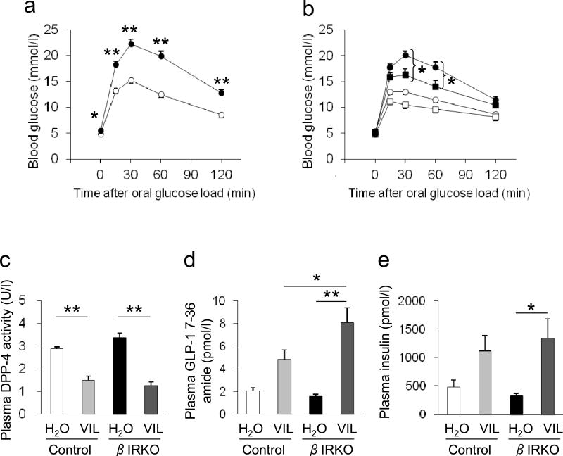
To explore interactions between insulin and GLP-1 signalling pathways in vivo, we compared the βIRKO mouse model with littermate control insulin-receptor-floxed mice [11]. βIRKO mice displayed glucose intolerance in response to oral glucose load following an OGTT (Fig. 1a). The GLP-1 peptide exhibits a short half-life in vivo because of its rapid degradation by DPP-4 [32]. To achieve sustained stimulation of GLP-1 signalling we elevated endogenous GLP-1 levels by treating mice with the DPP-4 inhibitor vildagliptin (0.5 mg/ml in drinking water, estimated dose >1mg/day per mouse) for 6 weeks. OGTTs performed on day 32 of treatment revealed significantly improved glucose tol erance in βIRKO mice (Fig. 1b). Although we noted a slight but insignificant increase in basal levels of DPP-4 in the βIRKO mice, at the end of the treatment period, a significantly reduced DPP-4 activity in the βIRKO and control groups confirmed successful inhibition of DPP-4 (Fig. 1c). Plasma levels of GLP-1, 5 min after an oral glucose load, were significantly higher in vildagliptin-treated βIRKO mice than in vehicle-treated βIRKO mice; GLP-1 levels in the former group were also significantly higher than in vildagliptin-treated control mice (Fig. 1d). Consistent with these findings, vildagliptin treatment significantly improved insulin secretion 5 min after oral glucose administration in βIRKO mice (Fig. 1e). In contrast, the improvement in insulin secretion observed in vildagliptin-treated control mice did not reach statistical significance.
Fig. 1.
Effects of vildagliptin treatment on glycaemic variables in mice. (a, b) OGTT before and after vildagliptin treatment in mice. Circles, non-treated mice; squares, vildagliptin-treated mice. White symbols, littermate insulin-receptor-floxed control mice; black symbols, βIRKO mice. In (a), n=23 in control group and n=38 in βIRKO group; in (b), n=11–19 in each group. Plasma (c) DPP-4 activity, (d) GLP-1 and (e) insulin 5 min after oral glucose load, after vildagliptin (VIL) treatment of mice (n=11–19). Mice were fasted for 16 h and glucose (1 g/kg body weight) was administered orally. Data are expressed as mean ± SEM. *p<0.05, **p<0.01, βIRKO vs control or as indicated
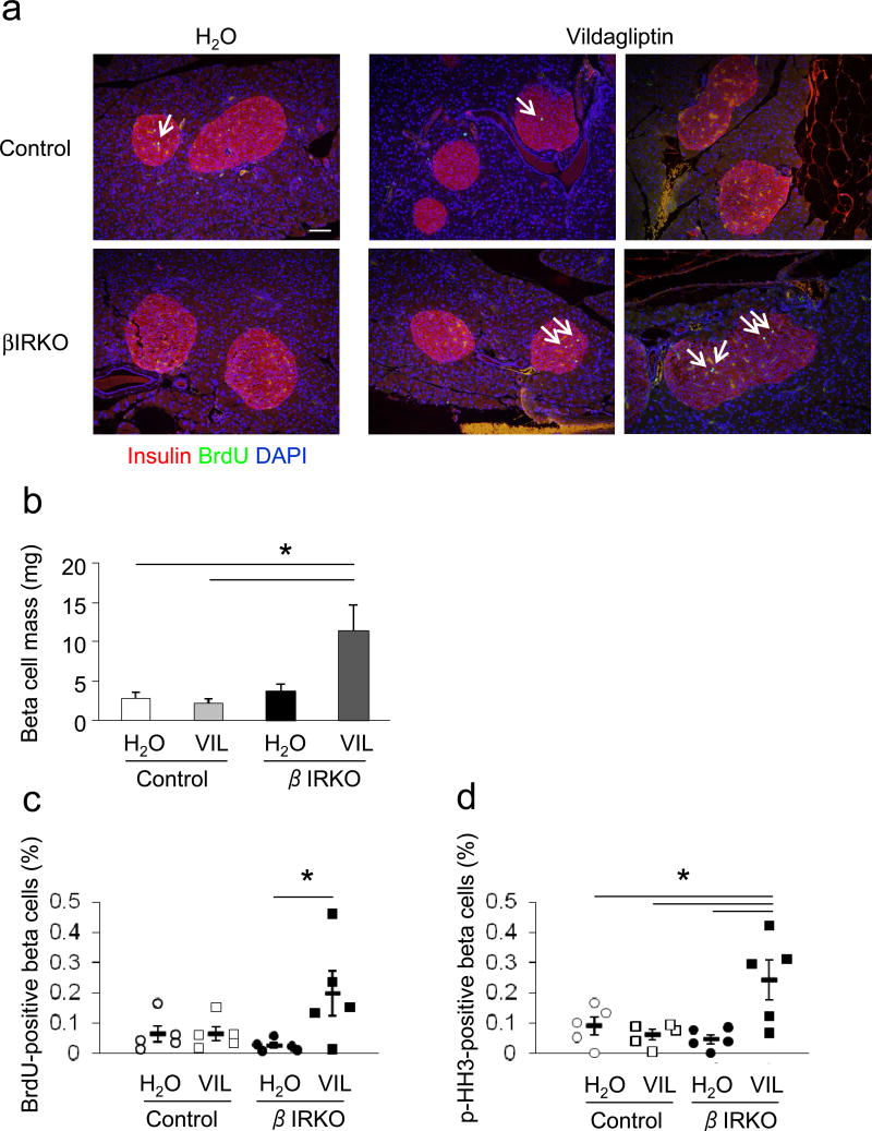
Histomorphometric analyses of pancreases (Fig. 2a), revealed an increase in the beta cell mass in vildagliptin-treated βIRKO mice (Fig. 2b). Consistently, the number of BrdU-positive and p-HH3-positive beta cells in vildagliptin-treated βIRKO mice displayed a robust increase (Fig. 2c,d), indicating that DPP-4 inhibitor therapy and consequent enhanced circulating GLP-1 promoted beta cell proliferation and significant increase in beta cell volume. Interestingly, in the vildagliptin-treated βIRKO group, only a subset of the mice exhibited a significant increase in beta cell proliferation (Fig. 2c), suggesting a variable response. On the other hand, we did not detect a significant increase in apoptotic beta cells in βIRKO mice, as assessed by TUNEL assay (data not shown). Vildagliptin treatment did not significantly alter random fed blood glucose levels in either group (ESM Table 3) but was associated with changes in body weight in the βIRKO mice (ESM Table 4).
Fig. 2.
Effects of vildagliptin treatment on beta cell mass and proliferation in mice. (a) Co-immunostaining of insulin (red) and BrdU (green) with DAPI (blue) in pancreas sections from βIRKO mice and littermate insulin-receptor-floxed control mice after vildagliptin treatment. A representative islet for each group at magnification ×20 is presented. Arrows indicate BrdU-positive beta cells. Scale bar, 50 µm. (b) Beta cell mass in mice (n=3–6 in each group). Data are expressed as mean ± SEM. (c) Ratio of BrdU-positive beta cells and (d) p-HH3-positive beta cells in pancreas samples (expressed as % of 5000 cells counted). Circles and squares indicate data of individual samples; horizontal bars indicate average of groups ± SEM. *p<0.05 as indicated. VIL, vildagliptin
The inhibition of DPP-4 also enhances another incretin, glucose-dependent insulinotropic peptide (GIP) [32]. Thus, to avoid the confounding effects on GIP and examine the direct effects of GLP-1, we treated glucose-intolerant βIRKO mice and control mice with GLP-1 (500 µmol/kg body weight) or saline (154 mmol/l NaCl) intraperitoneally, twice a day, for 20 consecutive days (ESM Methods, ESM Fig. 1a,b). The physiological effect of GLP-1 administration was confirmed by the significant blood-glucose-lowering effect observed 30 min after injection in both groups (ESM. Fig. 1c); βIRKO mice were significantly more responsive than control mice to GLP-1 administration. At the end of the 20 day treatment (day 21), morphological analyses of pancreas sections revealed a significant increase in proliferating beta cells in GLP-1-treated βIRKO mice as demonstrated by a greater number of p-HH3-positive beta cells (ESM Fig. 1d,e). There was no significant alteration in islet beta cell mass (data not shown), random fed blood glucose (ESM Table 5), body weight (ESM Table 6) or glucose tolerance (ESM Fig. 1b) in either group.
GLP-1 induces activation of Akt signalling in beta cell lines independent of insulin action
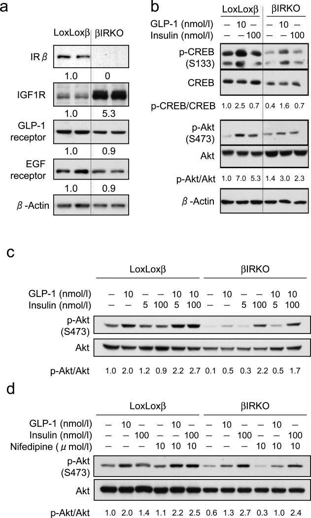
Next, to investigate the potential mechanism(s) underlying the in vivo effects of GLP-1 we used beta cell lines derived from βIRKO mice [29] which, as expected, exhibited a compensatory increase in the expression of IGF-1 receptors [29] but showed a normal complement of GLP-1 and EGF receptors compared with control beta cell lines from insulin-receptor-floxed mice (Fig. 3a).
Fig. 3.
Effects of GLP-1 on beta cell lines. (a) Western blotting for insulin receptor (IR), IGF-1 receptor (IGF1R), GLP-1 receptor, EGF receptor and actin. (b) Western blotting for total and p-CREB and total and p-Akt normalised for actin 15 min after GLP-1 or insulin stimulation. (c) Western blotting for total and p-Akt 15 min after GLP-1 and/or insulin stimulation. (d) Western blotting for total and p-Akt 15 min after GLP-1 or insulin stimulation with or without 16 h nifedipine treatment. Representative images of three independent experiments are shown in all panels. Numbers below bands represent relative values of the density quantification. LoxLoxβ, beta cells from littermate insulin-receptor-floxed control mice
Acute GLP-1 stimulation (10 nmol/l, 15 min) induced phosphorylation of CREB (Fig. 3b), indicating intact GLP-1 signalling, in both beta cell lines. GLP-1 treatment enhanced phosphorylation of Akt (Fig. 3b) and its downstream target p70S6K (data not shown) in both cell lines. The mild effects of exogenous insulin on Akt phosphorylation were likely due to the upregulation of IGF-1 receptors in βIRKO cells. Notably, in control beta cells, GLP-1-stimulated Akt phosphorylation was more robust compared with the effects of insulin stimulation alone, suggesting additional effects of direct GLP-1 action and local effects of secreted insulin.
To investigate the effects of GLP-1 and insulin on Akt phosphorylation, βIRKO and control cell lines were stimulated with either low (5 nmol/l) or high (100 nmol/l) concentrations of insulin for 15 min in the presence or absence of GLP-1 (Fig. 3c). In controls, 10 nmol/l GLP-1 stimulated Akt phosphorylation to a greater extent than 5 nmol/l insulin (the concentration of insulin found in culture media after stimulation with GLP-1 for 30 min; Fig. 3c and ESM Fig. 2a). Stimulation with GLP-1 (10 nmol/l) in the presence of increasing concentrations of insulin (5 or 100 nmol/l) further enhanced Akt phosphorylation when compared with the effects of either agent alone, suggesting that GLP-1 induces Akt phosphorylation independent of and distinct from the effects of exogenous insulin. We also observed that in βIRKO cells, the stimulation of Akt phosphorylation that was evident with 10 nmol/l GLP-1 was enhanced when the insulin dose was increased to 100 nmol/l, probably mediated by the upregulated IGF-1 receptor expression (Fig. 3c).
Next, to circumvent the effects of secreted insulin on Akt phosphorylation, we pre -treated cells with the L-type Ca2+ channel blocker, nifedipine, for 16 h to inhibit insulin exocytosis [33]. In non-nifedipine-treated control beta cells, GLP-1 stimulation resulted in greater Akt phosphorylation when compared with insulin stimulation (Fig. 3d). In nifedipine-treated cells, in which GLP-1-induced insulin exocytosis is suppressed thus limiting the effects of secreted insulin (ESM Fig. 2b), acute GLP-1 treatment induced Akt phosphorylation to a similar degree to that seen after exogenous insulin stimulation (Fig. 3d). In addition, in βIRKO cells, GLP-1 treatment stimulated Akt phosphorylation even in the presence of nifedipine (Fig. 3d).
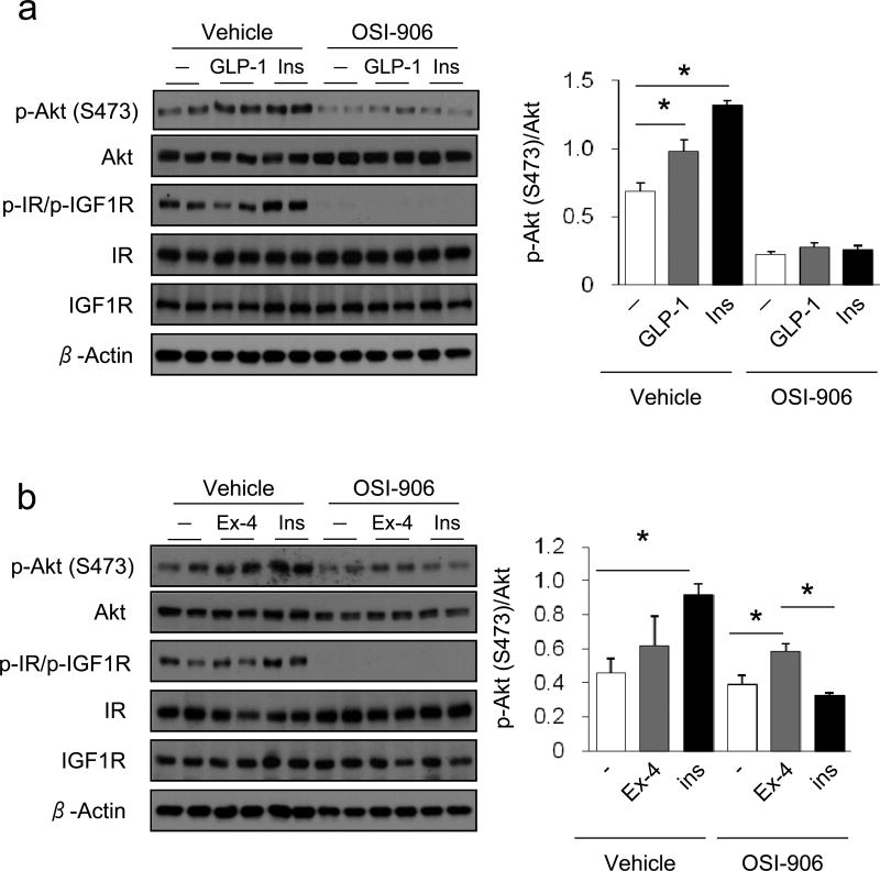
We also stimulated control beta cells with GLP-1 or insulin in the presence of a dual inhibitor for insulin and IGF-1 receptors (OSI-906) to assess the effect of secreted insulin induced by GLP-1 on insulin and IGF-1 receptor signalling. Treatment of control beta cells with OSI-906 completely blocked phosphorylation of insulin and IGF-1 receptors (Fig. 4). The upregulation of Akt phosphorylation induced by treatment with GLP-1 or insulin for 15 min was suppressed by the OSI-906 treatment (Fig. 4a). On the other hand, in the presence of OSI-906, 24 h stimulation with the GLP-1 receptor agonist exendin-4 (Ex-4) was still able to significantly upregulate Akt phosphorylation in contrast to the blunted effects of insulin (Fig. 4b).
Fig. 4.
Effects of GLP-1 on beta cell lines treated with insulin/IGF-1 receptor antagonist. Western blotting for total and p-Akt, total and p-insulin receptors (IR) and IGF-1 receptors (IGF1R) in beta cells from insulin-receptor-floxed control mice. The cells were stimulated with or without 10 nmol/l GLP-1 or 100 nmol/l insulin for (a) 15 min or (b) 24 h in the presence or absence of 200 nmol/l OSI-906. Histograms show relative phosphorylation of Akt, normalised by total Akt, in control beta cells in the absence or presence of OSI-906, after treatment with GLP-1 (a) or Ex-4 (b). All data are expressed as mean ± SEM. n=3 in each group. *p<0.05 as indicated
GLP-1 signalling upregulates multiple cyclins in beta cells lacking functional insulin receptors
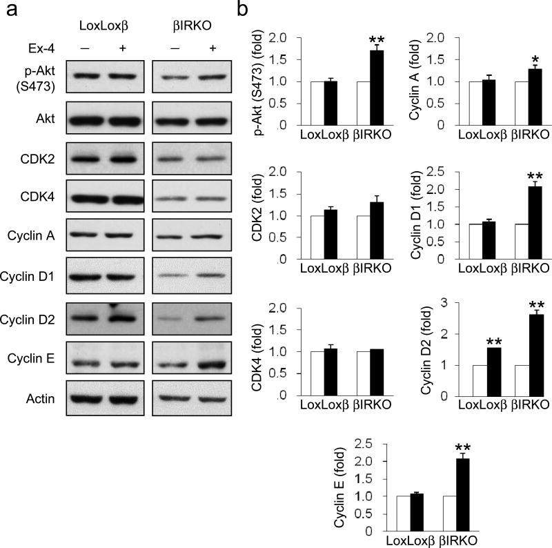
Since our in vivo data indicated that activation of GLP-1 signalling in βIRKO mice enhanced beta cell proliferation, we examined the effects of chronically activating GLP-1 signalling in vitro by Ex-4 (10 nmol/l, 24 h; Fig. 5a,b). Ex-4 treatment increased the expression of cyclin D2 in βIRKO beta cells and control insulin-receptor-floxed mouse beta cells while enhancing Akt phosphorylation only in the former. A concomitant significantly increased expression of cyclins A, D1 and E was also evident in βIRKO cells but not in control cells. Ex-4 treatment had no apparent effect on the expression of cyclin-dependent kinases (CDKs) 2 and 4 in either group. The basal expression of cyclin D2 was decreased in βIRKO cells (Fig. 5a).
Fig. 5.
Effects of chronic GLP-1 signalling activation on beta cell lines. (a) Western blotting or βIRKO mouse beta cells and control beta cells (LoxLoxβ) for total and p-Akt, CDK2, CDK4, cyclin A, cyclin D1, cyclin D2 and cyclin E with or without 24 h stimulation with 10 nmol/l Ex-4. Representative images of three independent experiments are shown. (b) Relative expression of proteins compared with non-stimulated state. Expression was normalised to actin except for p-Akt which was normalised to total Akt. White bars, control; black bars, Ex-4-treated. All data are expressed as mean ± SEM. n=3 in each group. *p<0.05, **p<0.01, Ex-4-treated vs control
Insulin receptor signalling modulates the protein expression of cyclin D2 in clonal beta cells
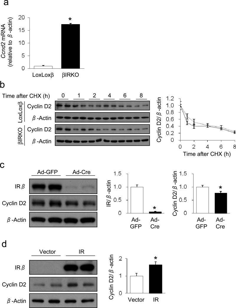
Next, we focused on the alteration in expression of cyclin D2 in βIRKO mouse beta cells. Cyclin D2 gene expression evaluated by real-time quantitative PCR displayed a robust increase in βIRKO cells (Fig. 6a) in contrast to the decrease in cyclin D2 protein. To further interrogate this disconnect between gene and protein expression we analysed protein stability of cyclin D2 by treating the cells with an inhibitor of protein biosynthesis, CHX. The degradation of cyclin D2 protein, however, was similar in control and βIRKO cells (Fig. 6b). We then sought to directly assess the impact of disruption of insulin signalling on cyclin D2 expression by knocking down the insulin receptor in control insulin receptor-floxed cells using Cre recombinase-expressing adenovirus. The virtual absence of insulin receptor expression following Cre expression was associated with a reduction in cyclin D2 protein (Fig. 6c). Conversely, reconstitution of human insulin receptor expression by lentiviral gene transfer in βIRKO cells significantly enhanced the expression of cyclin D2 protein (Fig. 6d).
Fig. 6.
Effects of insulin signalling on cyclin D2 expression in beta cell lines. (a) Gene expression of cyclin D2 (Ccnd2) evaluated by quantitative real-time PCR in βIRKO mouse beta cells and control beta cells (LoxLoxβ). n=4 in each group. (b) Western blotting for cyclin D2 at indicated time periods after CHX treatment and graph showing quantification. Solid line, control LoxLoxβ cells; dashed line, βIRKO cells. (c) Western blotting for insulin receptor (IR) and cyclin D2 after infection of control cells with adenoviruses carrying GFP or Cre recombinase. Graphs show quantification. (d) Western blotting for IR and cyclin D2 after lentiviral gene transfer of IR to βIRKO cells. All protein expression was normalised to actin expression. All data are expressed as mean ± SEM. n=3 in each group. *p<0.05 vs control condition
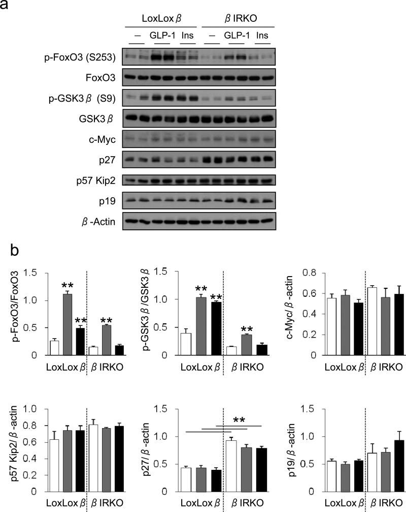
To dissect the mechanisms underlying the decrease in cyclin D2 expression in βIRKO cells, we analysed the downstream signalling proteins reported to be involved in cyclin D2 expression (Fig. 7a,b). The phosphorylation of FoxO3 and GSK3β in control insulin receptor-floxed cells was significantly enhanced by GLP-1 treatment. In the βIRKO cells, phosphorylation of these proteins was also elevated by GLP-1 but not by insulin treatment. However, the magnitude of phosphorylation in the βIRKO cells was generally lower than in control cells, suggesting an impairment of cyclin D2 expression. There was no change in c-Myc expression either between cells or following treatment. Among cell-cycle inhibitors, expression of p27 was significantly increased in βIRKO cells, while p57 Kip2 and p19 were not significantly altered (Fig. 7a,b).
Fig. 7.
Effects of GLP-1 and insulin on beta cell lines. (a) Western blotting for total and p-FoxO3, total and p-GSK3β, c-Myc, p27, p57 Kip2 and p19. βIRKO beta cells and control beta cells (LoxLoxβ) were treated with or without 10 nmol/l GLP-1 or 100 nmol/l insulin for 15 min. (b) Relative phosphorylation of FoxO3 and GSK3β normalised to respective total protein expression. Protein expression for c-Myc, p57 Kip2, p27 and p19 were normalised for actin expression. White bars, control untreated cells; grey bars, GLP-1-treated cells; black bars, insulin-treated cells. All data are expressed as mean ± SEM. n=3 in each group. **p<0.01 vs control condition, or as indicated
DPP-4 inhibitor treatment upregulates cyclin D1 in βIRKO mice
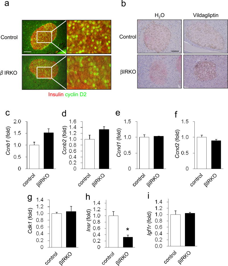
Consistent with in vitro data showing altered regulation of cyclins, histological analyses of βIRKO mouse pancreas sections revealed impaired cyclin D2 expression in beta cells (Fig. 8a). However, while the protein expression of cyclinD1 in the basal state was not reduced in islets from βIRKO mice when compared with islets from control insulin receptor-floxed mice, its expression appeared to be enhanced by vildagliptin treatment (Fig. 8b). This result is consistent with the increase in expression of the cyclins D1 and D2 in Ex-4-treated βIRKO cell lines (Fig. 5a). The mRNA expression of the cyclins D1 and D2 was not reduced in the islets from βIRKO mice vs control mice (Fig. 8e,f).
Fig. 8.
Effect of GLP-1 signalling activation on cyclin expression in mouse islets. (a) Co-immunostaining of pancreas sections from insulin-receptor-floxed control mice and βIRKO mice for insulin (red) and cyclin D2 (green). A representative islet is shown for each group. Magnification ×40; scale bar, 50 µm. (b) Immunostaining of pancreas sections from control and βIRKO mice for cyclin D1 (brown) with a nuclear counter stain. A representative islet is shown for each group. Magnification ×40; scale bar, 50 µm. (c–i) Gene expression in islets isolated from control and βIRKO mice. Expression was normalised to β-actin. n=3 in each group. *p<0.05 vs control
Discussion
In this study, we investigated the effects of incretins in a unique mouse model of diabetes that exhibits beta cell defects also evident in humans with impaired glucose tolerance. Enhancing the activity of GLP-1 in βIRKO mice led to an increase in beta cell proliferation secondary to an upregulation of cyclins, with subsequent promotion of secretory function and improvement in glucose tolerance. These findings have therapeutic implications for the use of incretins in improving beta cell mass and function in individuals with impaired glucose tolerance or type 2 diabetes who manifest defective growth factor signalling in beta cells.
GLP-1 has been reported to improve the function of beta cells by enhancing their proliferation in several rodent models of type 2 diabetes [34]. Though supraphysiological levels of incretins are necessary to enhance the insulin secretory response in individuals with type 2 diabetes, their effects on beta cell proliferation are less clear [35]. To investigate these effects, we chose the βIRKO mouse, a model that exhibits blunted acute phase insulin secretion and an age-dependent loss of beta cell mass [11]. This model also reflects features similar to the dysfunctional growth factor signalling found in beta cells of individuals with type 2 diabetes as a result of a downregulation in signalling proteins observed in [26, 27, 36].
In the current study, upregulation of beta cell mitosis was observed in βIRKO mice following injection of both DPP-4 inhibitor and GLP-1. A chronic endogenous increase in GLP-1, following treatment with vildagliptin, suppressed DPP-4 activity to an equal extent in βIRKO mice and littermate control insulin-receptor-floxed mice (Fig. 1c). The βIRKO mice, however, exhibited a greater increase in circulating GLP-1 and insulin and consequently a greater improvement in glycaemia. It is possible that the beneficial effects on beta cells also occur, in part, due to preserved circulating levels of GIP, another direct target of DPP-4 [34]. Indeed, it is reported that both GLP-1 and GIP receptors in beta cells mediate the beneficial effects of DPP-4 inhibitors [37]. On the other hand, direct GLP-1 monotherapy in βIRKO mice also improved glycaemia due, in part, to an increase in the proliferation of beta cells, emphasising the merits of enhancing the levels of GLP-1 in our current study (ESM Fig. 1), in agreement with reports in other models [4, 38, 39]. Our additional experiments using beta cell lines treated with GLP-1 and its analogue (Fig. 3 and Fig. 5) also support the in vivo data of GLP-1 effects on beta cell mitosis. Recent findings in a beta cell-specific GIP receptor-disrupted mouse model indicate that the effects of GIP on beta cells are mostly due to suppression of apoptosis and enhancement of insulin secretion [40]. Other considerations when examining the pleiotropic actions of DPP-4 and inhibiting its activity include: (1) a role for other peptides, including stromal cell-derived factor 1 (SDF-1), substance P and neuropeptide Y (NPY), which are known substrates of DPP-4 [41]; (2) data suggesting that in addition to its peptidase activity, DPP-4 binds to other proteins and is directly involved in immune stimulation, binding to extracellular matrix and lipid accumulation [42]; (3) reports that DPP-4 itself acts as an adipokine which impairs insulin sensitivity [43]. Thus, while it is possible that the effects of vildagliptin observed in βIRKO mice are due to the actions of both incretins, additional studies are necessary to ascertain whether the contributions of one incretin dominates the other on beta cell mitosis and whether the pleiotropic effects of DPP-4 inhibitors add or detract from the benefits of the incretins in the treatment of diabetes.
In vildagliptin-treated βIRKO mice, the plasma GLP-1 levels were significantly upregulated to a level that was higher than those in control mice treated similarly. While the pathways that promote higher levels of circulating GLP-1 in the βIRKO mice are not understood it is notable that elevated plasma DPP-4 activity has been reported in several studies, suggesting a relationship between impaired glucose tolerance, obesity and DPP-4 enzymatic activity [44–46]. Thus, it is possible that basal DPP-4 activity is upregulated in the glucose-intolerant βIRKO mice, leading to compensatory upregulation of GLP-1 secretion prior to treatment and that, subsequently, efficient DPP-4 inhibition by vildagliptin resulted in higher plasma GLP-1 values following oral glucose ingestion.
In addition to enhancing GSIS in beta cells, incretins exhibit anti-apoptotic activity [47]. In our study, although treatment of βIRKO mice with a DPP-4 inhibitor upregulated beta cell proliferation we did not detect significant changes in apoptosis. This may be related, in part, to the milder ‘glucose-intolerant’ phenotype in βIRKO mice in contrast to the severe hyperglycaemia typically observed in models of diabetes such as db/db mice [48].
The longer duration of treatment with vildagliptin (6 weeks), compared with acute GLP-1 treatment (3 weeks), may have also contributed to the improvement in glucose homeostasis in the former group. Indeed, a significant increase in beta cell mass was evident in vildagliptin-treated mice, while 3 weeks of GLP-1 treatment led to a significant increase in cellular proliferation markers but no discernible change in mass. These data indicate that in addition to acute stimulation of insulin secretion, the chronic effect of prolonged GLP-1 therapy is necessary to increase beta cell mass. The lack of effects of GLP-1 in control mice is consistent with a selective role for the incretin hormone in hyperglycaemia and underscores its significance as a therapeutic agent for type 2 diabetes [35]. The possibility that GLP-1 signalling is suppressed by the dominant effects of insulin signalling, to prevent excess stimulation and allow for beta cell rest in the normal state, requires further work.
In mice, cyclin D2 is a dominant determinant of beta cell-cycle progression [19], and disruption of cyclin D2 or its functional partner CDK4 leads to a diabetic phenotype due to severely impaired beta cell proliferation [18, 19]. In our studies, the low levels of cyclin D2 protein in beta cells are consistent with their slow growth and reduced mass in the βIRKO mice [10, 11]. The results from experiments following insulin receptor re-expression in βIRKO cells or acute ablation of insulin receptors in insulin receptor-floxed cells suggests that modulation of insulin receptors directly regulates cyclin D2 expression in beta cells. The increase in beta cell proliferation in βIRKO mice following vildagliptin treatment can be explained by the upregulation of cyclins A, D1, D2 and E, each of which is important for beta cell-cycle progression [17]. Indeed, elevated cyclin D1 promotes beta cell proliferation and an increase in islet mass in mice, while another study reported that Ex-4 treatment elevated cyclin A2 expression in beta cells in a knock-in mouse model, with upregulated cAMP signalling secondary to activated CREB [23, 24].
It has been reported that cyclin D2 is regulated at the level of protein stability rather than transcription [49]. The expression of cyclin D2 protein was significantly reduced in βIRKO cells; its mRNA expression was not decreased in islets from βIRKO mice and, rather, was increased in the βIRKO beta cell line. However, protein stability of cyclin D2 assessed by CHX assay was comparable between groups. Since p27 protein is degraded by cyclin D2-dependent translocation to cytosol during G0 to G1 cell-cycle entry [50], the enhanced expression of p27 in βIRKO cells could be linked to the reduced cyclin D2 activity. These findings warrant further investigation into the regulatory relationship between insulin receptor and cyclin D2 protein expression in beta cells.
In summary, promotion of GLP-1 signalling enhances beta cell proliferation by upregulating various cyclins and compensates for impaired proliferation of beta cells lacking insulin receptors. These findings suggest an important therapeutic role for GLP-1 and it analogues even in beta cells with dysfunctional signalling as occurs in individuals with type 2 diabetes.
Supplementary Material
Acknowledgments
We thank C. R. Kahn and R. Suzuki (Joslin Diabetes Center, Boston, MA, USA) for discussions, A. J. Kurpad (Joslin Diabetes Center) for technical assistance and H. Li, Z. Fu and G. Sankaranarayanan (Specialized Assay Core, DERC, Joslin Diabetes Center, Boston, MA, USA) for assays.
Funding
DK is the recipient of a Research Fellowship (Manpei Suzuki Diabetes Foundation, Japan) and a JDRF Post-doctoral Fellowship. JS is the recipient of a Research Fellowship (the Japan Society for the Promotion of Science [JSPS] and the Uehara Memorial Foundation, Japan). This work was supported in part by NIH DK67536 (RNK), NIH DK103215 (RNK), NIH DK55523 (RNK), 5P30DK36836 (Joslin DERC Specialized Assay and Advanced Microscopy Cores), K99DK090210 (CWL), R00DK090210 (CWL) and the Novartis Institutes for Biomedical Research Inc. (RNK). The study sponsors were not involved in the design of the study; the collection, analysis, and interpretation of data; writing the report; or the decision to submit the report for publication.
Abbreviations
- βIRKO
Beta cell-specific insulin receptor knockout
- CDK
Cyclin-dependent kinase
- CHX
Cycloheximide
- CREB
cAMP response-element binding protein
- DPP-4
Dipeptidyl peptidase-4
- Ex-4
Exendin-4
- GFP
Green fluorescent protein
- GIP
Glucose-dependent insulinotropic peptide
- GLP-1
Glucagon-like peptide-1
- GSIS
Glucose-stimulated insulin secretion
- HH3
Histone H3
- IRES
Internal ribosome entry site
- KRBB
Krebs-Ringer bicarbonate buffer
Footnotes
Data availability The datasets generated during and/or analysed during the current study are available from the corresponding author on reasonable request.
Duality of interest AD is an employee of the Novartis Institutes for Biomedical Research Inc. BB was an employee of the Novartis Institutes for Biomedical Research Inc. and is an employee of Zafgen Inc. All other authors declare that there is no duality of interest associated with their contribution to this manuscript.
Contribution statement DK, JS and RNK planned the experiments, researched data, wrote the manuscript and reviewed/edited the manuscript. CWL, JH and TM acquired and analysed the data. AD and BB provided materials and contributed to conception, study design, and interpretation of the data. DK and RNK are the guarantors of this work, had full access to all the data and take full responsibility for the integrity of data and the accuracy of data analysis. All authors approved the manuscript for publication.
References
- 1.Kahn CR, Weir GC, King GL, Jacobson AM, Moses AC, Smith RJ. Diabetes mellitus. Lippincott, Williams and Wilkins; New York, NY: 2005. [Google Scholar]
- 2.Butler AE, Janson J, Bonner-Weir S, Ritzel R, Rizza RA, Butler PC. Beta-cell deficit and increased beta-cell apoptosis in humans with type 2 diabetes. Diabetes. 2003;52:102–110. doi: 10.2337/diabetes.52.1.102. [DOI] [PubMed] [Google Scholar]
- 3.Drucker DJ. The biology of incretin hormones. Cell metabolism. 2006;3:153–165. doi: 10.1016/j.cmet.2006.01.004. [DOI] [PubMed] [Google Scholar]
- 4.Stoffers DA, Kieffer TJ, Hussain MA, et al. Insulinotropic glucagon-like peptide 1 agonists stimulate expression of homeodomain protein IDX-1 and increase islet size in mouse pancreas. Diabetes. 2000;49:741–748. doi: 10.2337/diabetes.49.5.741. [DOI] [PubMed] [Google Scholar]
- 5.Habener JF, Stoffers DA. A newly discovered role of transcription factors involved in pancreas development and the pathogenesis of diabetes mellitus. Proceedings of the Association of American Physicians. 1998;110:12–21. [PubMed] [Google Scholar]
- 6.Farilla L, Bulotta A, Hirshberg B, et al. Glucagon-like peptide 1 inhibits cell apoptosis and improves glucose responsiveness of freshly isolated human islets. Endocrinology. 2003;144:5149–5158. doi: 10.1210/en.2003-0323. [DOI] [PubMed] [Google Scholar]
- 7.Hui H, Nourparvar A, Zhao X, Perfetti R. Glucagon-like peptide-1 inhibits apoptosis of insulin-secreting cells via a cyclic 5'-adenosine monophosphate-dependent protein kinase A- and a phosphatidylinositol 3-kinase-dependent pathway. Endocrinology. 2003;144:1444–1455. doi: 10.1210/en.2002-220897. [DOI] [PubMed] [Google Scholar]
- 8.Jhala US, Canettieri G, Screaton RA, et al. cAMP promotes pancreatic β-cell survival via CREB-mediated induction of IRS2. Genes & development. 2003;17:1575–1580. doi: 10.1101/gad.1097103. [DOI] [PMC free article] [PubMed] [Google Scholar]
- 9.Ranta F, Avram D, Berchtold S, et al. Dexamethasone induces cell death in insulin-secreting cells, an effect reversed by exendin-4. Diabetes. 2006;55:1380–1390. doi: 10.2337/db05-1220. [DOI] [PubMed] [Google Scholar]
- 10.Assmann A, Hinault C, Kulkarni RN. Growth factor control of pancreatic islet regeneration and function. Pediatr Diabetes. 2009;10:14–32. doi: 10.1111/j.1399-5448.2008.00468.x. [DOI] [PMC free article] [PubMed] [Google Scholar]
- 11.Kulkarni RN, Bruning JC, Winnay JN, Postic C, Magnuson MA, Kahn CR. Tissue-specific knockout of the insulin receptor in pancreatic beta cells creates an insulin secretory defect similar to that in type 2. Cell. 1999;96:329–339. doi: 10.1016/s0092-8674(00)80546-2. [DOI] [PubMed] [Google Scholar]
- 12.Kulkarni RN, Holzenberger M, Shih DQ, et al. β-cell-specific deletion of the Igf1 receptor leads to hyperinsulinemia and glucose intolerance but does not alter β-cell mass. Nature genetics. 2002;31:111–115. doi: 10.1038/ng872. [DOI] [PubMed] [Google Scholar]
- 13.Xuan S, Kitamura T, Nakae J, et al. Defective insulin secretion in pancreatic β cells lacking type 1 IGF receptor. The Journal of clinical investigation. 2002;110:1011–1019. doi: 10.1172/JCI15276. [DOI] [PMC free article] [PubMed] [Google Scholar]
- 14.Shirakawa J, Fernandez M, Takatani T, et al. Insulin signaling regulates the FoxM1/PLK1/CENP-A pathway to promote adaptive pancreatic β cell proliferation. Cell Metab. 2017;25:868–882. doi: 10.1016/j.cmet.2017.02.004. [DOI] [PMC free article] [PubMed] [Google Scholar]
- 15.Cornu M, Modi H, Kawamori D, Kulkarni RN, Joffraud M, Thorens B. Glucagon-like peptide-1 increases β-cell glucose competence and proliferation by translational induction of insulin-like growth factor-1 receptor expression. The Journal of biological chemistry. 2010;285:10538–10545. doi: 10.1074/jbc.M109.091116. [DOI] [PMC free article] [PubMed] [Google Scholar]
- 16.Cornu M, Yang JY, Jaccard E, Poussin C, Widmann C, Thorens B. Glucagon-like peptide-1 protects β-cells against apoptosis by increasing the activity of an IGF-2/IGF-1 receptor autocrine loop. Diabetes. 2009;58:1816–1825. doi: 10.2337/db09-0063. [DOI] [PMC free article] [PubMed] [Google Scholar]
- 17.Cozar-Castellano I, Fiaschi-Taesch N, Bigatel TA, et al. Molecular control of cell cycle progression in the pancreatic β-cell. Endocrine reviews. 2006;27:356–370. doi: 10.1210/er.2006-0004. [DOI] [PubMed] [Google Scholar]
- 18.Georgia S, Bhushan A. β cell replication is the primary mechanism for maintaining postnatal β cell mass. The Journal of clinical investigation. 2004;114:963–968. doi: 10.1172/JCI22098. [DOI] [PMC free article] [PubMed] [Google Scholar]
- 19.Kushner JA, Ciemerych MA, Sicinska E, et al. Cyclins D2 and D1 are essential for postnatal pancreatic β-cell growth. Molecular and cellular biology. 2005;25:3752–3762. doi: 10.1128/MCB.25.9.3752-3762.2005. [DOI] [PMC free article] [PubMed] [Google Scholar]
- 20.Georgia S, Hinault C, Kawamori D, et al. Cyclin D2 is essential for the compensatory β-cell hyperplastic response to insulin resistance in rodents. Diabetes. 2010;59:987–996. doi: 10.2337/db09-0838. [DOI] [PMC free article] [PubMed] [Google Scholar]
- 21.Zhang X, Gaspard JP, Mizukami Y, Li J, Graeme-Cook F, Chung DC. Overexpression of cyclin D1 in pancreatic beta-cells in vivo results in islet hyperplasia without hypoglycemia. Diabetes. 2005;54:712–719. doi: 10.2337/diabetes.54.3.712. [DOI] [PubMed] [Google Scholar]
- 22.Friedrichsen BN, Neubauer N, Lee YC, et al. Stimulation of pancreatic β-cell replication by incretins involves transcriptional induction of cyclin D1 via multiple signalling pathways. The Journal of endocrinology. 2006;188:481–492. doi: 10.1677/joe.1.06160. [DOI] [PubMed] [Google Scholar]
- 23.Liu Z, Habener JF. Glucagon-like peptide-1 activation of TCF7L2-dependent Wnt signaling enhances pancreatic beta cell proliferation. The Journal of biological chemistry. 2008;283:8723–8735. doi: 10.1074/jbc.M706105200. [DOI] [PMC free article] [PubMed] [Google Scholar]
- 24.Song WJ, Schreiber WE, Zhong E, et al. Exendin-4 stimulation of cyclin A2 in β-cell proliferation. Diabetes. 2008;57:2371–2381. doi: 10.2337/db07-1541. [DOI] [PMC free article] [PubMed] [Google Scholar]
- 25.Xie T, Chen M, Zhang QH, Ma Z, Weinstein LS. β cell-specific deficiency of the stimulatory G protein α-subunit Gsα leads to reduced beta cell mass and insulin-deficient diabetes. Proceedings of the National Academy of Sciences of the United States of America. 2007;104:19601–19606. doi: 10.1073/pnas.0704796104. [DOI] [PMC free article] [PubMed] [Google Scholar]
- 26.Folli F, Okada T, Perego C, et al. Altered insulin receptor signalling and β-cell cycle dynamics in type 2 diabetes mellitus. PloS one. 2011;6:e28050. doi: 10.1371/journal.pone.0028050. [DOI] [PMC free article] [PubMed] [Google Scholar]
- 27.Gunton JE, Kulkarni RN, Yim S, et al. Loss of ARNT/HIF1beta mediates altered gene expression and pancreatic-islet dysfunction in human type 2 diabetes. Cell. 2005;122:337–349. doi: 10.1016/j.cell.2005.05.027. [DOI] [PubMed] [Google Scholar]
- 28.Winzell MS, Ahren B. The high-fat diet-fed mouse: a model for studying mechanisms and treatment of impaired glucose tolerance and type 2 diabetes. Diabetes. 2004;53(Suppl 3):S215–S219. doi: 10.2337/diabetes.53.suppl_3.s215. [DOI] [PubMed] [Google Scholar]
- 29.Assmann A, Ueki K, Winnay JN, Kadowaki T, Kulkarni RN. Glucose effects on β-cell growth and survival require activation of insulin receptors and insulin receptor substrate 2. Molecular and cellular biology. 2009;29:3219–3228. doi: 10.1128/MCB.01489-08. [DOI] [PMC free article] [PubMed] [Google Scholar]
- 30.Liu S, Okada T, Assmann A, et al. Insulin signaling regulates mitochondrial function in pancreatic β-cells. PLoS One. 2009;4:e7983. doi: 10.1371/journal.pone.0007983. [DOI] [PMC free article] [PubMed] [Google Scholar]
- 31.Kawamori D, Kaneto H, Nakatani Y, et al. The forkhead transcription factor Foxo1 bridges the JNK pathway and the transcription factor PDX-1 through its intracellular translocation. The Journal of biological chemistry. 2006;281:1091–1098. doi: 10.1074/jbc.M508510200. [DOI] [PubMed] [Google Scholar]
- 32.Drucker DJ, Nauck MA. The incretin system: glucagon-like peptide-1 receptor agonists and dipeptidyl peptidase-4 inhibitors in type 2 diabetes. Lancet. 2006;368:1696–1705. doi: 10.1016/S0140-6736(06)69705-5. [DOI] [PubMed] [Google Scholar]
- 33.Yang J, Robert CE, Burkhardt BR, et al. Mechanisms of glucose-induced secretion of pancreatic-derived factor (PANDER or FAM3B) in pancreatic β-cells. Diabetes. 2005;54:3217–3228. doi: 10.2337/diabetes.54.11.3217. [DOI] [PubMed] [Google Scholar]
- 34.Kim W, Egan JM. The role of incretins in glucose homeostasis and diabetes treatment. Pharmacological reviews. 2008;60:470–512. doi: 10.1124/pr.108.000604. [DOI] [PMC free article] [PubMed] [Google Scholar]
- 35.Holst JJ, Vilsboll T, Deacon CF. The incretin system and its role in type 2 diabetes mellitus. Molecular and cellular endocrinology. 2009;297:127–136. doi: 10.1016/j.mce.2008.08.012. [DOI] [PubMed] [Google Scholar]
- 36.Marchetti P, Lupi R, Del Guerra S, Bugliani M, Marselli L, Boggi U. The β-cell in human type 2 diabetes. Advances in experimental medicine and biology. 2010;654:501–514. doi: 10.1007/978-90-481-3271-3_22. [DOI] [PubMed] [Google Scholar]
- 37.Flock G, Baggio LL, Longuet C, Drucker DJ. Incretin receptors for glucagon-like peptide 1 and glucose-dependent insulinotropic polypeptide are essential for the sustained metabolic actions of vildagliptin in mice. Diabetes. 2007;56:3006–3013. doi: 10.2337/db07-0697. [DOI] [PubMed] [Google Scholar]
- 38.Buteau J, Foisy S, Joly E, Prentki M. Glucagon-like peptide 1 induces pancreatic β-cell proliferation via transactivation of the epidermal growth factor receptor. Diabetes. 2003;52:124–132. doi: 10.2337/diabetes.52.1.124. [DOI] [PubMed] [Google Scholar]
- 39.Li Y, Cao X, Li LX, Brubaker PL, Edlund H, Drucker DJ. β-Cell Pdx1 expression is essential for the glucoregulatory, proliferative, and cytoprotective actions of glucagon-like peptide-1. Diabetes. 2005;54:482–491. doi: 10.2337/diabetes.54.2.482. [DOI] [PubMed] [Google Scholar]
- 40.Campbell JE, Ussher JR, Mulvihill EE, et al. TCF1 links GIPR signaling to the control of beta cell function and survival. Nature medicine. 2016;22:84–90. doi: 10.1038/nm.3997. [DOI] [PubMed] [Google Scholar]
- 41.Drucker DJ. Dipeptidyl peptidase-4 inhibition and the treatment of type 2 diabetes: preclinical biology and mechanisms of action. Diabetes care. 2007;30:1335–1343. doi: 10.2337/dc07-0228. [DOI] [PubMed] [Google Scholar]
- 42.Itou M, Kawaguchi T, Taniguchi E, Sata M. Dipeptidyl peptidase-4: a key player in chronic liver disease. World journal of gastroenterology. 2013;19:2298–2306. doi: 10.3748/wjg.v19.i15.2298. [DOI] [PMC free article] [PubMed] [Google Scholar]
- 43.Lamers D, Famulla S, Wronkowitz N, et al. Dipeptidyl peptidase 4 is a novel adipokine potentially linking obesity to the metabolic syndrome. Diabetes. 2011;60:1917–1925. doi: 10.2337/db10-1707. [DOI] [PMC free article] [PubMed] [Google Scholar]
- 44.Ryskjaer J, Deacon CF, Carr RD, et al. Plasma dipeptidyl peptidase-IV activity in patients with type-2 diabetes mellitus correlates positively with HbAlc levels, but is not acutely affected by food intake. European journal of endocrinology. 2006;155:485–493. doi: 10.1530/eje.1.02221. [DOI] [PubMed] [Google Scholar]
- 45.Fadini GP, Albiero M, Menegazzo L, de Kreutzenberg SV, Avogaro A. The increased dipeptidyl peptidase-4 activity is not counteracted by optimized glucose control in type 2 diabetes, but is lower in metformin-treated patients. Diabetes Obes Metab. 2012;14:518–522. doi: 10.1111/j.1463-1326.2011.01550.x. [DOI] [PubMed] [Google Scholar]
- 46.Sell H, Bluher M, Kloting N, et al. Adipose dipeptidyl peptidase-4 and obesity: correlation with insulin resistance and depot-specific release from adipose tissue in vivo and in vitro. Diabetes care. 2013;36:4083–4090. doi: 10.2337/dc13-0496. [DOI] [PMC free article] [PubMed] [Google Scholar]
- 47.Shirakawa J, Amo K, Ohminami H, et al. Protective effects of dipeptidyl peptidase-4 (DPP-4) inhibitor against increased beta cell apoptosis induced by dietary sucrose and linoleic acid in mice with diabetes. The Journal of biological chemistry. 2011;286:25467–25476. doi: 10.1074/jbc.M110.217216. [DOI] [PMC free article] [PubMed] [Google Scholar]
- 48.Shirakawa J, Okuyama T, Kyohara M, et al. DPP-4 inhibition improves early mortality, β cell function, and adipose tissue inflammation in db/db mice fed a diet containing sucrose and linoleic acid. Diabetology & metabolic syndrome. 2016;8:16. doi: 10.1186/s13098-016-0138-4. [DOI] [PMC free article] [PubMed] [Google Scholar]
- 49.He LM, Sartori DJ, Teta M, et al. Cyclin D2 protein stability is regulated in pancreatic β-cells. Mol Endocrinol. 2009;23:1865–1875. doi: 10.1210/me.2009-0057. [DOI] [PMC free article] [PubMed] [Google Scholar]
- 50.Susaki E, Nakayama K, Nakayama KI. Cyclin D2 translocates p27 out of the nucleus and promotes its degradation at the G0-G1 transition. Molecular and cellular biology. 2007;27:4626–4640. doi: 10.1128/MCB.00862-06. [DOI] [PMC free article] [PubMed] [Google Scholar]
Associated Data
This section collects any data citations, data availability statements, or supplementary materials included in this article.


