



Abstract
Key points
The mucosa may have neuron‐like functions as urinary bladder mucosa releases bioactive substances that modulate sensory nerve activity as well as detrusor muscle contractility. However, such mucosal function in other visceral organs remains to be established.
The role of mucosa in generating spontaneous contractions in seminal vesicles (SVs), a paired organ in the male reproductive tract, was investigated.
The intact mucosa is essential for the generation of spontaneous phasic contractions of SV smooth muscle arising from electrical slow waves and corresponding increases in intracellular Ca2+. These spontaneous events primarily depend on Ca2+ handling by sarco‐endoplasmic reticulum Ca2+ stores.
A population of mucosal cells developed spontaneous rises in intracellular Ca2+ relying on sarco‐endoplasmic reticulum Ca2+ handling.
The spontaneously active cells in the SV mucosa appear to drive spontaneous activity in smooth muscle either by sending depolarizing signals and/or by releasing humoral substances.
Abstract
The role of the mucosa in generating the spontaneous activity of guinea‐pig seminal vesicle (SV) was explored. Changes in contractility, membrane potential and intracellular Ca2+ dynamics of SV smooth muscle cells (SMCs) were recorded using isometric tension recording, intracellular microelectrode recording and epi‐fluorescence Ca2+ imaging, respectively. Mucosa‐intact but not mucosa‐denuded SV preparations generated TTX‐ (1 μm) resistant spontaneous phasic contractions that were abolished by nifedipine (3 μm). Consistently, SMCs developed mucosa‐dependent slow waves (SWs) that triggered action potentials and corresponding Ca2+ flashes. Nifedipine (10 μm) abolished the action potentials and spontaneous contractions, while suppressing the SWs and Ca2+ flashes. Both the residual SWs and spontaneous Ca2+ transients were abolished by cyclopiazonic acid (CPA, 10 μm), a sarco‐endoplasmic reticulum Ca2+‐ATPase (SERCA) inhibitor. DIDS (300 μm) and niflumic acid (100 μm), blockers for Ca2+‐activated Cl− channels (CACCs), or low Cl− solution also slowed or prevented the generation of SWs. In SV mucosal preparations detached from the muscle layer, a population of mucosal cells generated spontaneous Ca2+ transients that were blocked by CPA but not nifedipine. These results suggested that spontaneous contractions and corresponding Ca2+ flashes in SV SMCs arise from action potential generation due to the opening of L‐type voltage‐dependent Ca2+ channels. Spontaneous Ca2+ transients appear to primarily result from Ca2+ release from sarco‐endoplasmic reticulum Ca2+ stores to activate CACCs to develop SWs. The mucosal cells firing spontaneous Ca2+ transients may play a critical role in driving spontaneous activity of SV smooth muscle either by sending depolarizing signals or by releasing humoral substances.
Keywords: intracellular Ca2+ release, mucosa, seminal vesicle, slow wave, spontaneous contraction
Key points
The mucosa may have neuron‐like functions as urinary bladder mucosa releases bioactive substances that modulate sensory nerve activity as well as detrusor muscle contractility. However, such mucosal function in other visceral organs remains to be established.
The role of mucosa in generating spontaneous contractions in seminal vesicles (SVs), a paired organ in the male reproductive tract, was investigated.
The intact mucosa is essential for the generation of spontaneous phasic contractions of SV smooth muscle arising from electrical slow waves and corresponding increases in intracellular Ca2+. These spontaneous events primarily depend on Ca2+ handling by sarco‐endoplasmic reticulum Ca2+ stores.
A population of mucosal cells developed spontaneous rises in intracellular Ca2+ relying on sarco‐endoplasmic reticulum Ca2+ handling.
The spontaneously active cells in the SV mucosa appear to drive spontaneous activity in smooth muscle either by sending depolarizing signals and/or by releasing humoral substances.
Abbreviations
- α‐SMA
α‐smooth muscle actin
- AMP
peak amplitude
- ANO1
anoctamin1
- AR
adrenoceptor
- BK channel
large conductance Ca2+‐activated K+ channel
- CACC
Ca2+‐activated Cl− channel
- COX‐2
cyclooxygenase‐2
- CPA
cyclopiazonic acid
- DAPI
4′6‐diamidino‐2‐phenylindole
- EFS
electrical field stimulation
- GI
gastrointestinal
- HW
half‐width
- ICC
interstitial cells of Cajal
- IR
immunoreactive
- LP
lamina propria
- LVDCC
L‐type voltage‐dependent Ca2+ channel
- M
muscular layer
- NKCC
Na+–K+–Cl− cotransporter
- PDGFRα
platelet‐derived growth factor receptor α
- x% PBSTNB
PBS containing x% TritonX, 0.1% NaN3 and 1% BSA
- PSS
physiological salt solution
- RMP
resting membrane potential
- ROI
region of interest
- SERCA
sarco‐endoplasmic reticulum Ca2+‐ATPase
- SK
small conductance Ca2+‐activated K+ channel
- SR/ER
sarcoplasmic/endoplasmic reticulum
- STD
spontaneous transient depolarization
- SV
seminal vesicle
- SW
slow wave
- TEA
tetraethylammonium
Introduction
Visceral smooth muscle organs develop spontaneous contractile activity, in addition to vigorously contracting in response to autonomic and/or intrinsic nervous input. Spontaneous contractions were originally considered myogenic in origin, i.e. generated by the smooth muscle cells themselves. However, growing evidence has revealed the role of distinct populations of cells driving the smooth muscle cells. In the gastrointestinal (GI) tract, it is well established that interstitial cells of Cajal (ICC) act as pacemaker cells to electrically drive the bulk of smooth muscle (Sanders et al. 2014). Anoctamin 1 (ANO1), a Ca2+‐activated Cl− channel (CACC), expressed in ICC plays a critical role in generating slow waves (SWs) in the GI tract (Sanders et al. 2014; Oh & Jung, 2016; Zhu et al. 2016). In the urinary tract, atypical smooth muscle cells in the renal pelvis (Lang et al. 2007a) and interstitial cells in the urethra (Sergeant et al. 2000) appear to function as pacemaker cells sending depolarizing signals to neighbouring smooth muscle cells. Recently, platelet‐derived growth factor receptor α (PDGFRα)‐positive cells, that were previously known as fibroblast‐like cells, have been reported to develop spontaneous Ca2+ transients and resultant hyperpolarizations to regulate smooth muscle contractility in the GI tract and the urinary bladder (Kurahashi et al. 2011; Baker et al. 2013; Lee et al. 2013).
In male reproductive organs, smooth muscles of vas deferens, prostate and seminal vesicles (SVs) contract during ejaculation upon sympathetic nerve excitation (Coolen et al. 2004). During inter‐ejaculatory storage phase, these organs do not remain quiescent, but rather generate spontaneous phasic contractions that do not rely on autonomic nervous activity. Stretch‐sensitive mechanisms may maintain spontaneous peristalsis in the vas deferens to transport sperm from epididymis towards the ampulla (Bruschini et al. 1977). Spontaneous contractions of the prostate appear to be driven by specialized pacemaker cells, i.e. interstitial cells (Dey et al. 2010; Lam et al. 2011; Lang et al. 2014). Spontaneous contractions of the SVs have also been reported, both in vivo (Melin, 1970; Hib et al. 1983) and in vitro (Ohkawa, 1973, 1981, 1982; Birowo et al. 2010) in several mammals including humans.
The mechanisms underlying spontaneous activity in SVs remain to be explored. We have recently demonstrated that distention of ‘isolated’ whole SV of the guinea pig shows spontaneous TTX‐insensitive, phasic contractions (Hayashi et al. 2016). This is inconsistent with a previous report in which circular muscle strips of the guinea pig SV do not exhibit spontaneous contractions but respond to sympathetic nerve stimulation (Kubota et al. 2003). Such inconsistency may be attributable to the presence or absence of the mucosal layer in the SV preparations, and thus it is envisaged that the mucosa may play a critical role in developing spontaneous activity of SVs. However, the function of the SV mucosa is largely unknown except for secretion of seminal fluids that may play a pivotal role in reproductive success (Gonzales, 2001; Bromfield, 2014). In the bladder, the mucosa functions as a local centre of sensing micturition desire depending on the interplay amongst ‘neuron‐like’ urothelium, afferent nerves and heterogeneous populations of interstitial cells. In addition, the mucosal signals may also modulate spontaneous contractility of detrusor smooth muscle (Kanai et al. 2007; Birder & Andersson, 2013; Kushida & Fry, 2016).
In the present study, the mechanisms underlying spontaneous activity of guinea pig SVs were investigated, particularly focusing on the role of the mucosa. The contractile, electrical and intracellular Ca2+ activity was compared in mucosa‐intact and mucosa‐denuded SV smooth muscle preparations. In addition, Ca2+ imaging of ‘isolated’ mucosa was carried out to explore if any mucosal cells generate spontaneous Ca2+ transients. The cells within the mucosa were also investigated using immunohistochemistry.
Methods
Ethical approval
Animal housing and the experimental procedures have been approved by the animal experimentation ethics committee at Kurume University School of Medicine and Nagoya City University Graduate School of Medical Sciences in accordance with the guidelines of the Journal of Physiology (Grundy, 2015).
Tissue preparation
Ninety adult male Hartley guinea pigs (Japan SLC, Inc., Hamamatsu, Japan) weighing 600–1100 g were used for this study. Guinea pigs were anaesthetized with 3% isoflurane and exsanguinated by decapitation. The pair of SVs was excised and their lumens were flushed with 20 ml of physiological salt solution (PSS; see below for composition) after cutting off their blind ends. Ring segments (approximately 4 mm wide) were dissected out and incubated in oxygenated PSS at room temperature. For isometric tension recordings, the ring segments were inverted and used as reversed ring preparations. For intracellular recordings and intracellular calcium imaging, the ring segments were cut open, and the outer muscular layers were removed leaving the inner circular muscle layer (2–3 mm × 2–3 mm). Mucosa‐denuded smooth muscle preparations were prepared by peeling the mucosa off using fine forceps. In a separate series of experiments, the ‘isolated’ mucosa detached from smooth muscle layers was used for intracellular Ca2+ imaging and immunohistochemistry.
Isometric tension recordings
For isometric tension recordings of the SV circular muscles, reversed ring preparations were transversely mounted in 3 ml organ baths and superfused with warmed (36°C) PSS at a constant flow rate (4 ml min−1). A pair of stainless steel needles (0.45 mm outer diameter) was inserted into the lumen; one needle was fixed to the bottom of the organ bath, while the other was connected to an isometric force transducer connected to a bridge amplifier. An initial tension of 1 g was applied to the ring preparations. Isometric tension changes were digitized using the PowerLab 26T software program (ADInstruments, Bella Vista, NSW, Australia) and stored on a personal computer for later analysis.
In some experiments, ring preparations were initially adjusted to 0.2 g and electrical field stimulation (EFS) was applied after a minimum equilibration period of 30 min by passing brief square pulses (5 V, 0.1 ms) between a pair of the stainless needle stimulation electrodes every 4 min. The maximum contractions were evoked at frequencies above 80 Hz, and the test frequency was set at 20 or 30 Hz that evoked approximately 50% of the maximum contractions. The relative peak amplitudes evoked by the stimuli in the mucosa‐intact and mucosa‐denuded preparations were 63 ± 14% (50–86%, n = 5) and 58 ± 9% (50–68%, n = 5, P = 0.48) of the maximum response, respectively. The neuronal selectivity of EFS was confirmed when the evoked responses were abolished by 0.06–1 μm TTX.
Intracellular recordings
For intracellular recordings, inner circular muscle strips were firmly pinned, mucosal side down, on a silicone elastomer Sylgard (Dow Corning Corp., Midland, MI, USA) plate at the bottom of a 1 ml chamber. Preparations were superfused with warmed (36°C) PSS at a constant flow rate (2 ml min−1). After equilibration for more than 30 min, smooth muscle cells were impaled with glass capillary microelectrodes filled with 0.5 m KCl (tip resistance, 60–180 MΩ). 3 m KCl–agar salt bridges were used as a reference electrode. Membrane potential changes were recorded using a high input impedance amplifier (Axoclamp‐2A, Axon Instruments, Inc., Foster City, CA, USA) and displayed on a cathode‐ray oscilloscope (VC‐10, Nihon‐Kohden, Tokyo, Japan). After low‐pass filtering (cut‐off frequency 1 kHz), membrane potential changes were digitized using a Digidata 1322A interface (Axon Instruments) and stored on a personal computer for later analysis.
Intracellular calcium imaging
For intracellular calcium imaging, inner circular muscle strips or dissected mucosa were pinned out on a silicone elastomer Sylgard plate at the bottom of the recording chamber (volume, approximately 1 ml), epithelial side down. Preparations were superfused with warmed (36°C) PSS at a constant flow rate (2 ml min−1) and equilibrated for more than 30 min. To visualize intracellular calcium dynamics, preparations were incubated in low Ca2+ PSS ([Ca2+]o = 0.1 mm) containing 10 μm Cal‐520 acetoxymethyl ester (AAT Bioquest, Sunnyvale, CA, USA) and cremphor EL (0.01%, Sigma, St. Louis, MO, USA) for 60 min at 35°C. Following incubation, the recording chamber was mounted on the stage of an upright epifluorescence microscope (BX51WI, Olympus, Tokyo, Japan) equipped with a back‐thinned electron multiplying CCD camera (C9100‐13, Hamamatsu Photonics, Hamamatsu, Japan). Preparations were superfused with dye‐free PSS, viewed with a water immersion objective (UMPlanFLx20 or LUMPlanFL x40, x60, Olympus) and illuminated at 495 nm. Fluorescence was captured through a barrier filter above 515 nm, and images were obtained every 107–137 ms (frame interval) with an exposure time of 30–70 ms using a micro‐photoluminescence measurement system (AQUACOSMOS, Hamamatsu Photonics). Relative changes in ([Ca2+]i) were expressed as ∆F t /F 0 = (F t − F 0)/F 0, where F t is the fluorescence generated by an event, and F 0 is the basal fluorescence.
Scanning electron microscopy
To confirm the successful removal of the mucosa in the reversed ring preparations, after a brief isometric tension recording in normal PSS, some mucosa‐intact and mucosa‐denuded ring preparations were fixed in the organ bath by replacing the solution with Karnovsky fixative for 2 h. Preparations were post‐fixed in 1% osmium tetroxide solution for 1 h, dehydrated through a graded series of acetone, and freeze dried in t‐butylalcohol. They were then sputter coated with gold, and examined in a Hitachi S‐800 scanning electron microscope (Tokyo, Japan) operated at an accelerating voltage of 20 kV.
Light microscopy
To verify the removal of the mucosa, mucosa‐intact and mucosa‐denuded smooth muscle preparations were fixed for more than 48 h in 10% neutral buffered formalin (Wako Pure Chemical Industries, Osaka, Japan) and washed with PBS (pH 7.4). The preparations were embedded in paraffin blocks following dehydration with ethanol. Sections of the paraffin‐embedded specimens were cut at a thickness of 4 μm, adhered to charged slides and stained with haematoxylin and eosin and observed with a light microscope (BZ‐X710, KEYENCE, Osaka, Japan).
Immunohistochemistry
For whole mount immunostaining, the muscle‐free mucosa preparations were obtained in the same manner as for the intracellular calcium measurement. Specimens were fixed with acetone at −20°C for 20 min for c‐Kit, and fixed with Zamboni solution (0.1 m phosphate buffer: pH 7.3, 2% paraformaldehyde, 1.5% saturated picric acid) for 2 h at room temperature for PDGFRα and vimentin. After washing in PBS, specimens were blocked with 10% normal donkey serum in PBS containing 0.5% TritonX, 0.1% NaN3 and 1% BSA (0.5% PBSTNB, pH 7.5) for 12 h at 4°C, and then incubated in 0.5% PBSTNB for 72 h at 4°C with three combinations of primary antibodies for double labelling as follows: (1) chicken anti‐vimentin (1:2000; Abcam, Cambridge, UK) and mouse anti‐pancytokeratin (1:2000; Sigma), (2) rabbit anti‐PDGFRα (1:500; Cell Signaling Technology, Danvers, MA, USA) and mouse anti‐pancytokeratin (1:2000; Sigma), and (3) rat anti‐c‐Kit (1:100; LSBio, Seattle, WA, USA) and mouse anti‐α‐smooth muscle actin (α‐SMA, 1:2000; Sigma). The specimens were visualized by incubation with Alexa Fluor 488‐conjugated anti‐chicken IgG (1:1000; Abcam), Alexa Fluor 488‐conjugated anti‐rabbit or rat IgG (1:1000; Thermo Fisher Scientific, Waltham, MA, USA), Alexa Fluor 568‐conjugated anti‐mouse IgG (1:1000; Thermo Fisher Scientific) and 4′6‐diamidino‐2‐phenylindole (DAPI, 1:5000; Wako Pure Chemical Industries) for 12 h at 4°C. For washing both the primary and the secondary antibodies, PBS containing 0.05% Tween20 was used. The preparations were examined from the basolateral (subepithelial) side with a confocal laser scanning microscope (FV1000, Olympus). Serial images were obtained from the surface to the depth of several micrometres and processed using Olympus confocal imaging software.
In some experiments, c‐Kit or ANO1 immunoreactivity in the SV wall was compared with that in smooth muscle layer of the guinea pig stomach (positive control) using the same protocol as follows: coronal cryosections (5 μm thick) were mounted on the glass slides were fixed with acetone at −20°C for 20 min. The sections were blocked with 10% normal donkey serum in 0.1% PBSTNB (0.1% TritonX, pH 7.5) for 15 min and incubated with the primary antibody: rat anti‐c‐Kit (1:100) or rabbit anti‐ANO1 (1:100; Abcam), and double‐immunolabelled with mouse anti‐α‐SMA (1:2000) in 0.1% PBSTNB for 1 h. The sections were visualized via incubation with Alexa Fluor 488 anti‐rat IgG for c‐Kit, Alexa Fluor 488 anti‐rabbit IgG for ANO1 and the Alexa Fluor 568 anti‐mouse IgG for α‐SMA (1:1000; Thermo Fisher Scientific) for 30 min. Nuclei were stained with DAPI (1:5000). PBS was used for washing both primary and secondary antibodies. The preparations were examined with a confocal laser scanning microscope (FV1000) or a fluorescence microscope (Axioskop, Zeiss, Germany).
Solutions
The composition of PSS was (in mm): Na+ 143.2, K+ 5.9, Ca2+ 2.5, Mg2+ 1.2, HCO3 − 25, H2PO4 − 1.2, Cl− 130.3 and glucose 11. Low Cl− solution ([Cl−]o = 13.3 mm) was prepared by replacing NaCl with equimolar sodium isethionate. Perfusate was continuously bubbled with 95% O2 and 5% CO2. The pH of bubbled solutions was approximately 7.4 at 36°C. Drugs used were ATP, carbenoxolone disodium salt, cyclopiazonic acid (CPA), nifedipine, niflumic acid, DIDS, phenylephrine hydrochloride (from Sigma), TTX (from Nacalai Tesque, Kyoto, Japan) and T16Ainh‐A01 (from Cayman Chemical, Ann Arbor, MI, USA). Drugs were dissolved in distilled water, except for CPA, DIDS, nifedipine, niflumic acid and T16Ainh‐A01, which were dissolved in DMSO. The final concentration of these solvents in the PSS did not exceed 1:1000.
Data analysis
The following parameters of spontaneous events were measured: peak amplitude, measured as the value from the resting level to the peak of events; half‐width, measured as the time between 50% of peak amplitude on the rising and falling phases; and maximum rising slope, measured as the maximum rate of rise of the peak calculated as an average of five events. The frequency (min−1) was calculated as an average over 1–5 min of recording. The parameters of the isometric tension were analysed using the LabChart software package (ADInstruments). The parameters of the membrane potential and [Ca2+]i, were measured using the Clampfit 10 software (Axon Instruments‐Molecular Devices, Union City, CA, USA). For statistical analysis of all data, SigmaPlot11 software (Hulinks Inc. ,Tokyo, Japan) was used. Measured values are expressed as means ± SD. Statistical significance was tested using Student's t test (paired or unpaired), and probabilities of less than 5% (P < 0.05) were considered significant; n denotes the number of the observations within a single experiment and equals the number of animals used.
Results
Morphological properties of mucosa‐intact and mucosa‐denuded SV preparations
Prior to the functional investigation of the mucosa, the successful removal of mucosa was verified by histological examinations.
Scanning electron microscopy clearly revealed the intact mucosal structure on the luminal surface of reversed ring preparations used for contractile study (n = 3; Fig. 1 A, left). In mucosa‐denuded ring preparations (n = 3, Fig. 1 A, right) the mucosal layer was almost completely removed without any obvious damage on the smooth muscle layer.
Figure 1. Morphological properties of mucosa‐intact and mucosa‐denuded SV smooth muscle.

A, scanning electron micrographs of the mucosal surface of the mucosa‐intact [Mucosa (+)] and mucosa‐denuded [Mucosa (−)] reversed ring SV preparations. Folding of the intact mucosa is evident in mucosa (+) but not mucosa (−) preparations. B, light micrographs of haematoxylin‐eosin‐stained coronal sections of reversed ring preparations of SV. Mucosa (+) preparation consists of inner mucosa, thin lamina propria (LP) and external muscular layer (M). In the mucosa (−) preparation, only loose lamina propria and muscular layer were left. In both mucosa (+) and mucosa (−) preparations, circular muscle layer is obvious just beneath the lamina propria.
The intact mucosa was also validated in cross sections of the SV preparations stained with haematoxylin and eosin under light microscopy (n = 3; Fig. 1 B, left). In the cross sections, the circular muscle layer was separated from the mucosal layer by a loose lamina propria. In mucosa‐denuded preparations (n = 3, Fig. 1 B, right) the mucosal layer was completely dissected away, while the smooth muscle layer was well preserved.
Mucosa dependence of spontaneous contractions in SV
Stretching of the SV ring preparations induced spontaneous phasic contractions (Fig. 2 A, upper trace) that had a peak amplitude of 0.32 ± 0.26 g (n = 8) and a frequency of 4.9 ± 1.2 min−1. Spontaneous contractions of the mucosa‐intact preparations were abolished by nifedipine (3 μm), a blocker for L‐type voltage‐dependent Ca2+ channels (LVDCCs) within 9.4 ± 4.5 min (n = 5, Fig. 2 B).
Figure 2. Mucosa dependence of spontaneous contractions in SV.
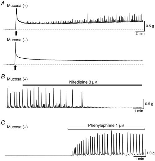
A, spontaneous phasic contractions (upward deflections) were periodically developed in mucosa‐intact [Mucosa (+)] but not mucosa‐denuded [Mucosa (−)] SV preparations. Black arrows indicate the timing of initial stretching to 1 g. Dotted base lines indicate 0 g. B, nifedipine (3 μm) abolished the spontaneous contractions in the mucosa‐intact SV. C, in mucosa‐denuded SV that did not generate spontaneous contractions, bath‐applied phenylephrine (1 μm) induced large oscillatory contractions.
In eight mucosa‐denuded preparations, stretching the preparations invariably failed to induce spontaneous phasic contractions (Fig. 2 A, lower trace). Despite the lack of spontaneous contractions, all mucosa‐denuded preparations were capable of generating oscillatory contractions associated with a rise in the basal tone of 2.00 ± 0.66 g (n = 8) in response to bath‐applied noradrenaline (3 μm), indicating that removal of the mucosa did not impair the contractility of SV smooth muscle. Noradrenaline‐induced oscillatory contractions occurred at a frequency of 19.1 ± 3.3 min−1 and had a peak amplitude of 0.90 ± 0.47 g (n = 8). Phenylephrine (1 μm), an α1‐adrenoceptor (AR) agonist, also developed oscillatory contractions in the mucosa‐denuded quiescent SV preparations associated with a rise in the basal tone of 0.14 ± 0.10 g (n = 5, Fig. 2 C). Phenylephrine‐induced oscillatory contractions occurred at a frequency of 4.8 ± 1.7 min−1 and had a peak amplitude of 2.56 ± 1.25 g (n = 5).
Nerve‐evoked contractions of SV did not require intact mucosa
To further confirm the viability of mucosa‐denuded SVs, nerve‐evoked contractions were compared between mucosa‐intact and mucosa‐denuded preparations.
In the mucosa‐intact preparations, EFS (20 or 30 Hz for 3 s) evoked phasic contractions that had a peak amplitude of 0.99 ± 0.47 g and a half‐width of 3.4 ± 0.7 s (n = 5, Fig. 3 A). TTX (1 μm, n = 5) abolished the EFS‐induced contraction without affecting spontaneous contractions (peak amplitude: 0.17 ± 0.13 g in control, 0.18 ± 0.13 g in TTX, n = 5, P = 0.202; frequency: 5.5 ± 0.7 min−1 in control, 5.5 ± 0.5 min−1 in TTX, n = 5, P = 0.883, Fig. 3 A).
Figure 3. Spontaneous and nerve‐evoked contractions in SV.

A, in mucosa‐intact SV that exhibited spontaneous contractions, electrical field stimulation (EFS) triggered TTX‐ (1 μm) sensitive, nerve‐evoked contractions (asterisks). Lower traces show expanded recordings in control (a) and in TTX (1 μm, b). Traces a and b were obtained at the timings indicated by the corresponding characters in the upper trace. B, in mucosa‐denuded SV that did not generate spontaneous contractions, EFS triggered nerve‐evoked contractions (asterisks) that were reversibly blocked by TTX (0.1 μm). Trains of EFS (5 V, 0.1 ms, 30 Hz, 3 s) indicated by black bars were applied at 4 min intervals in A and B.
In the quiescent mucosa‐denuded preparations, EFS also evoked TTX‐sensitive phasic contractions that had a peak amplitude of 2.28 ± 1.16 g and a half‐width of 4.7 ± 2.7 s (n = 5, Fig. 3 B). Thus, the peak amplitude of nerve‐evoked contractions in the mucosa‐denuded preparations (n = 5) was even larger than that in the mucosa‐intact preparations (n = 5, P = 0.049), indicating that the nerve‐dependent contractility of the mucosa‐denuded smooth muscle was well preserved.
Mucosa dependence of spontaneous action potentials and slow waves in SV
Since the spontaneous contractions of the SV appeared to require an intact mucosa, we explored whether spontaneous electrical activity of SV smooth muscle may also be mucosa‐dependent.
During preliminary experiments, it was found that stable impalements of the intracellular microelectrode were fairly difficult due to the spontaneous phasic contraction of intact SV preparations. Therefore, we used ‘reduced’ SV preparations in which most of the muscular layer had been removed to minimize tissue distortion due to muscle contractions (Fig. 4 A). The ‘reduced’ SV preparations generated much weaker spontaneous contractions than full thickness SV, while their frequency of contraction was not altered.
Figure 4. Mucosa dependence of slow waves and action potentials in SV smooth muscle.

A, light micrographs of haematoxylin‐eosin‐stained coronal sections of the inner circular smooth muscle preparations of SV with and without intact mucosa similar to those used for recordings of membrane potentials or intracellular Ca2+ dynamics. In the right photograph of the mucosa‐denuded preparation [Mucosa (−)], some mucosa was left attached to indicate the mucosal side (arrow). B, in a mucosa‐intact preparation, SV smooth muscle developed spontaneous slow waves (SWs) with superimposed action potentials. Lower trace shows an SW with an expanded time scale to show measured parameters: RMP, resting membrane potential; AMP (SW), peak amplitude of SW; HW, half‐width of SW. C, in a mucosa‐denuded preparation, SV smooth muscle did not generate SWs nor spontaneous action potentials. D, in a ‘quiescent’ mucosa‐denuded SV smooth muscle, depolarizing current injection (200 pA for 400 ms) evoked several action potentials.
In mucosa‐intact preparations, SV smooth muscle had a resting membrane potential (RMP) of −61.2 ± 2.9 mV and exhibited SWs that triggered superimposed action potentials (Fig. 4 B, upper trace). SWs were generated at a mean frequency of 5.3 ± 1.0 min−1 and had a peak amplitude (AMP) of 39.4 ± 6.7 mV and a half‐width (HW) of 0.49 ± 0.24 s (n = 10, Fig. 4 B, lower trace). The peak membrane potential of the ‘superimposed’ action potentials reached absolute values of 0.8 ± 11.3 mV (n = 10).
Mucosa‐denuded preparations did not develop either SWs or spontaneous action potentials (n = 5, Fig. 4 C). However, depolarizing current injected through the recording electrode successfully evoked nifedipine‐sensitive action potentials (n = 5, Fig. 4 D), indicating that the membrane excitability was preserved in mucosa‐denuded preparations.
Besides SWs and superimposed action potentials, irregularly occurring small transient depolarizations were occasionally recorded in both mucosa‐intact and mucosa‐denuded preparations. Since these events were not associated with muscle contractions, their properties were not investigated further.
Role of LVDCC in generating SWs and action potentials in SV
Since the mucosa‐dependent spontaneous contractions of SV were abolished by nifedipine, the contribution of LVDCC to action potentials and SWs was investigated.
Nifedipine (1 μm) abolished action potentials and greatly suppressed associated spontaneous contraction without changing the ‘intrinsic’ frequency of SWs (n = 5, Fig. 5 A, D), suggesting that spontaneous contractions arise from LVDCC‐dependent action potentials but not SWs. Nifedipine (1 μm) also caused a small depolarization of the membrane (3.4 ± 2.4 mV, n = 5, Fig. 5 A). In nifedipine‐ (1 μm) treated preparations, ‘weakened’ spontaneous contractions could be visually observed, suggesting that 1 μm nifedipine was not sufficient to completely block LVDCCs as in the other urogenital tissues (Lam et al. 2011; Lang et al. 2007a, b).
Figure 5. Effects of nifedipine on action potentials and SWs in mucosa‐intact SV smooth muscle.

A, in a mucosa‐intact SV preparation, nifedipine (1 μm) abolished superimposed action potentials, leaving SWs. Lower traces show SWs in control (a) and in nifedipine (1 μm, b) with an expanded time scale. Traces a and b were obtained at the timings indicated by the corresponding characters in the upper trace. B, in another mucosa‐intact SV preparation, nifedipine (10 μm) abolished superimposed action potentials, reduced the amplitude of SWs and also depolarized the membrane by 10 mV. Lower traces show SWs in control (a) and in nifedipine (10 μm, b) with an expanded time scale. Traces a and b were obtained at the timings indicated by the corresponding characters in the upper trace. Dotted line indicates RMP in control. Summarized data of the effects of nifedipine (1 and 10 μm) on the peak amplitude [AMP (SW), C] and the frequency (D) of the SWs. The closed circles connected by a line indicate data obtained from identical cells.
A higher concentration of nifedipine (10 μm) abolished the action potentials and associated spontaneous contractions (Fig. 5 B). Nifedipine (10 μm) also reduced SW amplitudes to 37 ± 11% of control values (38.6 ± 8.3 mV in control, 14.8 ± 6.6 mV in nifedipine, P < 0.001, n = 5, Fig. 5 C) and prolonged SW half‐width to 500 ± 328% (0.58 ± 0.31 s in control, 2.21 ± 0.88 s in nifedipine, P = 0.030, n = 5) without changing the frequency of SWs (5.5 ± 1.0 min−1 in control, 5.5 ± 1.3 min−1 in nifedipine, P = 0.907, n = 5, Fig. 5 D). Thus, SWs appear to consist of LVDCC‐dependent and LVDCC‐independent components. Nifedipine (10 μm) also caused a larger depolarization of 8.3 ± 2.5 mV (from −61.5 ± 1.3 to −53.2 ± 1.5 mV, n = 5).
Role of LVDCC in generating mucosa‐dependent spontaneous Ca2+ flashes
To further explore the mechanisms underlying ‘nifedipine‐resistant’ SWs in SVs, the intracellular Ca2+ dynamics in SV smooth muscle were investigated.
In mucosa‐intact SV smooth muscle, spontaneous Ca2+ flashes were almost synchronously generated within the field of view, resulting in spontaneous contractions (Fig. 6 A). Spontaneous Ca2+ flashes had a frequency of 5.5 ± 1.4 min−1, peak amplitude of 2.3 ± 0.6 ∆F t /F 0 and a half‐width of 1.16 ± 0.60 s (n = 5, controls in Fig. 6 B, C).
Figure 6. Mucosa dependence of spontaneous Ca2+activity in SV smooth muscle.

A, sequential Cal‐520 fluorescence images (frame interval;107 ms) demonstrate spontaneous Ca2+ flashes in the mucosa‐intact SV. Scale bars = 30 μm. Spontaneous Ca2+ flashes were generated almost synchronously across SV smooth muscle cells. Middle traces show that Ca2+ flashes recorded from three regions of interest (ROIs: indicated by circles in upper right panel) were generated synchronously. Nifedipine (10 μm) greatly suppressed spontaneous Ca2+ flashes. Lower traces were obtained in control (a) and in nifedipine (10 μm, b) with an expanded time scale. Traces a and b were obtained at the timings indicated by the corresponding characters in the middle trace. The numbers of the traces correspond to the number of ROIs in the upper panel. Note that synchrony was preserved for the nifedipine‐resistant spontaneous Ca2+ transients. Effects of nifedipine (10 μm) on the peak amplitude (B) and frequency (C) of spontaneous Ca2+ flashes in mucosa‐intact SVs were summarized (n = 5). D, in a mucosa‐denuded SV that did not generate spontaneous Ca2+ transients, phenylephrine (1 μm) evoked synchronous oscillatory Ca2+ transients.
Nifedipine (10 μm) largely suppressed the peak amplitude of Ca2+ flashes to 16% of control (0.32 ± 0.12 ∆F t /F 0, P = 0.004, n = 5, Fig. 6 A, B) associated with a prolongation of the half‐width to 180% of control (2.08 ± 1.60 s, P = 0.116, n = 5). The residual spontaneous Ca2+ transients were still generated synchronously and their frequency was not significantly changed (4.4 ± 0.1 min−1, P = 0.127, n = 5, Fig. 6 A, C), suggesting that spontaneous Ca2+ transients trigger SW generation.
Mucosa‐denuded SV smooth muscle preparations invariably failed to develop spontaneous Ca2+ flashes, but were capable of responding to phenylephrine (1 μm) by generating oscillatory Ca2+ transients associated with a rise in basal Ca2+ level by 1.6 ± 0.5 ∆F t /F 0 (n = 5, Fig. 6 D). Phenylephrine‐induced Ca2+ oscillations were also synchronously generated within the field of view at a frequency of 6.4 ± 2.1 min−1 and had a peak amplitude of 2.4 ± 0.7 ∆F t /F 0 and a half‐width of 0.95 ± 0.40 s (n = 5).
Role of intracellular Ca2+ stores in generating SWs and spontaneous Ca2+ transients
The role of sarcoplasmic/endoplasmic reticulum (SR/ER) Ca2+ stores in the generation of SWs and corresponding Ca2+ transients was further investigated.
In preparations that had been pretreated with 10 μm nifedipine, CPA (10 μm), an inhibitor for sarco‐endoplasmic reticulum Ca2+‐ATPase (SERCA), abolished SWs (peak amplitude: 14.7 ± 5.6 mV; frequency: 4.2 ± 1.0 min−1, n = 5) within 5 min and also depolarized the membrane by 6.8 ± 1.1 mV (n = 5, Fig. 7 A). The generation of SWs was restored after 15–40 min wash out of CPA (n = 5).
Figure 7. Blockade of SERCA abolished nifedipine‐resistant SWs and spontaneous Ca2+ transients in mucosa‐intact SV smooth muscle.

A, in a nifedipine‐ (10 μm) pretreated preparation that exhibited SWs, CPA (10 μm) depolarized the membrane by about 5 mV and prevented the generation of SWs. B, in another nifedipine‐ (10 μm) pretreated preparation that developed spontaneous Ca2+ transients, CPA (10 μm) increased the basal Ca2+ level and abolished Ca2+ transients. Dotted line in A indicates RMP.
Consistent with the above electrophysiological findings, ‘nifedipine‐resistant’ spontaneous Ca2+ transients were also abolished by CPA (10 μm) associated with a rise in the basal Ca2+ level by 0.23 ± 0.12 ∆F t /F 0 (n = 5, Fig. 7 B).
Role of CACC in generating mucosa‐dependent SWs in SV
Since SR/ER Ca2+ handing appears to play a critical role in generating SWs, the involvement of CACCs in SW generation was investigated.
In mucosa‐intact SVs that had been pretreated with nifedipine (10 μm), the generation of the SWs (peak amplitude: 18.2 ± 2.4 mV, frequency: 3.8 ± 0.7 min−1 in control, n = 5) was prevented by reducing [Cl−]o from 130.3 to 13.3 mm, and restored within 4–6 min after switching from low Cl− to normal PSS (Fig. 8 A). Note that artificial changes in the membrane potential were generated by liquid junction potentials upon changing [Cl−]o: ‘hyperpolarization’ by 4.8 ± 0.7 mV and ‘depolarization’ by 3.8 ± 1.9 mV (n = 5), indicated by upward and downward arrows in Fig. 8 A, respectively. After subtracting the liquid junction potential measured with the recording electrode dipped in the bath solution (negative shift by 4.8 ± 0.5 mV in the low Cl− PSS, n = 3), switching from normal to low Cl− PSS in fact depolarized the membrane by 4.2 ± 2.4 mV (from −55.9 ± 1.6 to −51.7 ± 3.6 mV, P = 0.017, n = 5).
Figure 8. Role of CACCs in the generation of SWs in mucosa‐intact SV.
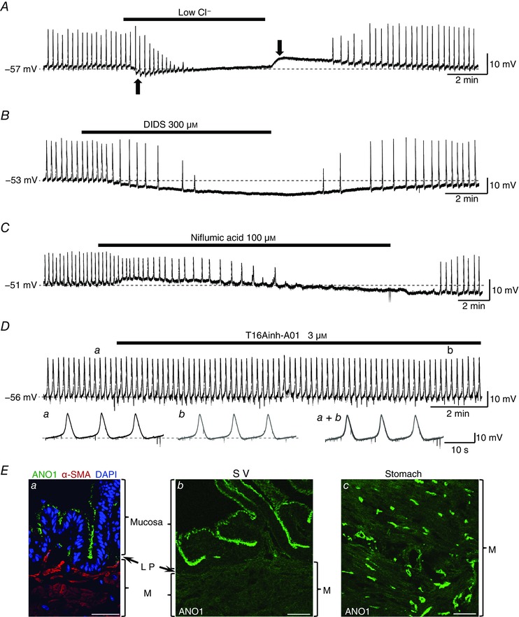
A, in a nifedipine‐ (10 μm) pretreated preparation that exhibited SWs, lowering [Cl−]o prevented the generation of SWs. Negative and positive shifts of the baseline upon the reduction and restoration of [Cl−]o, respectively (black arrows), were due to liquid junction potential. Subtraction of the measured liquid junction potential revealed that low Cl− solution depolarized the membrane by 4.2 mV. B, in another nifedipine‐ (10 μm) pretreated preparation, DIDS (300 μm) hyperpolarized the membrane and abolished SWs. C, in a different nifedipine‐ (10 μm) pretreated preparation, niflumic acid (100 μm) caused a transient depolarization that was followed by a hyperpolarization and abolished SWs. D, T16Ainh‐A01 (3 μm), an ANO1 inhibitor, did not affect either the generation of the nifedipine‐ (10 μm) resistant SWs or the RMP. Lower traces show SWs in control (a) and in T16Ainh‐A01 (3 μm, b) with an expanded time scale. Traces a and b were obtained at the timings indicated by the corresponding characters in the upper trace. Superimposed traces (a + b) show T16Ainh‐A01 (3 μm) reduced neither the amplitude nor the rising slope of the SWs. Dotted lines indicate RMP. Ea, in the coronal section of guinea pig SV, the apical side of the mucosa, but not lamina propria (LP) or musclular layer (M), was immunopositive for ANO1. The sections of another guinea pig SV (b) and gastric antrum (c) were immunolabelled with anti‐ANO1 antibody using the same protocol. In the SV, ANO1 immunoreactivity localized in the apical side of the mucosa (b), while ANO1‐immunoreactive cells were detected in the muscular layer of the stomach (c, positive control). All scale bars in E = 40 μm.
DIDS (300 μm), a known CACC inhibitor, also abolished SWs (peak amplitude: 14.9 ± 6.9 mV, frequency: 4.5 ± 0.7 min−1 in control, n = 5) with a hyperpolarization of the membrane by 12.5 ± 2.9 mV (n = 5, Fig. 8 B) within 4–12 min. The generation of SWs was restored after 2–4 min wash out of DIDS (n = 5).
Niflumic acid (100 μm), which is also known to inhibit CACC, reversibly abolished SWs (peak amplitude: 15.1 ± 9.3 mV, frequency: 4.3 ± 0.6 min−1 in control, n = 5) in 4 out of 5 preparations (Fig. 8 C). In the remaining single preparation, the frequency of the SWs was largely reduced from 3.6 to 0.5 min−1 during exposure to niflumic acid (100 μm) for 35 min. The peak amplitude and the maximum rising slope of the residual SWs in niflumic acid were also decreased (mean peak amplitude: 29.7 ± 0.2 mV in control, 22.1 ± 0.3 mV in niflumic acid, P < 0.001; mean maximum rising slope: 47.9 ± 2.6 mV s–1 in control, 20.7 ± 2.6 mV s−1 in niflumic acid, P < 0.001). Niflumic acid (100 μm) caused a transient depolarization of 1.6 ± 0.8 mV (n = 5) that was followed by a hyperpolarization of 2.8 ± 2.3 mV (n = 5, Fig. 8 C).
To further explore the role of CACC, the effects of T16Ainh‐A01 (3 μm), an ANO1 blocker (Scudieri et al. 2011), on SV SWs were also investigated.
T16Ainh‐A01 (3 μm) failed to inhibit the SWs (peak amplitude: 20.3 ± 8.6 mV in control, 20.4 ± 8.0 mV in T16Ainh‐A01; maximum rising slope: 25.4 ± 18.7 mV s−1 in control, 27.9 ± 16.8 mV s−1 in T16Ainh‐A01; frequency: 4.9 ± 0.7 min−1 in control, 4.7 ± 0.6 min−1 in T16Ainh‐A01, n = 4) in the SV smooth muscle (Fig. 8 D). The resting membrane potential was not affected by T16Ainh‐A01 (3 μm).
Since T16Ainh‐A01 unexpectedly failed to inhibit the SWs, ANO1 immunoreactivity in SV cross sections was examined. The apical side of the epithelial cells expressed immunoreactivity against ANO1 antibody, while ANO1‐immunoreactivity was hardly detected in either the subepithelial (lamina propria) cells or smooth muscle cells (Fig. 8 Ea, b). Consistent with previous reports (Baker et al. 2013; Sanders et al. 2014), the ANO1 antibody specifically stained ICC in the smooth muscle layer of the guinea pig stomach (Fig. 8 Ec), and thus its viability and specificity were verified.
Role of gap junctions in generating SWs in SV smooth muscle
To explore the communication pathway between the mucosa and smooth muscle layer, the role of gap junctions in the generation of the mucosa‐dependent SWs was investigated. In mucosa‐intact SV smooth muscle that had been pretreated with 10 μm nifedipine, carbenoxolone (100 μm), a known gap junction inhibitor, reversibly abolished the SWs (peak amplitude: 16.7 ± 8.6 mV, frequency: 4.0 ± 0.5 min−1, n = 4, Fig. 9) within 5–13 min. Carbenoxolone (100 μm) also depolarized the membrane by 7.6 ± 3.3 mV (from −54.5 ± 1.5 to −46.9 ± 3.7 mV, n = 4, Fig. 9).
Figure 9. Blockade of gap junctions abolished CACC‐dependent SWs in mucosa‐intact SV smooth muscle.

In a nifedipine‐ (10 μm) pretreated preparation that exhibited SWs, carbenoxolone (100 μm) depolarized the membrane and prevented the generation of SWs. Lower traces were obtained in control (a), in carbenoxolone (100 μm, b) and after recovery (c) with an expanded time scale. Traces a–c were obtained at the timings indicated by the corresponding characters in the upper trace.
Spontaneous Ca2+ transients of mucosal cells
Since the intact mucosa appears to be fundamental in generating spontaneous activity of SV smooth muscle, intracellular Ca2+ imaging was carried out using ‘isolated’ SV mucosal preparations detached from the smooth muscle layer to investigate if there may be mucosal cells generating spontaneous Ca2+ transients.
In the basal surface of ‘isolated’ mucosal preparations that had been pretreated with nifedipine (10 μm), a population of cells developed spontaneous Ca2+ transients (Fig. 10 Aa). Unlike spontaneous Ca2+ flashes or Ca2+ transients in the mucosa‐intact smooth muscle that were generated synchronously as well as periodically, individual mucosal cells exhibited spontaneous Ca2+ transients independently of each other (Fig. 10 B, n = 6). Spontaneous Ca2+ transients were generated at a frequency of 1.3 ± 0.7 min−1 (46 cells, n = 6), and had a peak amplitude of 0.76 ± 0.27 ∆F t /F 0 and a half‐width of 5.4 ± 2.8 s (46 cells, n = 6).
Figure 10. Spontaneous and ATP‐induced Ca2+ transients in SV submucosal cells.

A, in a mucosal preparation that had been pretreated with nifedipine (10 μm), a population of cells in the basal surface of the mucosa generated the spontaneous Ca2+ transients (a, yellow arrows). Subsequent ATP (100 μm) evoked a massive increase in the intracellular Ca2+ not only in spontaneously active but also in previously quiescent cells, and clearly visualized their irregularly shaped cell bodies extending several short processes (b). Scale bar = 30 μm. B, asynchronous spontaneous Ca2+ transients recorded from six submucosal cells in another nifedipine‐ (10 μm) pretreated preparation. Individual submucosal cells generated ‘irregularly occurring’ Ca2+ transients independently of each other. CPA (10 μm) increased the basal Ca2+ level and abolished the Ca2+ transients. Dotted line indicates basal Ca2+ level of the submucosal cells before the application of CPA.
Spontaneous Ca2+ transients in mucosal cells were abolished by CPA (10 μm) associated with a rise in the basal Ca2+ level by 0.57 ± 0.14 ∆F t /F 0 (n = 3, Fig. 10 B), indicating ER Ca2+ handling plays a fundamental role in generating their spontaneous Ca2+ transients as in the case of SWs and spontaneous Ca2+ flashes or transients in SV smooth muscle.
Bath‐applied ATP (100 μm) evoked intense Ca2+ transients in not only spontaneously active but also previously quiescent mucosal cells. In the presence of ATP, numerous cells that had an irregular‐shaped cell body with several short processes were clearly visualized (Fig. 10 Ab, Supplementary Movie S1), indicating that ATP greatly facilitates the ability of the mucosal cells to generate Ca2+ transients. The density of the ATP‐sensitive mucosal cells was 34.1 ± 5.3 cells per 100 μm2 (11 preparations, n = 4), and they had a length of 17.4 ± 2.3 μm and a width of 8.0 ± 1.5 μm (38 cells, n = 4).
Distribution of interstitial cells in SV mucosa
To identify the mucosal cells generating spontaneous Ca2+ transients, double‐immunolabelling with several cell markers was performed using ‘isolated’ mucosa preparations. The serial images were obtained from the basolateral side of the mucosa preparations (Fig. 11 A).
Figure 11. Distribution of the interstitial cells in the subepithelium of SV mucosa preparations.

A, an illustration of dissected SV mucosa from the muscular layer used for the whole mount preparation in B, C and F. Serial images were obtained from the basal (subepithelial) side of the mucosa preparations via confocal microscopy. B, serial images of a whole mount SV mucosa preparation immunolabelled with anti‐pancytokeratin (red: a marker of epithelial cell) and vimentin (green: a marker of interstitial cell). ‘a + 5 μm’ in the right image indicates that the image was obtained 5 μm from the basal side of ‘a’. Vimentin‐immunoreactive (IR) cells located beneath the epithelial layer. C, double immunolabelling with anti‐c‐Kit and α‐SMA antibodies in another whole mount SV mucosa preparation. α‐SMA‐IR (red) cells were not found except for the blood vessels, and a few oval‐shaped c‐Kit‐IR (green) cells are observed. Double immunolabelled cross‐sections of whole tissue of SV (D) and muscular layer of gastric antrum (E) produced under the same protocol. In SV wall, non‐significant signals of c‐Kit (green) were observed, while interstitial cells of Cajal immunolabelled with c‐Kit antibody were observed in the stomach. LP, lamina propria; M, musclular layer. F, serial images of a whole mount SV mucosa preparation. PDGFRα‐IR (green) cells are found beneath the epithelial layer (a + 4.2 μm, a + 6.2 μm). Scale bars = 40 μm.
Vimentin‐immunoreactive (IR), ‘slender’ interstitial cells were distributed at the basolateral side of the pancytokeratin‐IR epithelial cells, indicating the interstitial cells were distributed just beneath the epithelial layer (Fig. 11 B).
As candidates for the spontaneously active mucosal cells, the distribution of c‐Kit‐positive interstitial cells and α‐SMA‐positive smooth muscle cells was examined. In the SV mucosa preparations, α‐SMA‐IR cells were not observed, except for vascular smooth muscle cells of the blood vessels (Fig. 11 C). A few c‐Kit‐IR cells that had an oval‐shaped cell body, presumably mast cells, were sparsely scattered (Fig. 11 C). To verify the viability and specificity of the c‐Kit antibody, the sections of the guinea pig stomach and the SV were also immunolabelled with the antibody using the same protocol. c‐Kit immunoreactivity was observed in ICC distributed in the muscular layer of stomach, (Fig. 11 E: positive control), whereas c‐Kit‐IR cells were not detected in the mucosa, lamina propria or musclular layer of SV (Fig. 11 D).
PDGFRα‐IR interstitial cells that had long cytoplasmic processes were observed beneath the epithelial layer in SV mucosa preparations (Fig. 11 F). Both vimentin‐IR and PDGFRα‐IR cells in the subepithelial layer were larger than the pancytokeratin‐IR epithelial cells in the basolateral side of the mucosa preparations (Fig. 11 B, F).
Discussion
This study has demonstrated for the first time that the mucosa of SV plays a critical role in generating spontaneous phasic contractions of SV smooth muscle. Mucosa‐intact SV smooth muscle developed spontaneous contractions arising from action potentials triggered by slow waves and associated Ca2+ flashes. However, quiescent mucosa‐denuded SV smooth muscle readily contracted upon nerve excitation or α1‐AR stimulation. This may well explain previous controversies in terms of spontaneous electrical activity and associated contractions in guinea pig SV smooth muscle (Ohkawa, 1982; Kubota et al. 2003). A population of mucosal cells exhibited spontaneous Ca2+ transients, and thus may drive spontaneous activity in mucosa‐intact smooth muscle.
Role of LVDCC in generating spontaneous activity
In the mucosa‐intact SV smooth muscle, nifedipine (1 μm) abolished superimposed action potentials without preventing the generation of SWs (Fig. 5). A higher concentration of nifedipine (10 μm) reduced the SW amplitude, indicating that the activation of LVDCCs contributes to the configuration of SWs. This is in contrast to gastric smooth muscle where the configuration of the SWs is not changed upon blockade of LVDCCs (Sanders et al. 2014). In the prostate of the guinea pig and the urethra of the rabbit and guinea pig, SW duration is shortened by blockade of LVDCCs, while their amplitude is little affected (Hashitani et al. 1996; Hashitani & Edwards, 1999; Shigemasa et al. 2014). Thus, the contribution of LVDCCs to SW configuration appears to be varied amongst different smooth muscle tissues.
Nifedipine also depolarized the membrane of SV smooth muscle. Since tetraethylammonium (TEA)‐sensitive, Ca2+‐dependent K+ current has been recorded in the isolated guinea pig SV smooth muscle cells (Sadraei & Beech, 1995), ‘nifedipine‐induced’ depolarization may result from the reduced activation of large conductance Ca2+‐activated K+ (BK) channels that could be activated upon Ca2+ entry through LVDCCs. Nifedipine‐induced prolongation of the half‐width of SWs or Ca2+ flashes (transients) may also be explained by this diminished BK activity.
The blockade of LVDCCs by nifedipine (10 μm) strongly suppressed the spontaneous Ca2+ flashes and abolished the spontaneous phasic contractions leaving synchronous Ca2+ transients (Fig. 6). These results indicate that the Ca2+ influx through LVDCCs during action potential firing is fundamental for SV spontaneous contractions. In contrast, nifedipine did not change the frequency of the residual SWs or spontaneous Ca2+ transients, suggesting that LVDCCs are not involved in generating intrinsic periodicity of SV smooth muscle. Synchronous Ca2+ transients were generated across SV smooth muscle cells even after the complete blockade of LVDCCs (Fig. 6). This is in marked contrast to the spontaneous Ca2+ transients in atypical smooth muscle cells of the renal pelvis (Lang et al. 2007a) or suburothelial venular smooth muscle cells (Hashitani et al. 2011, 2012) in which LVDCCs play a critical role in maintaining their synchrony.
Role of intracellular Ca2+ stores and CACCs in generating spontaneous activity in SV smooth muscle
In mucosa‐intact SV smooth muscle, spontaneous Ca2+ transients and SWs were abolished by CPA (Fig. 7), suggesting that both events depend on Ca2+ handling by the SR/ER. Similar mechanisms termed ‘cytosolic Ca2+ oscillators’, i.e. the cycle of Ca2+ uptake by SERCA and Ca2+ release, driving spontaneous transient depolarizations (STDs) or SWs have been reported in smooth muscle of other visceral organs (Sergeant et al. 2001; Craven et al. 2004; Lang et al. 2007b; Sanders et al. 2014).
CPA also caused membrane depolarization and a rise of basal Ca2+ level (Fig. 7). Since CPA is known to cause a sustained rise in the intracellular Ca2+ due to the blockade of SERCA as well as subsequent activation of store‐operated Ca2+ channels (Wayman et al. 1996; Noble et al. 2014), a similar mechanism may be involved in the depolarization in SV smooth muscle.
In ICC in the GI tract and interstitial cells in the urinary tract, ‘cytosolic Ca2+ oscillators’ drive the spontaneous electrical activity by activating CACCs (Sergeant et al. 2000; Sanders et al. 2014). In those cells, opening of Cl− channels causes Cl− efflux resulting in depolarization as [Cl−]i is maintained relatively high by active transport of Cl− via the Na+–K+–Cl− cotransporter (NKCC1; Sanders et al. 2014; Zhu et al. 2016). In the GI ICC, ANO1 channels function as a CACC to develop SWs (Sanders et al. 2014; Oh & Jung, 2016; Zhu et al. 2016).
In the present study, the SWs of SV smooth muscle were abolished by lowering [Cl−]o (Fig. 8 A). SWs were also inhibited by known CACC blockers but not by an ANO1 inhibitor (Fig. 8 B–D), suggesting that CACCs other than ANO1 contribute to the generation of SWs in SV. However, it should be noted that CACC blockers used at the high concentrations could modulate functions of some other proteins (Scudieri et al. 2011). Since ANO1 expression was restricted to the apical surface of the epithelial cells, ANO1 may contribute to secretion of Cl− into seminal fluid but not the generation of SWs (Levine et al. 1975; Scudieri et al. 2011; Oh & Jung, 2016).
Communication between mucosa and smooth muscle
Since intact mucosa was required for generating CACC‐dependent SWs and corresponding spontaneous Ca2+ transients in SV smooth muscle (Figs 4 and 6), it is possible that both events are driven by spontaneously active cells in mucosa. In accordance with this notion, a population of cells was distributed in the basolateral (submucosal) side of the dissected SV mucosa that generated spontaneous Ca2+ transients (Fig. 10).
Since the SWs in SV smooth muscle were abolished by carbenoxolone (Fig. 9), the spontaneous Ca2+ and/or electrical signals could be transmitted from the mucosal cells to smooth muscle via gap junctions. Indeed, both mucosal Ca2+ transients and the SWs and Ca2+ flashes (transients) in smooth muscle primarily rely on ER/SR Ca2+ handing. However, the characteristics of the spontaneous Ca2+ transients in SV mucosa were quite different from those in SV smooth muscle in terms of their synchrony and periodicity. In addition, the mucosal spontaneous Ca2+ transients lasted about five times longer than those in smooth muscle (Fig. 10).
One explanation for the different characteristics of the mucosal and smooth muscle Ca2+ transients is that smooth muscle syncytium may act as a low‐resistance pathway that electrically couples neighbouring mucosal cells to maintain their synchrony. Alternatively, the mucosal cells may act as ‘point sources’ of excitation. In the latter case, the periodicity of SWs and Ca2+ flashes (transients) may be determined by the ‘Ca2+‐dependent’ refractory period of SV smooth muscle cells, e.g. SR store refilling or BK channel activation.
Another possibility is that the Ca2+ transients in the mucosal cells may be associated with the release of humoral factors to enhance subthreshold ‘autorthythmicity’ in the SV smooth muscle by stimulating InsP3 production to facilitate ‘cytosolic Ca2+ oscillators’ (van Helden & Imtiaz, 2003). This notion was supported by the finding that the ‘quiescent’ mucosa‐denuded SV smooth muscle invariably generated oscillatory contractions and Ca2+ transients upon the activation of α1‐ARs (Figs 2 C and 6 D). Since the diffusion of humoral factors is likely to be restricted within a short distance, SWs originating in the smooth muscle cells adjacent to the submucosa may propagate to outer smooth muscle via gap junctions. In the guinea pig bladder, urothelium‐derived humoral factors such as ATP appear to enhance the spontaneous contractions (Kushida & Fry, 2016), while endogenous prostaglandins play a fundamental role in generating pyeloureteric contractions of several species (Lang et al. 2002). In the rabbit corpus cavernosum, cyclooxygenase‐2 (COX‐2)‐dependent prostaglandins may be released by intramuscular interstitial cells to develop phasic spontaneous activity (Hashitani et al. 2005). Since the inhibitors for α‐ARs or muscarinic receptors did not prevent the spontaneous contraction of the isolated guinea pig SVs (Ohkawa, 1973, 1981; Hayashi et al. 2016), neither noradrenaline nor ACh are the mucosa‐derived factor.
Interestingly, nerve‐evoked contractions in the mucosa‐denuded SV preparations were even larger than those in the mucosa‐intact SV preparations, suggesting that the mucosa may also release some inhibitory factors to attenuate SV muscle contractility.
Origin of spontaneous Ca2+ transients in the mucosa
Immunohistochemical examinations in the dissected SV mucosa failed to identify either c‐Kit‐positive interstitial cells or α‐SMA‐positive atypical smooth muscle cell acting as ‘pacemaker cells’ in the GI tract and renal pelvis, respectively (Fig. 11 C). Thus, the mucosal cells firing spontaneous Ca2+ transients appear to be distinct from known pacemaker cells in other visceral organs.
An electron microscopic study of guinea pig SV revealed spindle‐shaped fibroblasts located just beneath the basement membrane of the tall columnar epithelial cells (Tse & Wong, 1980). Consistent with this finding, we demonstrated vimentin‐IR interstitial cells situated beneath the epithelial cells (Fig. 11 B). In the same layer of the mucosa, PDGFRα‐positive interstitial cells were also distributed (Fig. 11 F). In the GI tract or the urinary bladder, PDGFRα‐positive ‘fibroblast‐like’ cells have been reported to express small conductance Ca2+‐activated K+ channels (SK3) and develop spontaneous Ca2+ transients (Baker et al. 2013), resulting in spontaneous transient outward currents (Kurahashi et al. 2011; Lee et al. 2013). In SV mucosa, different subpopulations of PDGFRα‐positive cells may express CACC but not SK to generate depolarizing signals. The PDGFRα‐positive cells in the GI tract or the urinary bladder have also been shown to generate SK3‐dependent outward currents upon purinergic receptor (P2Y1) activation (Kurahashi et al. 2011; Lee et al. 2014). The SV mucosal cells were also capable of developing Ca2+ transients in response to bath‐applied ATP. However, the morphology of the mucosal cells firing spontaneous Ca2+ transients was not similar to that of PDGFRα‐positive interstitial cells, although neither Ca2+ fluorescence nor immunohistochemistry fully visualized their actual cell morphology.
Physiological significance of spontaneous and nerve‐evoked contraction in SV
The SVs contract predominantly under control of sympathetic nerve excitation to expel a major component of seminal fluid (Aumüller & Riva, 1992; Coolen et al. 2004). Silodosin, a selective α1A‐AR antagonist commonly used for the treatment of benign prostate hyperplasia, often causes ejaculation disorders (Hisasue et al. 2006; Capogrosso et al. 2015; Hayashi et al. 2016), suggesting that activation of α1A‐ARs plays a primary role in SV neuromuscular transmission.
During inter‐ejaculatory phase, SVs store secretory fluids consisting of fructose, prostaglandins, antioxidant agents and bioactive proteins that are required for male fertility. The secretion of SVs contributes to the promotion of sperm motility, increases stability of sperm chromatin and modulates immune activity in the female reproductive tract (Aumüller & Riva, 1992; Gonzales, 2001; Bromfield, 2014). Thus, it is reasonable to assume that spontaneous contractions of SVs during the storage phase play a role in maintaining the quality of these secretory fluids. In the urinary bladder, spontaneous contractions of the detrusor during the filling phase are associated with no or very little change in the intraluminal pressure, and thus are considered to maintain a minimum volume‐to‐surface ratio (Drake et al. 2003; Brading, 2006). In contrast, whole SV preparations of guinea pig spontaneously contract and produce a transient increase of the intraluminal pressure (Hayashi et al. 2016). Such spontaneous constrictions of SV may have a ‘mixing function’ of the highly viscous seminal fluid (Gonzales, 2001), and thus improve the fluidity of the semen during the storage phase. Therefore, diminished SV spontaneous contractions may result in a decrease in bioactivity of any factors in the seminal fluid that may affect fertility.
Conclusions
Besides the secretory role of the mucosa of guinea pig SV that is essential for fertility, the mucosa is also fundamental in generating spontaneous contractions. The mucosal cells firing spontaneous Ca2+ transients may drive CACC‐dependent SWs by sending depolarizing signals or releasing humoral substances to trigger the opening of LVDCC in the smooth muscle layer.
Additional information
Competing interests
The authors declare no competing interests.
Author contributions
MT*, HH and MT conceived and designed the study. MT*, HH, TH, RH and KN collected the data. MT* and HH analysed the data. MT*, HH, TH, RH, KN and MT interpreted the data. MT*, HH, TH, KN and MT drafted the article, and all authors revised it critically and approved the final version. (MT* and MT indicate the first and the last author, respectively.)
Funding
This work was supported by JSPS KAKENHI, Grant‐in‐Aid for Scientific Research (B) (24300145, 16H05124) to MT, Scientific Research (C) (15K10635) to TH and Challenging Exploratory Research (26670705) to HH.
Supporting information
Disclaimer: Supporting information has been peer‐reviewed but not copyedited.
Movie S1: In a nifedipine‐ (10 μM) treated SV mucosal preparation in which submucosal cells generated asynchronous spontaneous Ca2+‐transients, subsequent ATP (100 μM) evoked a massive increase in the intracellular Ca2+ in not only spontaneously active but also quiescent cells.
Acknowledgements
The authors wish to thank Dr Richard Lang (Monash University) for critical reading of the manuscript and Ms Satoko Yamada (Kurume University) for excellent technical support. The authors also wish to thank Dr Dirk van Helden (University of Newcastle), Dr Retsu Mitsui (Nagoya City University), Dr Eiichiro Tanaka, Dr Yoshinaka Murai and Dr Noriyuki Nakashima (Kurume University) for helpful suggestions.
Linked articles This article is highlighted by a Hollywood et al. To read this Perspective, visit https://doi.org/10.1113/JP274499.
References
- Aumüller G & Riva A (1992). Morphology and functions of the human seminal vesicle. Andrologia 24, 183–196. [DOI] [PubMed] [Google Scholar]
- Baker SA, Hennig GW, Salter AK, Kurahashi M, Ward SM & Sanders KM (2013). Distribution and Ca2+ signalling of fibroblast‐like (PDGFRα+) cells in the murine gastric fundus. J Physiol 591, 6193–6208. [DOI] [PMC free article] [PubMed] [Google Scholar]
- Birder L & Andersson KE (2013). Urothelial signaling. Physiol Rev 93, 653–680. [DOI] [PMC free article] [PubMed] [Google Scholar]
- Birowo P, Ückert S, Kedia GT, Sonnenberg JE, Sandner P, Thon WF, Scheller F, Rahardjo D & Kuczyk MA (2010). Exposure of human seminal vesicle tissue to phosphodiesterase (PDE) inhibitors antagonizes the contraction induced by norepinephrine and increases production of cyclic nucleotides. Urology 76, 1518.e1–6. [DOI] [PubMed] [Google Scholar]
- Brading AF (2006). Spontaneous activity of lower urinary tract smooth muscles: correlation between ion channels and tissue function. J Physiol 570, 13–22. [DOI] [PMC free article] [PubMed] [Google Scholar]
- Bromfield JJ (2014). Seminal fluid and reproduction: much more than previously thought. J Assist Reprod Genet 31, 627–636. [DOI] [PMC free article] [PubMed] [Google Scholar]
- Bruschini H, Schmidt RA & Tanagho EA (1977). Studies on the neurophysiology of the vas deferens. Invest Urol 15, 112–116. [PubMed] [Google Scholar]
- Capogrosso P, Serino A, Ventimiglia E, Boeri L, Dehò F, Damiano R, Briganti A, Montorsi F & Salonia A (2015). Effects of silodosin on sexual function – realistic picture from the everyday clinical practice. Andrology 3, 1076–1081. [DOI] [PubMed] [Google Scholar]
- Coolen LM, Allard J, Truitt WA & McKenna KE (2004). Central regulation of ejaculation. Physiol Behav 83, 203–215. [DOI] [PubMed] [Google Scholar]
- Craven M, Sergeant GP, Hollywood MA, McHale NG & Thornbury KD (2004). Modulation of spontaneous Ca2+‐activated Cl– currents in the rabbit corpus cavernosum by the nitric oxide‐cGMP pathway. J Physiol 556, 495–506. [DOI] [PMC free article] [PubMed] [Google Scholar]
- Dey A, Kusljic S, Lang RJ & Exintaris B (2010). Role of connexin 43 in the maintenance of spontaneous activity in the guinea pig prostate gland. Br J Phramacol 161, 1692–1707. [DOI] [PMC free article] [PubMed] [Google Scholar]
- Drake MJ, Harvey IJ & Gillespie JI (2003). Autonomous activity in the isolated guinea pig bladder. Exp Physiol 88, 19–30. [DOI] [PubMed] [Google Scholar]
- Gonzales GF (2001). Function of seminal vesicles and their role on male fertility. Asian J Androl 3, 251–258. [PubMed] [Google Scholar]
- Grundy D (2015). Principles and standards for reporting animal experiments in The Journal of Physiology and Experimental Physiology . J Physiol 593, 2547–2549. [DOI] [PMC free article] [PubMed] [Google Scholar]
- Hashitani H & Edwards FR (1999). Spontaneous and neurally activated depolarizations in smooth muscle cells of the guinea‐pig urethra. J Physiol 514, 459–470. [DOI] [PMC free article] [PubMed] [Google Scholar]
- Hashitani H, Mitsui R, Shimizu Y, Higashi R & Nakamura K (2012). Functional and morphological properties of pericytes in suburothelial venules of the mouse bladder. Br J Pharmacol 167, 1723–1736. [DOI] [PMC free article] [PubMed] [Google Scholar]
- Hashitani H, Takano H, Fujita K, Mitsui R & Suzuki H (2011). Functional properties of suburothelial microvessels in the rat bladder. J Urol 185, 2382–2391. [DOI] [PubMed] [Google Scholar]
- Hashitani H, van Helden DF & Suzuki H (1996). Properties of spontaneous depolarizations in circular smooth muscle cells of rabbit urethra. Br J Pharmacol 118, 1627–1632. [DOI] [PMC free article] [PubMed] [Google Scholar]
- Hashitani H, Yanai Y, Shirasawa N, Soji T, Tomita A, Kohri K & Suzuki H (2005). Interaction between spontaneous and neurally mediated regulation of smooth muscle tone in the rabbit corpus cavernosum. J Physiol 569, 723–735. [DOI] [PMC free article] [PubMed] [Google Scholar]
- Hayashi T, Takeya M, Nakamura K & Matsuoka K (2016). Effects of silodosin and tamsulosin on the seminal vesicle contractile response. Lower Urin Tract Symptoms 8, 55–61. [DOI] [PubMed] [Google Scholar]
- Hib J, Ponzio R & Vilar O (1983). In vivo recording of contractile activity of pelvic urethra and seminal vesicle in rats. Effects of electrical stimulations and neurohypophysial hormones. Andrologia 15, 480–485. [DOI] [PubMed] [Google Scholar]
- Hisasue S, Furuya R, Itoh N, Kobayashi K, Furuya S & Tsukamoto T (2006). Ejaculatory disorder caused by alpha‐1 adrenoceptor antagonists is not retrograde ejaculation but a loss of seminal emission. Int J Urol 13, 1311–1316. [DOI] [PubMed] [Google Scholar]
- Kanai A, Roppolo J, Ikeda Y, Zabbarova I, Tai C, Birder L, Griffiths D, de Groat W & Fry C (2007). Origin of spontaneous activity in neonatal and adult rat bladders and its enhancement by stretch and muscarinic agonists. Am J Physiol Renal Physiol 292, F1065–1072. [DOI] [PMC free article] [PubMed] [Google Scholar]
- Kubota Y, Hashitani H, Fukuta H, Sasaki S, Kohri K & Suzuki H (2003). Mechanisms of excitatory transmission in circular smooth muscles of the guinea pig seminal vesicle. J Urol 169, 390–395. [DOI] [PubMed] [Google Scholar]
- Kurahashi M, Zheng H, Dwyer L, Ward SM, Koh SD & Sanders KM (2011). A functional role for the ‘fibroblast‐like cells’ in gastrointestinal smooth muscles. J Physiol 589, 697–710. [DOI] [PMC free article] [PubMed] [Google Scholar]
- Kushida N & Fry CH (2016). On the origin of spontaneous activity in the bladder. BJU Int 117, 982–992. [DOI] [PubMed] [Google Scholar]
- Lam M, Shigemasa Y, Exintaris B, Lang RJ & Hashitani H (2011). Spontaneous Ca2+ signaling of interstitial cells in the guinea pig prostate. J Urol 186, 2478–2486. [DOI] [PubMed] [Google Scholar]
- Lang RJ, Davidson ME & Exintaris B (2002). Pyeloureteral motility and ureteral peristalsis: essential role of sensory nerves and endogenous prostaglandins. Exp Physiol 87, 129–146. [DOI] [PubMed] [Google Scholar]
- Lang RJ, Hashitani H, Tonta MA, Parkington HC & Suzuki H (2007a). Spontaneous electrical and Ca2+ signals in typical and atypical smooth muscle cells and interstitital cell of Cajal‐like cells of mouse renal pelvis. J Physiol 583, 1049–1068. [DOI] [PMC free article] [PubMed] [Google Scholar]
- Lang RJ, Hashitani H, Tonta MA, Suzuki H & Parkington HC (2007b). Role of Ca2+ entry and Ca2+ stores in atypical smooth muscle cell autorhythmicity in the mouse renal pelvis. Br J Pharmacol 152, 1248–1259. [DOI] [PMC free article] [PubMed] [Google Scholar]
- Lang RJ, Tonta MA, Takano H & Hashitani H (2014). Voltage‐operated Ca2+ currents and Ca2+‐activated Cl– currents in single interstitial cells of the guinea‐pig prostate. BJU Int 114, 436–446. [DOI] [PubMed] [Google Scholar]
- Lee H, Koh BH, Peri LE, Sanders KM & Koh SD (2013). Functional expression of SK channels in murine detrusor PDGFRα+ cells. J Physiol 591, 503–513. [DOI] [PMC free article] [PubMed] [Google Scholar]
- Lee H, Koh BH, Peri LE, Sanders KM & Koh SD (2014). Purinergic inhibitory regulation of murine detrusor muscles mediated by PDGFRα+ interstitial cells. J Physiol 592, 1283–1293. [DOI] [PMC free article] [PubMed] [Google Scholar]
- Levine N, Rinaldo JE & Schultz SG (1975). Active chloride secretion by in vitro guinea‐pig seminal vesicle and its possible relation to vesicular function in vivo . J Physiol 246, 197–211. [DOI] [PMC free article] [PubMed] [Google Scholar]
- Melin P (1970). In vivo recording of contractile activity of male accessory genital organs in rabbits. Acta Physiol Scand 79, 109–113. [DOI] [PubMed] [Google Scholar]
- Noble D, Borysova L, Wray S & Burdyga T (2014). Store‐operated Ca2+ entry and depolarization explain the anomalous behavior of myometrial SR: effects of SERCA inhibition on electrical activity, Ca2+ and force. Cell Calcium 56, 188–194. [DOI] [PMC free article] [PubMed] [Google Scholar]
- Oh U & Jung J (2016). Cellular functions of TMEM16/anoctamin. Pflugers Arch 468, 443–453. [DOI] [PMC free article] [PubMed] [Google Scholar]
- Ohkawa H (1973). Further evidences for the cholinergic and adrenergic mechanisms in the isolated hypogastric nerve‐seminal vesicle preparation of the guinea‐pig. Bull Yamaguchi Med Sch 20, 73–84. [Google Scholar]
- Ohkawa H (1981). Evidence for adrenergic transmission in the circular smooth muscle of the guinea‐pig seminal vesicle. Tohoku J Exp Med 134, 141–158. [DOI] [PubMed] [Google Scholar]
- Ohkawa H (1982). Excitatory junction potentials recorded from the circular smooth muscles of the guinea‐pig seminal vesicle. Tohoku J Exp Med 136, 89–102. [DOI] [PubMed] [Google Scholar]
- Sadraei H & Beech DJ (1995). Ionic currents and inhibitory effects of glibenclamide in seminal vesicle smooth muscle cells. Br J Pharmacol 115, 1447–1454. [DOI] [PMC free article] [PubMed] [Google Scholar]
- Sanders KM, Ward SM & Koh SD (2014). Interstitial cells: regulators of smooth muscle function. Physiol Rev 94, 859–907. [DOI] [PMC free article] [PubMed] [Google Scholar]
- Scudieri P, Sondo E, Ferrera L & Galietta JV (2011). The anoctamin family: TMEM16A and TMEM16B as calcium‐activated chloride channels. Exp Physiol 97, 177–183. [DOI] [PubMed] [Google Scholar]
- Sergeant GP, Hollywood MA, McCloskey KD, McHale NG & Thornbury KD (2001). Role of IP3 in modulation of spontaneous activity in pacemaker cells of rabbit urethra. Am J Physiol Cell Physiol 280, C1349–1356. [DOI] [PubMed] [Google Scholar]
- Sergeant GP, Hollywood MA, McCloskey KD, Thornbury KD & McHale NG (2000). Specialised pacemaking cells in the rabbit urethra. J Physiol 526, 359–366. [DOI] [PMC free article] [PubMed] [Google Scholar]
- Shigemasa Y, Lam M, Mitsui R & Hashitani H (2014). Voltage dependence of slow wave frequency in the guinea pig prostate. J Urol 192, 1286–1292. [DOI] [PubMed] [Google Scholar]
- Tse MKW & Wong YC (1980). Structural study of the involution of the seminal vesicles of the guinea pig following orchiectomy. Acta Anat 108, 68–78. [DOI] [PubMed] [Google Scholar]
- van Helden DF & Imtiaz MS (2003). Ca2+ phase waves: a basis for cellular pacemaking and long‐range synchronicity in the guinea‐pig gastric pylorus. J Physiol 548, 271–296. [DOI] [PMC free article] [PubMed] [Google Scholar]
- Wayman CP, McFadzean I, Gibson A & Tucker JF (1996). Two distinct membrane currents activated by cyclopiazonic acid‐induced calcium store depletion in single smooth muscle cells of the mouse anococcygeus. Br J Pharmacol 117, 566–572. [DOI] [PMC free article] [PubMed] [Google Scholar]
- Zhu MH, Sung TS, Kurahashi M, O'Kane LE, O'Driscoll K, Koh SD & Sanders KM (2016). Na+–K+–Cl– cotransporter (NKCC) maintains the chloride gradient to sustain pacemaker activity in interstitial cells of Cajal. Am J Physiol Gastrointest Liver Physiol 311, G1037–1046. [DOI] [PMC free article] [PubMed] [Google Scholar]
Associated Data
This section collects any data citations, data availability statements, or supplementary materials included in this article.
Supplementary Materials
Disclaimer: Supporting information has been peer‐reviewed but not copyedited.
Movie S1: In a nifedipine‐ (10 μM) treated SV mucosal preparation in which submucosal cells generated asynchronous spontaneous Ca2+‐transients, subsequent ATP (100 μM) evoked a massive increase in the intracellular Ca2+ in not only spontaneously active but also quiescent cells.