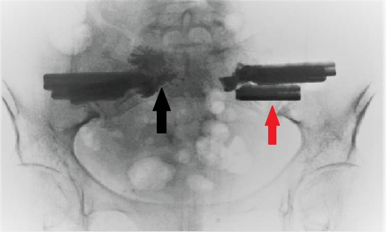
Figure 4. Intraoperative C-arm fluoroscopic radiograph of the pelvis and sacrum.
The figure shows three triangular implants (red arrow) placed bilaterally through the ilium and sacral ala across the SI joint. Also seen is the polymethylmethacrylate (PMMA) cement (black arrow) inserted into the SI joint bilaterally.

