

Abstract
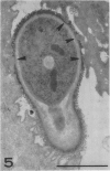
The colonization and invasion of various animal oral mucosae by Candida albicans were examined in an organ culture model. Scanning and transmission electron microscopy of the oral epithelium between 12 and 30 h after inoculation with the fungus revealed the morphological relationships between host and parasite. Examination of the fungi in thin sections showed five distinct layers in the cell wall of C. albicans within the epithelium, but changes were evident in the organization and definition of the outer cell wall layers in budding hyphae and in hyphae participating in colonization and invasion of the epithelial cells. Adherence of the fungus to the superficial cells of the oral mucosa appeared to involve intimate contact between the epithelial cell surface and the deeper layers of the fungal cell wall. During invasion a close seal was maintained between the invading hyphae and the surrounding epithelial cell envelope, there being no other evidence of damage to the host cell surface except at the site of entry. Within the epithelial cells there was only occasional loss of cytoplasmic components in the vicinity of the invading hyphae. These findings would suggest that enzymatic lysis associated with the invasive process is localized and that the mechanical support provided by surface adherence and the intimate association between the fungus and the epithelial cell envelope may permit growth of Candida on through the epithelium.
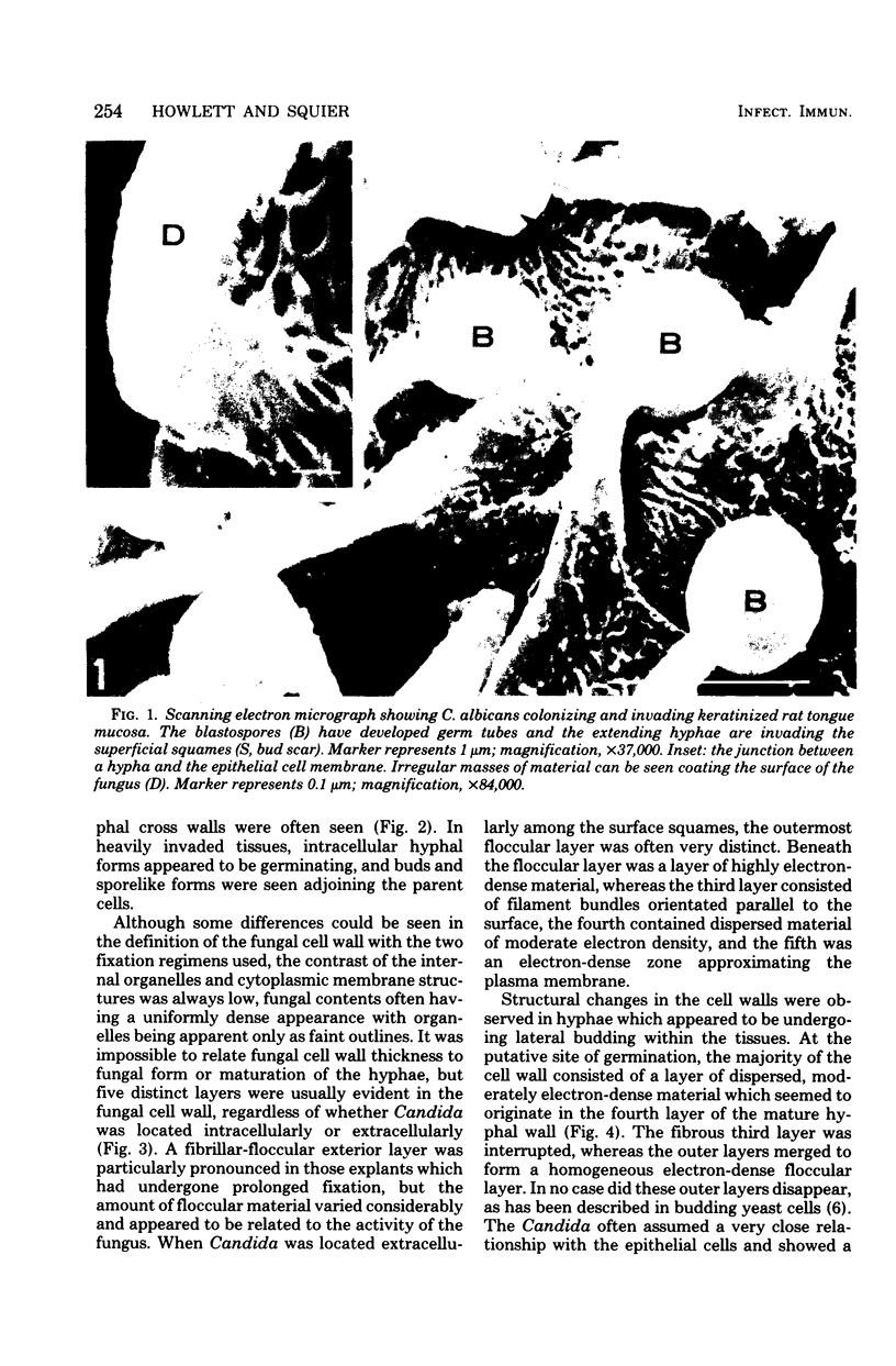
Full text
PDF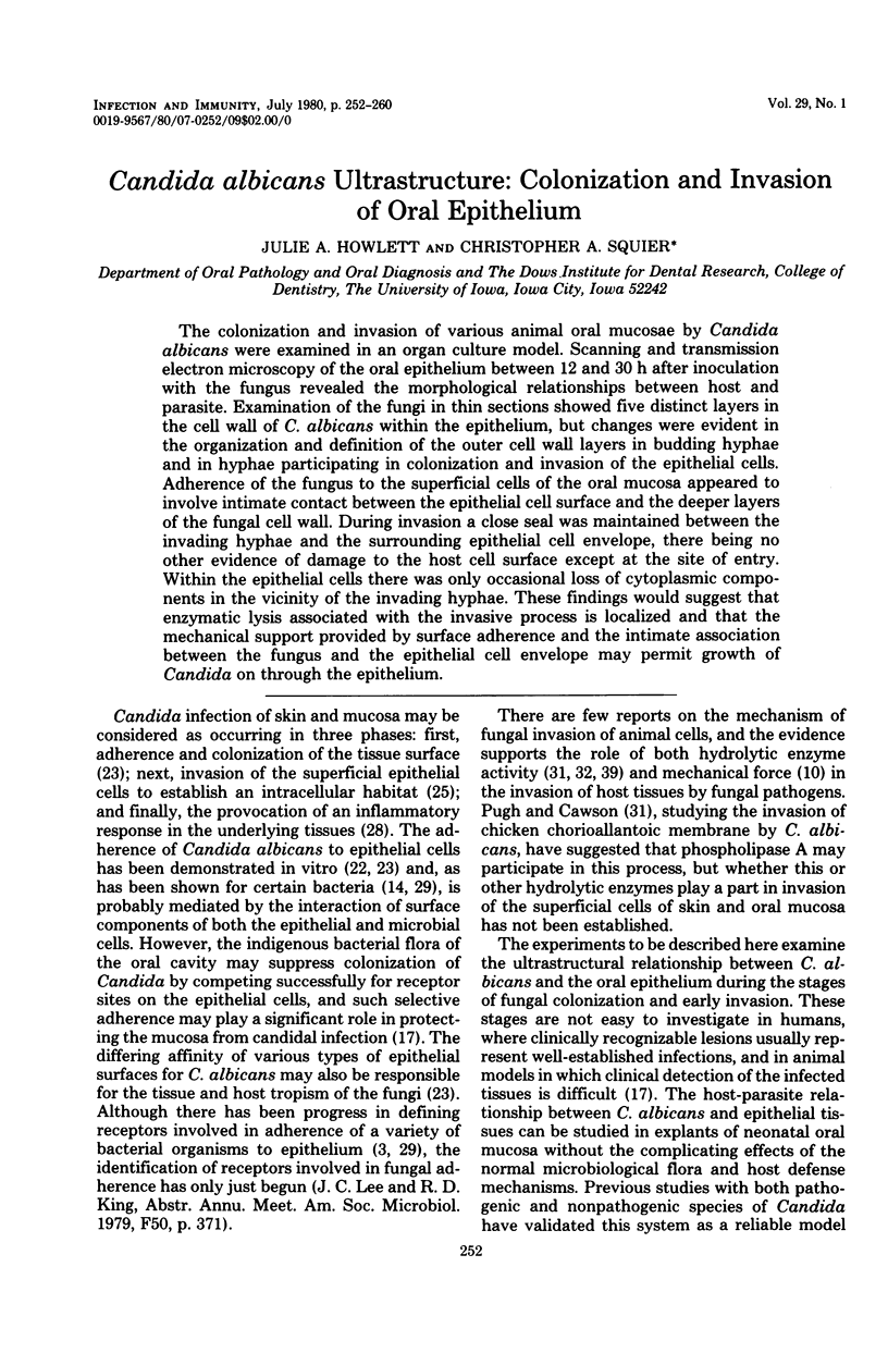


Images in this article


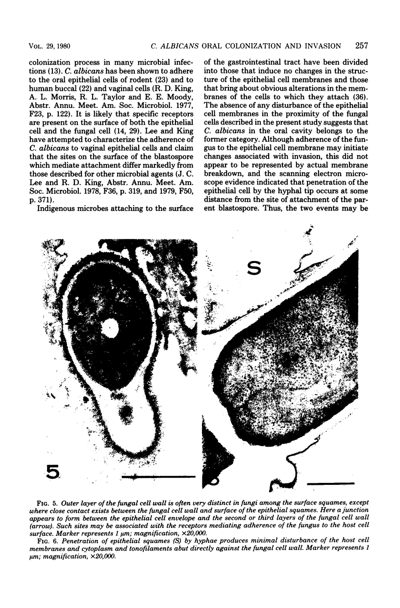
Selected References
These references are in PubMed. This may not be the complete list of references from this article.
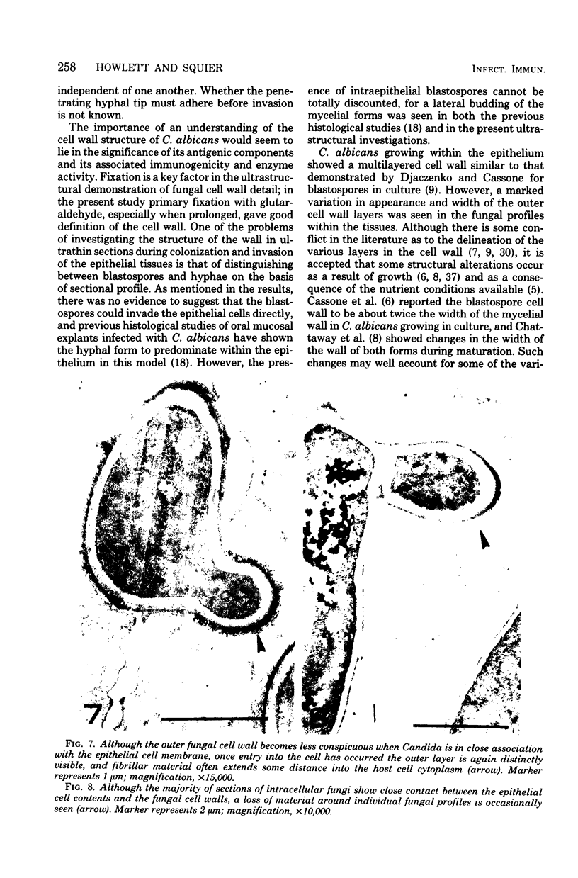
- Arai T., Mikami Y., Yokoyama K. Phagocytosis of Candida albicans by rabbit alveolar macrophages and guinea pig neutrophils. Sabouraudia. 1977 Jul;15(2):171–177. [PubMed] [Google Scholar]
- Bartelt M. A., Duncan J. L. Adherence of group A streptococci to human epithelial cells. Infect Immun. 1978 Apr;20(1):200–208. doi: 10.1128/iai.20.1.200-208.1978. [DOI] [PMC free article] [PubMed] [Google Scholar]
- Belcher R. W., Carney J. F., Monahan F. G. An electron microscopic study of phagocytosis of Candida albicans by polymorphonuclear leukocytes. Lab Invest. 1973 Dec;29(6):620–627. [PubMed] [Google Scholar]
- Cassone A., Kerridge D., Gale E. F. Ultrastructural changes in the cell wall of Candida albicans following cessation of growth and their possible relationship to the development of polyene resistance. J Gen Microbiol. 1979 Feb;110(2):339–349. doi: 10.1099/00221287-110-2-339. [DOI] [PubMed] [Google Scholar]
- Cassone A., Simonetti N., Strippoli V. Ultrastructural changes in the wall during germ-tube formation from blastospores of Candida albicans. J Gen Microbiol. 1973 Aug;77(2):417–426. doi: 10.1099/00221287-77-2-417. [DOI] [PubMed] [Google Scholar]
- Cawson R. A., Rajasingham K. C. Ultrastructural features of the invasive phase of Candida albicans. Br J Dermatol. 1972 Nov;87(5):435–443. doi: 10.1111/j.1365-2133.1972.tb01591.x. [DOI] [PubMed] [Google Scholar]
- Chattaway F. W., Shenolikar S., O'Reilly J., Barlow A. J. Changes in the cell surface of the dimorphic forms of Candida albicans by treatment with hydrolytic enzymes. J Gen Microbiol. 1976 Aug;96(2):335–347. doi: 10.1099/00221287-95-2-335. [DOI] [PubMed] [Google Scholar]
- Djaczenko W., Cassone A. Visulization of new ultrastructural components in the cell wall of Candida albicans with fixatives containing TAPO. J Cell Biol. 1972 Jan;52(1):186–190. doi: 10.1083/jcb.52.1.186. [DOI] [PMC free article] [PubMed] [Google Scholar]
- Frithiof L. Ultrastructural changes in the plasma membrane in human oral epithelium. J Ultrastruct Res. 1970 Jul;32(1):1–17. doi: 10.1016/s0022-5320(70)80033-8. [DOI] [PubMed] [Google Scholar]
- Garrison R. G., Boyd K. S. Aspects of the dimorphism of Histoplasma farciminosum: a light and electron microscopic study. Sabouraudia. 1975 Jul;13(2):174–184. doi: 10.1080/00362177585190321. [DOI] [PubMed] [Google Scholar]
- Gibbons R. J., van Houte J. Selective bacterial adherence to oral epithelial surfaces and its role as an ecological determinant. Infect Immun. 1971 Apr;3(4):567–573. doi: 10.1128/iai.3.4.567-573.1971. [DOI] [PMC free article] [PubMed] [Google Scholar]
- Gray G. M. Phosphatidyl-(N-acyl)-ethanolamine. A lipid component of mammalian epidermis. Biochim Biophys Acta. 1976 Apr 22;431(1):1–8. doi: 10.1016/0005-2760(76)90253-8. [DOI] [PubMed] [Google Scholar]
- Gray G. M., Yardley H. J. Different populations of pig epidermal cells: isolation and lipid composition. J Lipid Res. 1975 Nov;16(6):441–447. [PubMed] [Google Scholar]
- Helstrom P. B., Balish E. Effect of oral tetracycline, the microbial flora, and the athymic state on gastrointestinal colonization and infection of BALB/c mice with Candida albicans. Infect Immun. 1979 Mar;23(3):764–774. doi: 10.1128/iai.23.3.764-774.1979. [DOI] [PMC free article] [PubMed] [Google Scholar]
- Howlett J. A. Candidal infection of the oral mucosa: an in vitro model. Proc R Soc Med. 1976 Oct;69(10):766–770. doi: 10.1177/003591577606901020. [DOI] [PMC free article] [PubMed] [Google Scholar]
- Howlett J. A. The infection of rat tongue mucosa in vitro with five species of Candida. J Med Microbiol. 1976 Aug;9(3):309–316. doi: 10.1099/00222615-9-3-309. [DOI] [PubMed] [Google Scholar]
- Iwata K. Fungal toxins as a parasitic factor responsible for the establishment of fungal infections. Mycopathologia. 1978 Dec 18;65(1-3):141–154. doi: 10.1007/BF00447185. [DOI] [PubMed] [Google Scholar]
- Jessen H., Peters P. D., Hall T. A. Sulphur in different types of keratohyalin granules: a quantitative assay by X-ray microanalysis. J Cell Sci. 1974 Jul;15(2):359–377. doi: 10.1242/jcs.15.2.359. [DOI] [PubMed] [Google Scholar]
- Kimura L. H., Pearsall N. N. Adherence of Candida albicans to human buccal epithelial cells. Infect Immun. 1978 Jul;21(1):64–68. doi: 10.1128/iai.21.1.64-68.1978. [DOI] [PMC free article] [PubMed] [Google Scholar]
- Liljemark W. F., Gibbons R. J. Suppression of Candida albicans by human oral streptococci in gnotobiotic mice. Infect Immun. 1973 Nov;8(5):846–849. doi: 10.1128/iai.8.5.846-849.1973. [DOI] [PMC free article] [PubMed] [Google Scholar]
- Mohamed A. M. Ultrastructural aspects of chronic oral candidosis. J Oral Pathol. 1975 Oct-Nov;4(4):180–194. doi: 10.1111/j.1600-0714.1975.tb01741.x. [DOI] [PubMed] [Google Scholar]
- Montes L. F., Wilborn W. H. Ultrastructural features of host-parasite relationship in oral candidiasis. J Bacteriol. 1968 Oct;96(4):1349–1356. doi: 10.1128/jb.96.4.1349-1356.1968. [DOI] [PMC free article] [PubMed] [Google Scholar]
- Moulder J. W. Intracellular parasitism: life in an extreme environment. J Infect Dis. 1974 Sep;130(3):300–306. doi: 10.1093/infdis/130.3.300. [DOI] [PubMed] [Google Scholar]
- Myerowitz R. L. Ultrastructural observations in disseminated candidiasis. Arch Pathol Lab Med. 1978 Oct;102(10):506–511. [PubMed] [Google Scholar]
- Ofek I., Mirelman D., Sharon N. Adherence of Escherichia coli to human mucosal cells mediated by mannose receptors. Nature. 1977 Feb 17;265(5595):623–625. doi: 10.1038/265623a0. [DOI] [PubMed] [Google Scholar]
- Poulain D., Tronchin G., Dubremetz J. F., Biguet J. Ultrastructure of the cell wall of Candida albicans blastospores: study of its constitutive layers by the use of a cytochemical technique revealing polysaccharides. Ann Microbiol (Paris) 1978 Feb-Mar;129(2):141–153. [PubMed] [Google Scholar]
- Pugh D., Cawson R. A. The cytochemical localization of acid hydrolases in four common fungi. Cell Mol Biol Incl Cyto Enzymol. 1977;22(1):125–132. [PubMed] [Google Scholar]
- Pugh D., Cawson R. A. The cytochemical localization of phospholipase in Candida albicans infecting the chick chorio-allantoic membrane. Sabouraudia. 1977 Mar;15(1):29–35. [PubMed] [Google Scholar]
- Pugh D., Cawson R. A. The surface layer of Candida albicans. Microbios. 1978;23(91):19–23. [PubMed] [Google Scholar]
- REYNOLDS E. S. The use of lead citrate at high pH as an electron-opaque stain in electron microscopy. J Cell Biol. 1963 Apr;17:208–212. doi: 10.1083/jcb.17.1.208. [DOI] [PMC free article] [PubMed] [Google Scholar]
- Rooney L., Moens P. B. Nuclear divisions at meiosis in the ascomycetous yeast Wickerhamia fluorescens. Can J Microbiol. 1973 Nov;19(11):1383–1387. doi: 10.1139/m73-223. [DOI] [PubMed] [Google Scholar]
- Scherwitz C., Martin R., Ueberberg H. Ultrastructural investigations of the formation of Candida albicans germ tubes and septa. Sabouraudia. 1978 Jun;16(2):115–124. [PubMed] [Google Scholar]
- Schnell J. D., Voigt W. H. Das Verhalten von Sprosspilzen am nicht verhornenden Plattenepithel. Arch Gynakol. 1974;217(4):377–382. doi: 10.1007/BF00668025. [DOI] [PubMed] [Google Scholar]
- Wolff K., Schreiner E. Aufnahme, intracellulärer Transport und Abbau exogenen Proteins in Keratinocyten. Eine elektronenmikroskopisch-cytochemische Studie mit Peroxidase als Markierungssubstanz. Arch Klin Exp Dermatol. 1969;235(2):203–220. [PubMed] [Google Scholar]