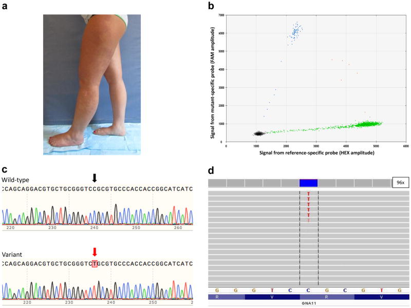
Figure 1.
GNA11 mutation in Participant 5. (a) Photograph depicting the patient’s capillary malformation and hypertrophy of her left lower extremity. (b) Results from a ddPCR reaction displaying the presence of the GNA11 c.547C>T (p.ArgR183Cys) mutation with a mutant allele frequency of ~5%. (c) Sanger sequencing of PCR-amplimer-subclones showing (Top) a clone with the wild-type sequence and (Bottom) another with the mutation at position 547 having a T (red arrow) instead of a C (black arrow). (d) Integrative Genomic Viewer screen-shot depicting MIP-seq coverage at the site of the mutation (c.547C>T) with ~5% mutant allele frequency. 96x indicates the depth of coverage at position 547.

