

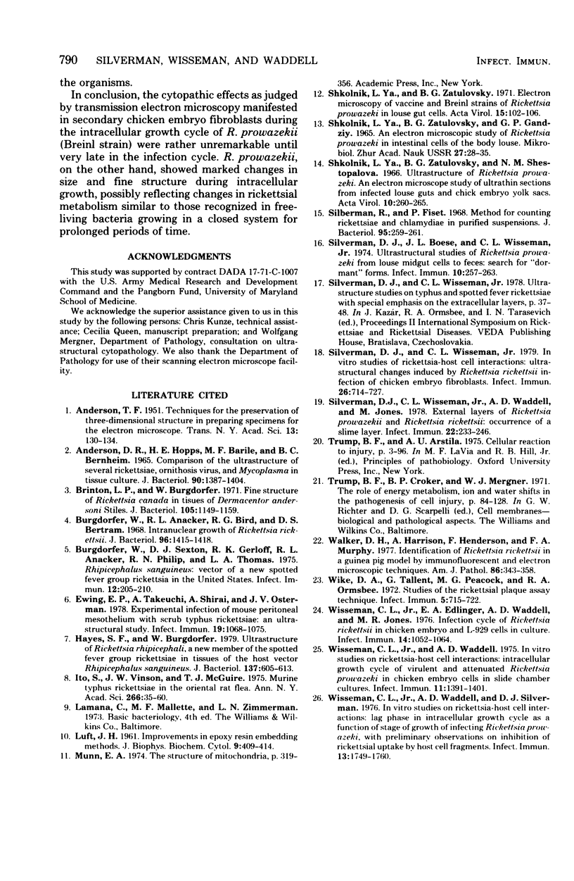
Abstract
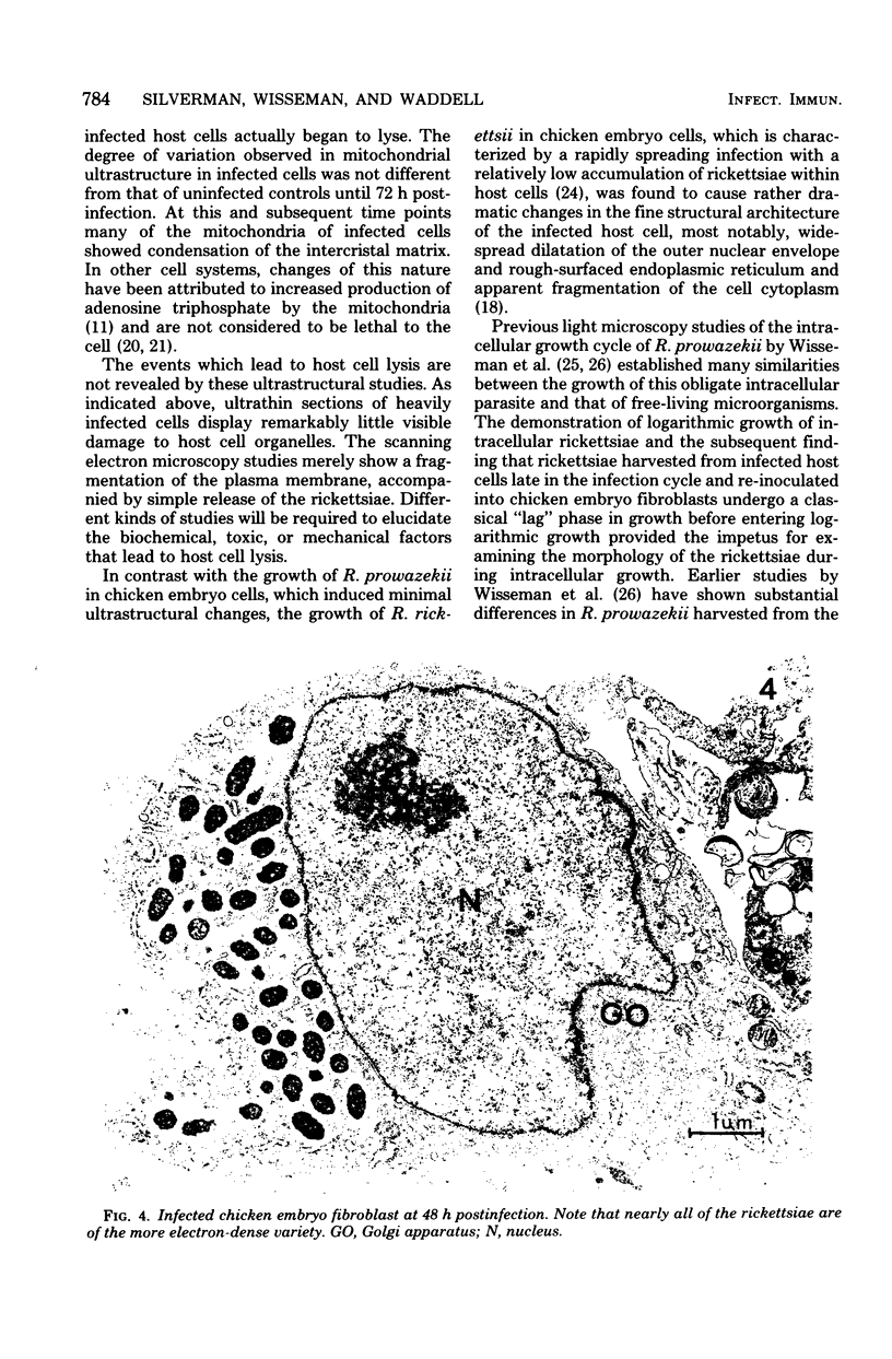
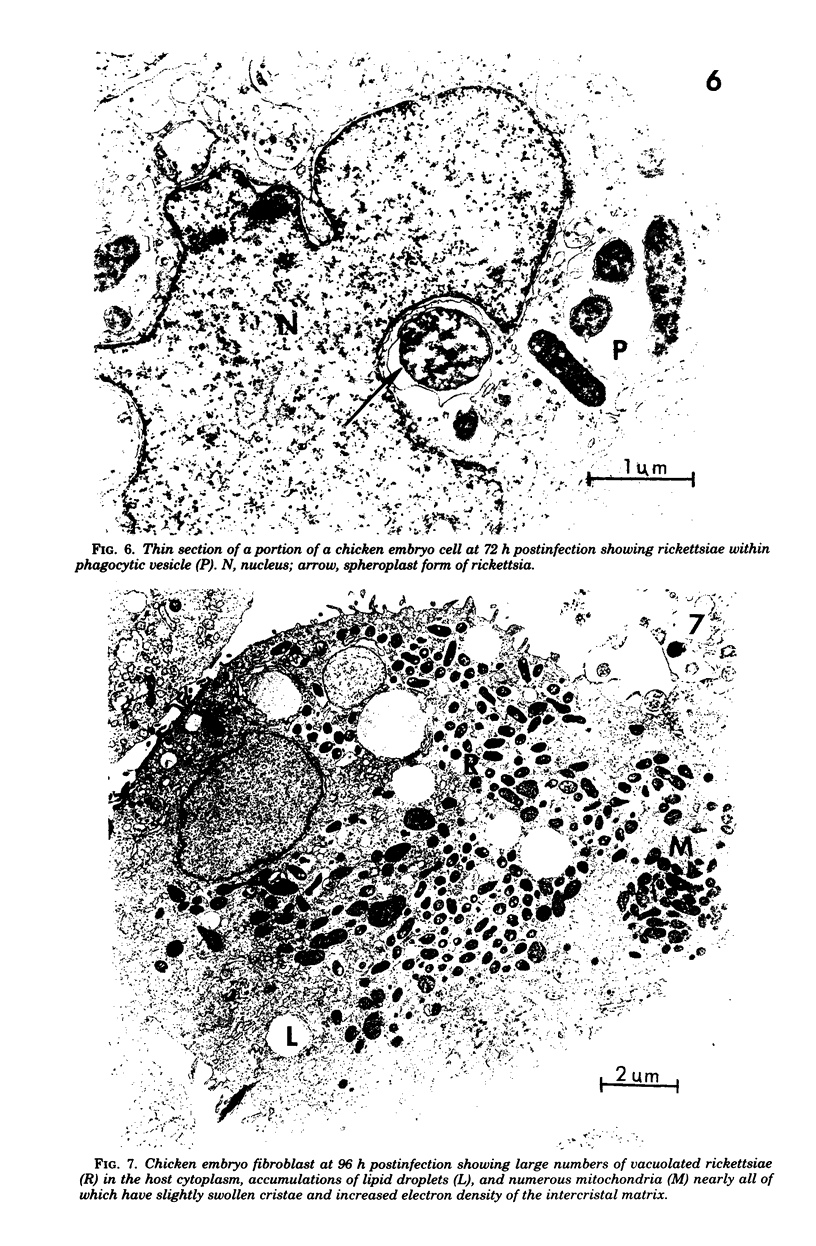
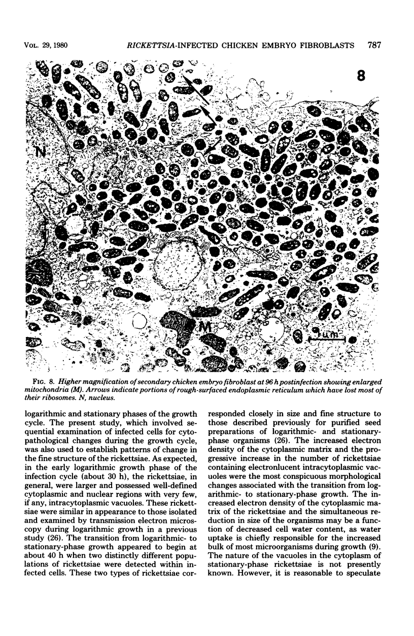
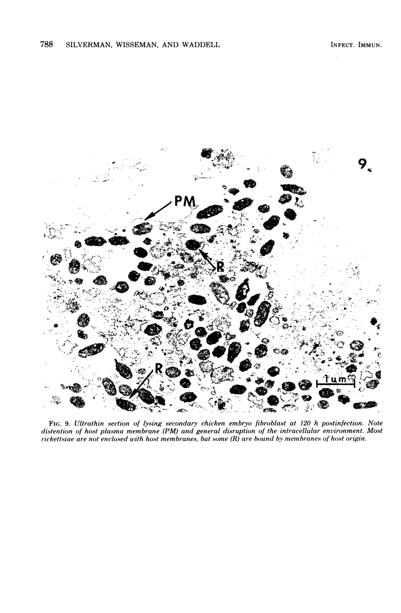
Secondary chicken embryo fibroblasts infected in suspension with the Breinl strain of Rickettsia prowazekii and grown in monolayer culture were examined by both transmission and scanning electron microscopy at specific intervals after infection to study the effects of prolonged intracellular growth on the fine structure of the host cell and the rickettsiae. Cytopathological changes in the infected host cells were not apparent until late in the intracellular growth cycle when the cells began to rupture as a result of a large rickettsial burden. The only recognizable changes in heavily infected cells before lysis were the condensation of the intercristal matrix of some mitochondria and the apparent dissociation of ribosomes from the rough-surfaced endoplasmic reticulum. Although the effects of intracellular growth of rickettsiae on the fine structure of the host cell were rather unremarkable when compared with those imposed by Rickettsia rickettsii in a similar cell system, noticeable morphological changes in the rickettsiae were recognized during the intracellular growth cycle. These changes first became apparent about 40 h postinfection and consisted primarily of an increased electron density of the rickettsiae, the appearance of numerous vacuoles in the rickettsial cytoplasm, and a slight reduction in size of the rickettsiae. Changes of this nature may reflect transitional phases of growth characteristically seen in free-living bacterial cell systems.
Full text
PDF


Images in this article


Selected References
These references are in PubMed. This may not be the complete list of references from this article.
- Anderson D. R., Hopps H. E., Barile M. F., Bernheim B. C. Comparison of the ultrastructure of several rickettsiae, ornithosis virus, and Mycoplasma in tissue culture. J Bacteriol. 1965 Nov;90(5):1387–1404. doi: 10.1128/jb.90.5.1387-1404.1965. [DOI] [PMC free article] [PubMed] [Google Scholar]
- Brinton L. P., Burgdorfer W. Fine structure of Rickettsia canada in tissues of Dermacentor andersoni Stiles. J Bacteriol. 1971 Mar;105(3):1149–1159. doi: 10.1128/jb.105.3.1149-1159.1971. [DOI] [PMC free article] [PubMed] [Google Scholar]
- Burgdorfer W., Anacker R. L., Bird R. G., Bertram D. S. Intranuclear growth of Rickettsia rickettsii. J Bacteriol. 1968 Oct;96(4):1415–1418. doi: 10.1128/jb.96.4.1415-1418.1968. [DOI] [PMC free article] [PubMed] [Google Scholar]
- Burgdorfer W., Sexton D. J., Gerloff R. K., Anacker R. L., Philip R. N., Thomas L. A. Rhipicephalus sanguineus: vector of a new spotted fever group rickettsia in the United States. Infect Immun. 1975 Jul;12(1):205–210. doi: 10.1128/iai.12.1.205-210.1975. [DOI] [PMC free article] [PubMed] [Google Scholar]
- Ewing E. P., Jr, Takeuchi A., Shirai A., Osterman J. V. Experimental infection of mouse peritoneal mesothelium with scrub typhus rickettsiae: an ultrastructural study. Infect Immun. 1978 Mar;19(3):1068–1075. doi: 10.1128/iai.19.3.1068-1075.1978. [DOI] [PMC free article] [PubMed] [Google Scholar]
- Hayes S. F., Burgdorfer W. Ultrastructure of Rickettsia rhipicephali, a new member of the spotted fever group rickettsiae in tissues of the host vector Rhipicephalus sanguineus. J Bacteriol. 1979 Jan;137(1):605–613. doi: 10.1128/jb.137.1.605-613.1979. [DOI] [PMC free article] [PubMed] [Google Scholar]
- Ito S., Vinson J. W., McGuire T. J., Jr Murine typhus Rickettsiae in the Oriental rat flea. Ann N Y Acad Sci. 1975;266:35–60. doi: 10.1111/j.1749-6632.1975.tb35087.x. [DOI] [PubMed] [Google Scholar]
- LUFT J. H. Improvements in epoxy resin embedding methods. J Biophys Biochem Cytol. 1961 Feb;9:409–414. doi: 10.1083/jcb.9.2.409. [DOI] [PMC free article] [PubMed] [Google Scholar]
- Shkolnik L. Y., Zatulovsky B. G. Electron microscopy of vaccine and Breinl strains of Rickettsia prowazeki in louse gut cells. Acta Virol. 1971 Jan;15(1):102–106. [PubMed] [Google Scholar]
- Shkolnik L. Y., Zatulovsky B. G., Shestopalova N. M. Ultrastructure of Rickettsia prow azeki. An electron microscope study of ultrathin sections from infected louse guts and chick embryo yolk sacs. Acta Virol. 1966 May;10(3):260–265. [PubMed] [Google Scholar]
- Silberman R., Fiset P. Method for counting Rickettsiae and Chlamydiae in purified suspensions. J Bacteriol. 1968 Jan;95(1):259–261. doi: 10.1128/jb.95.1.259-261.1968. [DOI] [PMC free article] [PubMed] [Google Scholar]
- Silverman D. J., Boese J. L., Wisseman C. L., Jr Ultrastructural studies of Rickettsia prowazeki from louse midgut cells to feces: search for "dormant" forms. Infect Immun. 1974 Jul;10(1):257–263. doi: 10.1128/iai.10.1.257-263.1974. [DOI] [PMC free article] [PubMed] [Google Scholar]
- Silverman D. J., Wisseman C. L., Jr In vitro studies of rickettsia-host cell interactions: ultrastructural changes induced by Rickettsia rickettsii infection of chicken embryo fibroblasts. Infect Immun. 1979 Nov;26(2):714–727. doi: 10.1128/iai.26.2.714-727.1979. [DOI] [PMC free article] [PubMed] [Google Scholar]
- Silverman D. J., Wisseman C. L., Jr, Waddell A. D., Jones M. External layers of Rickettsia prowazekii and Rickettsia rickettsii: occurrence of a slime layer. Infect Immun. 1978 Oct;22(1):233–246. doi: 10.1128/iai.22.1.233-246.1978. [DOI] [PMC free article] [PubMed] [Google Scholar]
- Walker D. H., Harrison A., Henderson F., Murphy F. A. Identification of Rickettsia rickettsii in a guinea pig model by immunofluorescent and electron microscopic techniques. Am J Pathol. 1977 Feb;86(2):343–358. [PMC free article] [PubMed] [Google Scholar]
- Wike D. A., Tallent G., Peacock M. G., Ormsbee R. A. Studies of the rickettsial plaque assay technique. Infect Immun. 1972 May;5(5):715–722. doi: 10.1128/iai.5.5.715-722.1972. [DOI] [PMC free article] [PubMed] [Google Scholar]
- Wisseman C. L., Jr, Edlinger E. A., Waddell A. D., Jones M. R. Infection cycle of Rickettsia rickettsii in chicken embryo and L-929 cells in culture. Infect Immun. 1976 Oct;14(4):1052–1064. doi: 10.1128/iai.14.4.1052-1064.1976. [DOI] [PMC free article] [PubMed] [Google Scholar]
- Wisseman C. L., Jr, Waddell A. D. In vitro studies on rickettsia-host cell interactions: intracellular growth cycle of virulent and attenuated Rickettsia prowazeki in chicken embryo cells in slide chamber cultures. Infect Immun. 1975 Jun;11(6):1391–1404. doi: 10.1128/iai.11.6.1391-1401.1975. [DOI] [PMC free article] [PubMed] [Google Scholar]
- Wisseman C. L., Jr, Waddell A. D., Silverman D. J. In vitro studies on Rickettsia-host cell interactions: lag phase in intracellular growth cycle as a function of stage of growth of infecting Rickettsia prowazeki, with preliminary observations on inhibition of rickettsial uptake by host cell fragments. Infect Immun. 1976 Jun;13(6):1749–1760. doi: 10.1128/iai.13.6.1749-1760.1976. [DOI] [PMC free article] [PubMed] [Google Scholar]