Abstract
Objective
To determine the accuracy of intraoperative computed tomography (iCT) in localizing deep brain stimulation (DBS) electrodes by comparing this modality with postoperative magnetic resonance imaging (MRI).
Background
Optimal lead placement is a critical factor for the outcome of DBS procedures and preferably confirmed during surgery. iCT offers 3-dimensional verification of both microelectrode and lead location during DBS surgery. However, accurate electrode representation on iCT has not been extensively studied.
Methods
DBS surgery was performed using the Leksell stereotactic G frame. Stereotactic coordinates of 52 DBS leads were determined on both iCT and postoperative MRI and compared with intended final target coordinates. The resulting absolute differences in X (medial-lateral), Y (anterior-posterior), and Z (dorsal-ventral) coordinates (ΔX, ΔY, and ΔZ) for both modalities were then used to calculate the euclidean distance.
Results
Euclidean distances were 2.7 ± 1.1 and 2.5 ± 1.2 mm for MRI and iCT, respectively (p = 0.2).
Conclusion
Postoperative MRI and iCT show equivalent DBS lead representation. Intraoperative localization of both microelectrode and DBS lead in stereotactic space enables direct adjustments. Verification of lead placement with postoperative MRI, considered to be the gold standard, is unnecessary.
Keywords: Intraoperative computed tomography, Deep brain stimulation, Stereotactic neurosurgery, Movement disorders
Introduction
Deep brain stimulation (DBS) has become a widely performed surgical procedure for patients with movement disorders such as Parkinson disease, dystonia, and essential tremor [1,2,3]. Accurate lead placement in the intended target is of outmost importance for achieving maximal symptom suppression and minimizing potential stimulation induced side effects [4,5]. Advances in imaging have enhanced direct target planning and postoperative lead localization, but intraoperative verification of the (micro-)electrode position in relation to intended target coordinates remains challenging.
Currently anteroposterior (AP) radiography and lateral fluoroscopy are widely implemented during DBS surgery in order to localize both microelectrode and DBS lead [6]. Intracerebral structures are not shown and converting these two-dimensional modalities to euclidean space necessitates software that is not available in every center [7]. Ideally, three-dimensional (3D) imaging is done after each DBS lead placement with the patient still in the operating room so that any necessary electrode repositioning can be performed immediately. In recent years, intraoperative magnetic resonance imaging (MRI) and computed tomography (CT) have become available in some operating room suites.
In our institution, the transportable flat-panel CT imaging system Medtronic O-arm (Medtronic Inc., Minneapolis, MN, USA) is used. The O-arm is essentially a C-arm with a telescoping gantry and has been used in spinal surgery to obtain intraoperative CT (iCT) imaging for a longer period of time [8]. Whether O-arm iCT offers accurate lead representation is open for debate. Two studies compared both modalities in frame-based DBS surgery and showed that iCT and postoperative MRI have highly similar lead representation. However, euclidean distances differed which hampers definitive conclusions [9,10].
To address this issue and examine whether iCT can provide useful data during DBS surgery, lead tip coordinates on iCT were determined and compared with those on corresponding postoperative MRI, which is considered to be the gold standard for assessment of electrode location.
Methods
Patients
During the period from August 2011 to February 2013, information was collected from all patients who underwent microelectrode recording (MER)-guided DBS implantation using the Medtronic O-arm at the Rush University Medical Center, Chicago, IL, USA. This study was approved by the institutional review board.
Surgical Procedure
The surgical procedure of DBS placement is described in detail elsewhere [11]. The same team of movement disorder neurologist (L.V.M.) and neurosurgeon (R.B.) determined preoperatively for each individual case if iCT would be implemented during surgery. In staged bilateral procedures, preference was occasionally given to usage of AP radiography [6].
After placement of the final DBS electrode an iCT was performed, and in bilateral cases an iCT was performed following placement in each hemisphere. In order to monitor microelectrode positioning, iCT was also performed after the first MER trajectory was completed. When additional MER trajectories were made, an iCT was also performed after each trajectory in order to confirm intended movement in the vast majority of cases.
The iCT was positioned concentric with the patient's head after the Alpha Omega (Alpha Omega, Nazareth, Israel) system was set up to perform the first MER. Concentric positioning is of importance in order to obtain the highest possible resolution of the entire skull. All cranial bone structures are to be displayed so that subsequent co-registration reaches optimal accuracy. After transferring iCT images to the Stealthstation (Medtronic Inc.) co-registration with preoperative stereotactic MRI was performed. When this did not result in sufficient imaging overlap or electrode representation was of poor quality, for example due to movement artefact, a new iCT was acquired. Anatomical and stereotactic evaluation of electrode artefact was done for the entire trajectory course. Throughout surgery, the iCT scanner remained in the same position. Once the final lead was placed and iCT confirmed adequate placement, the device was removed from the operative field. We used “standard mode” of the O-arm with 120 kVp and 128 mAs. Contrast resolution with these settings is insufficient for full view of intracranial structures as brain parenchyma and ventricles [12].
The O-arm itself emits an effective radiation dose of 0.5 mSv per standard 3D spin for a small head protocol, which is the protocol used in our frame-based DBS procedures. We applied a maximum of 5 O-arm iCTs per patient, therefore, the maximum total radiation dose will not exceed 2.5 mSv. This total radiation dose is less then when using lateral fluoroscopy or AP radiography [13].
Lead Tip on MRI and iCT
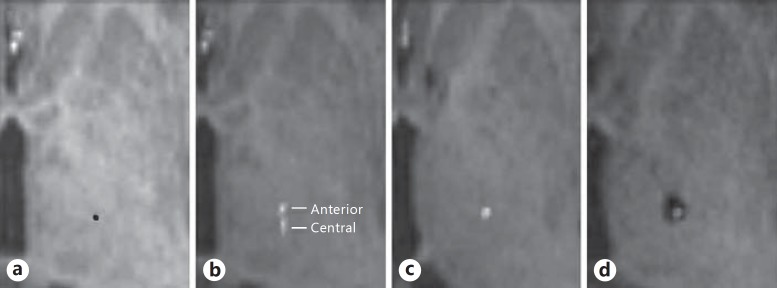
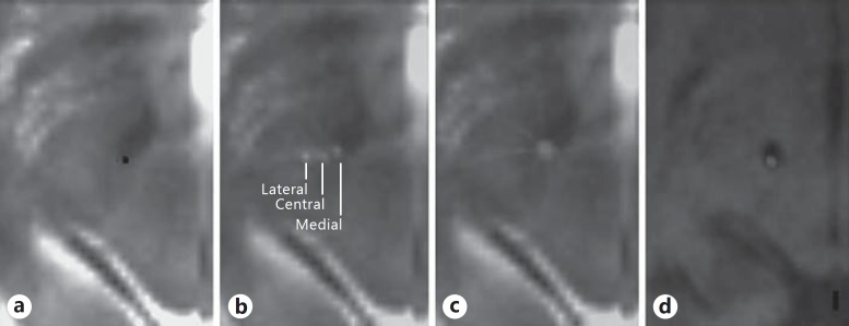
Both iCT and postoperative MRI were co-registered to preoperative stereotactic MRI in order to register in cartesian coordinates (Fig. 1). Lead tip coordinates were determined on both modalities in order to evaluate placement accuracy. A lead artefact appears differently on MRI compared to iCT, and therefore, each requires a different approach in lead tip determination. In vitro and in vivo MRI DBS lead measurements have shown that the ellipsoid shaped artefact extends approximately 1.4 mm over the proximal limits of the contact [14]. The actual beginning of contact 0 is characterized by a discrete round signal void and is best used for DBS lead measurements [15]. Flat panel iCT (such as O-arm) offers high spatial resolution; the lead artefact appears as a clear hyperdense signal in the obscured (after windowing) intracranial space. The bottom of contact 0 is identified at the most caudal point of lead artefact. Usage of 3 different planes (axial, sagittal, and coronal) and “trajectory view” allowed for a good geometric approximation of the actual electrode tip for both modalities. By using the “look ahead” view on the Framelink software (Framelink 5.1, Medtronic Inc.), a clear point of lead beginning can be determined by sliding into the artefact by using submillimetric steps. In both MRI and iCT, the center of artefact was chosen due to the fact that in a vast majority of cases the artefact is concentrically shaped. Microelectrode tip coordinates were determined on iCT in order to evaluate both placement accuracy and intended movement of microelectrodes during surgery (Fig. 2). Placement accuracy was evaluated by analyzing the first central microelectrode pass in each hemisphere (not any simultaneous placed channels). Movement was evaluated by analyzing microelectrode tips of second, third, and fourth microelectrode passes (if performed). The influence of subdural air volumes on lead placement accuracy was evaluated. Subdural air volumes were measured by usage of volume of interest. An overlay was created for every slice containing subdural air, resulting in a 3D volume of interest which represented total volume of air in milliliters. These measurements were done using a free software available online (MRIcro, www.mricro.com).
Fig. 1.
DBS electrode tip is displayed on axial orientated MRI and iCT. The right thalamic region is displayed on axial orientated imaging parallel to the line interconnecting the anterior and posterior commissure. a Stereotactic 1.5-Tesla T1 MRI at target depth, the red dot indicates the planned target point for right VIM. The stereotactic coordinates of the target point were 14.6 mm lateral, 5.0 mm posterior, and 0.6 mm dorsal relative to the midcommisural point. b Stereotactic 1.5-Tesla T1 MRI co-registered with iCT at target depth. Two hyperdense (white) dots represent the artefacts of the microelectrode channels as they appear on iCT. The lower dot represents the central microelectrode channel, the upper dot the anterior channel. The anterior channel is located 2 mm anterior to the central channel. c Stereotactic 1.5-Tesla T1 MRI co-registered with iCT. The hyperdense (white) dot represents the artefact of the DBS lead as it appears on iCT. The central channel was chosen for definite lead placement. d Postoperative 1.5-Tesla T1 MRI and iCT co-registered with stereotactic 1.5-Tesla T1 MRI (not shown). The electrode artefact as it appears on MRI (round signal void) coincides with the iCT electrode artefact. The stereotactic coordinates of the final electrode on MRI were 14.1 mm lateral, 4.6 mm posterior, and 1.1 mm dorsal to the midcommisural point, confirming that the central channel was used.
Fig. 2.
DBS electrode tip is displayed on axial orientated MRI and iCT. The left subthalamic region is displayed on axial orientated imaging parallel to the line interconnecting the anterior and posterior commissure. a Stereotactic 1.5-Tesla T2 MRI at target depth, the red dot indicates the planned target point for the subthalamic nucleus. Stereotactic coordinates of the target point were 12.3 mm lateral, 3.0 mm posterior, and 4.1 mm ventral relative to the midcommisural point. b Stereotactic 1.5-Tesla T2 MRI co-registered with iCT at the target depth. Three hyperdense (white) dots represent the artefacts of the microelectrode channels as they appear on iCT. The central dot represents the central microelectrode channel, the right dot the medial channel, and the left dot the lateral channel. The medial and lateral channels are located 2 mm apart from the central channel. The stereotactic coordinates of the central microelectrode channel were 14.0 mm lateral, 3.0 mm posterior, and 4.1 mm ventral relative to the midcommisural point, indicating a slight lateral displacement. c Stereotactic 1.5-Tesla T1 MRI co-registered with iCT. The hyperdense (white) dot represents the artefact of the DBS lead as it appears on iCT. The medial channel was chosen for definite lead placement. d Postoperative 1.5-Tesla T1 MRI and iCT co-registered with stereotactic 1.5-Tesla T1 MRI (not shown). The electrode artefact as it appears on MRI (round signal void) coincides with the iCT electrode artefact. Stereotactic coordinates of the final electrode on MRI imaging were 12.1 mm lateral, 3 mm posterior, and 4.8 mm ventral to the midcommisural point, confirming that the medial channel was used. DBS, deep brain stimulation; MRI, magnetic resonance imaging; iCT, intraoperative computed tomography.
Statistical Analysis
For both postoperative MRI and iCT absolute, directional and euclidean distances between intraoperative expected target coordinates and measured lead tip coordinates were calculated. For iCT absolute, directional and euclidean distances were also calculated for microelectrode placement. The euclidean distance,
is the total distance in 3D space and represents our main outcome measure. The calculated differences (euclidean, absolute, and directional) were compared using the Student t test. Numbers are given with standard deviation, and statistical significance was defined as p < 0.05. In order to determine a possible influence of intraoperative subdural air volume on placement accuracy the Pearson correlation was used.
Results
Lead Placements and MER Trajectories
A total of 52 leads were placed in 32 patients. There were 19 male and 13 female patients, and their average age at surgery was 61 years. Twenty patients underwent bilateral DBS placement, 12 patients underwent unilateral placement. The left side was operated on first in 10 bilateral cases. Target structure was subthalamic nucleus in 34 placements, globus pallidus internus (GPi) in 12 placements, and ventral intermediate nucleus of the thalamus (VIM) in 6 placements. Of the evaluated MER trajectories, 50 were first passes, 29 were subsequent passes (second, third, or fourth).
Lead Representation on MRI and iCT
The mean euclidean distance measured with postoperative MRI was 2.7 ± 1.1 versus 2.5 ± 1.2 mm using iCT (p = 0.2). No statistical difference was found between left- and right-sided placements.
The absolute mean X, Y, and Z distances measured on MRI were 1.3 ± 1.1, 1.0 ± 1.0, and 1.7 ± 1.1 mm, respectively. The absolute mean X, Y, and Z distances measured on iCT were 1.3 ± 1.0, 1.3 ± 1.1, and 1.3 ± 0.9 mm, respectively. Only the difference found in the Z distance was statistically significant (p = 0.01).
By comparing directional distances of the 2 modalities, we were able to determine if a systematic directional difference occurred in lead representation. The average distances measured on MRI for X, Y, and Z were 0.7 ± 1.5 mm medial, 0.5 ± 1.3 mm posterior, and 1.5 ± 1.3 mm ventral, respectively. The average directional distances on iCT for X, Y, and Z were 0.8 ± 1.4 mm medial, 1.0 ± 1.4 mm posterior, and 0.7 ± 1.4 mm ventral, respectively. Differences in the AP and dorsoventral direction were statistically significant (p < 0.01).
Microelectrode Representation on iCT
The mean euclidean distance of the first (central) microelectrode trajectory was 1.7 ± 0.8 mm. This was significantly smaller than euclidean distances calculated with final lead placement for both iCT and MRI (p < 0.01). There was no difference between left- and right-sided trajectories.
A total of 29 iCTs were made in order to evaluate intraoperative microelectrode movement. A second iCT for microelectrode evaluation was made in 22 placements, a third in 5 placements and a fourth in 2 placements. When a 2-mm microelectrode movement was made using a 5-channel micro-drive (not changing frame settings), which was performed 27 times, an average step of 1.9 ± 0.7 mm was measured. The direction of intended movement (in the medial-lateral and anterior-posterior plane) was confirmed by iCT in all cases.
Subdural Air Volume, Number of MERs and Accuracy of Lead Placement
In 24 patients, subdural air volumes were measured. The average subdural air volumes after DBS placement were 11.6 ± 9.6 and 7.9 ± 6.3 mL for right and left, respectively. The euclidean and absolute distances measured on MRI and iCT after lead placement did not significantly correlate with air volume. Subdural air increased with each subsequent microelectrode track, with the largest infusion of air seen after the first track. The mean increase in air volume (of all patients) measured was 11.0 ± 6.8 mL after the first microelectrode trajectory, 3.0 ± 4.5 mL after the second, and 2.4 ± 2.8 mL after the third. In 6 cases, subdural air collections could not be determined due to suboptimal representation of patient cranium on iCT. The frontal bone (cavity of frontal sinus) was not entirely displayed in these cases. In 2 cases, iCT could not be correctly displayed (loaded) in the free online software (MRIcro) that was used.
Discussion
Comparison of Postoperative MRI and iCT
We compared lead representation on iCT with that on postoperative MRI and found comparable euclidean distances. This indicates that iCT provides reliable information regarding lead placement. When the desired placement is confirmed with iCT, the need for an postoperative MRI is obviated.
Absolute and directional mean differences in lead representation in our study were ≤0.5 and ≤0.8 mm for the X, Y, and Z coordinates, respectively. Placement error of the Leksell system exceed these; hence, we feel that differences in lead representation on MRI and iCT are not clinically relevant [11,15]. The differences were largest in the dorsoventral plane where a more dorsal lead tip localization on iCT was noted. Determination of the lead tip in this plane is usually most challenging, especially on MRI which in general produced a more gradual beginning of an electrode tip. As stated in the method section, MRI creates an ellipsoid-shaped artefact which may have contributed to differences found in the dorsoventral plane.
Intraoperative Subdural Air and Stereotactic Accuracy
No correlation was found between subdural air volume and placement accuracy (both euclidean and absolute) of lead trajectory. This in is agreement with Shahlaie et al. [9] and Sharma and Deogaonkar [16]. Both groups have also used the O-arm for determination of intraoperative pneumocephaly. This suggests that accuracy of lead placement, when measured intraoperatively, is not (or not significantly) influenced by subdural air. Possible influence of subdural air volume on brain shift during DBS procedures has been described extensively [17,18,19,20]. Although postoperative MRI indicates shift of both anterior and posterior commissure, exact influence on accuracy of lead placement is less clear. Subdural air is more clearly delineated on iCT compared to MRI. However, intracerebral structures as the anterior and posterior commissure cannot be determined on iCT which makes measuring shift of subcortical structures not feasible. We cannot exclude that air volumes larger than those measured in our series could be of direct influence on lead placement.
iCT and DBS in the Literature
One paper described the use of O-arm during frame-based DBS surgery and compared its lead representation with that of postoperative MRI. Shahlaie et al. [9] compared euclidean and absolute differences (calculated differences between iCT and MRI lead tip localization) of 24 lead placements. They measured an average euclidean difference of 1.7 mm and concluded this to be statistically different from zero. Had they calculated euclidean distances of lead placement for both iCT and MRI and subsequently compared these, then possibly no significant difference would have been found. In addition, their absolute differences between both modalities did not differ significantly and were small enough (less than 0.5 mm) to be considered not clinically relevant [11,15].
Mirzadeh et al. [10] described the use of an intraoperative multidetector CT (not the O-arm) for 48 lead placements. For MRI and iCT, they found euclidean distances of 1.6 and 1.1 mm, respectively. The authors stated that the difference found is largely attributable to error in measurement by the observer.
Although the radiation dose using O-arm is estimated to be lower compared to fluoroscopy, the radiation applied is not benign. Care must be taken to limit the number of passes performed, thereby reducing effective radiation exposure [13].
In agreement with our findings, both groups consider iCT as a possible replacement for postoperative MRI for lead evaluation of lead placement [9,10]. Several advantages and disadvantages, considered by our group, using O-arm during DBS procedures are shown in Table 1.
Table 1.
Advantages and disadvantages of using the O-arm during DBS procedures
| Advantages | |
| – | The O-arm is a C-arm-based flat panel volume CT and combines both CT and fluoroscopy function. This is an advantage compared to intraoperative multidetector systems, as millimetric adjustments in the dorsoventral plane could be verified by using fluoroscopy (which is more easily done than using repetitive iCT). |
| – | Image acquisition, for 3D, is around 13 s, so that the surgical time is not notably prolonged. Acquisition time is considerably faster compared to intraoperative MRI [20]. |
| – | High spatial resolution of flat-panel iCT enables direct evaluation of microelectrode trajectories. While this was not the primary goal of our study, we found the possibility of evaluating both microelectrode placement and movement highly useful. |
| – | Using a high-definition optical enhancement mode enables detecting occurrence of a (small) hematoma in the trajectory course. |
| – | Different position settings can be saved by the O-arm, which allows simple switching between the established position for surgical proceedings versus that for performing imaging. |
| – | Although not implemented by our group, an interesting application of iCT is to replace conventional preoperative stereotactic CT. The field of view is modified in the latest version of the O-arm so that performing registration series using the Leksell frame is enabled. |
| Disadvantages | |
| – | Contrast resolution with standard settings is insufficient for full view of intracranial structures as brain parenchyma and ventricles which can be used to evaluate co-registration or brain shift. |
| – | The O-arm is quite large, this requires careful planning to maintain field sterility and provide adequate surgical access. |
Study Limitations
Occurrence of possible lead repositioning though iCT findings and prevention of reimplementation surgery was not noted. Lead localization was evaluated only, not any possible influence on (long-term) patient outcome (for example, measured using UPDRS scores). Findings during MER and/or test stimulation were not compared to anatomical findings displayed by iCT co-registered to MRI. Both patient outcome and anatomical findings would provide important insight for possible future omitting of MER and/or test stimulation through implementation of iCT.
Conclusion
Postoperative MRI and iCT show equivalent DBS lead representation. Intraoperative localization of microelectrode and DBS lead in stereotactic space enables direct adjustments. Verification of lead placement with postoperative MRI, considered to be the gold standard, is unnecessary.
Disclosure Statement
Dr. Verhagen has received compensation from Medtronic for educational activities. M. Bot received travel grants from Medtronic. The other authors do not have any personal or institutional financial interest in any drugs, materials, or devices described in this submission.
Acknowledgments
This study was supported by an investigator-initiated grant from Medtronic, Parkinson's Foundation the Netherlands, Vreedefonds Foundation, Drie Lichten Foundation, Noorthey Foundation, Amsterdam Foundation for Promoting Neurosurgical Development, and The Parkinson Disease Foundation.
References
- 1.Odekerken VJ, van Laar T, Staal MJ, Mosch A, Hoffmann CF, Nijssen PC, Beute GN, van Vugt JP, Lenders MW, Contarino MF, Mink MS, Bour LJ, van den Munckhof P, Schmand BA, de Haan RJ, Schuurman PR, de Bie RM. Subthalamic nucleus versus globus pallidus bilateral deep brain stimulation for advanced Parkinson's disease (NSTAPS study): a randomised controlled trial. Lancet Neurol. 2013;12:37–44. doi: 10.1016/S1474-4422(12)70264-8. [DOI] [PubMed] [Google Scholar]
- 2.Volkmann J, Mueller J, Deuschl G, Kuhn AA, Krauss JK, Poewe W, Timmermann L, Falk D, Kupsch A, Kivi A, Schneider GH, Schnitzler A, Sudmeyer M, Voges J, Wolters A, Wittstock M, Muller JU, Hering S, Eisner W, Vesper J, Prokop T, Pinsker M, Schrader C, Kloss M, Kiening K, Boetzel K, Mehrkens J, Skogseid IM, Ramm-Pettersen J, Kemmler G, Bhatia KP, Vitek JL, Benecke R, DBS Study Group for Dystonia Pallidal neurostimulation in patients with medication-refractory cervical dystonia: a randomised, sham-controlled trial. Lancet Neurol. 2014;13:875–884. doi: 10.1016/S1474-4422(14)70143-7. [DOI] [PubMed] [Google Scholar]
- 3.Schuurman PR, Bosch DA, Merkus MP, Speelman JD. Long-term follow-up of thalamic stimulation versus thalamotomy for tremor suppression. Mov Disord. 2008;23:1146–1153. doi: 10.1002/mds.22059. [DOI] [PubMed] [Google Scholar]
- 4.Richardson RM, Ostrem JL, Starr PA. Surgical repositioning of misplaced subthalamic electrodes in Parkinson's disease: location of effective and ineffective leads. Stereotact Funct Neurosurg. 2009;87:297–303. doi: 10.1159/000230692. [DOI] [PubMed] [Google Scholar]
- 5.Ellis TM, Foote KD, Fernandez HH, Sudhyadhom A, Rodriguez RL, Zeilman P, Jacobson CE, Okun MS. Reoperation for suboptimal outcomes after deep brain stimulation surgery. Neurosurgery. 2008;63:754–760. doi: 10.1227/01.NEU.0000325492.58799.35. discussion 760-751. [DOI] [PubMed] [Google Scholar]
- 6.Verhagen Metman L, Pilitsis JG, Stebbins GT, Bot M, Bakay RA. Intraoperative X-ray to measure distance between DBS leads: a reliability study. Mov Disord. 2012;27:1056–1059. doi: 10.1002/mds.25056. [DOI] [PubMed] [Google Scholar]
- 7.Bjartmarz H, Rehncrona S. Comparison of accuracy and precision between frame-based and frameless stereotactic navigation for deep brain stimulation electrode implantation. Stereotact Funct Neurosurg. 2007;85:235–242. doi: 10.1159/000103262. [DOI] [PubMed] [Google Scholar]
- 8.Oertel MF, Hobart J, Stein M, Schreiber V, Scharbrodt W. Clinical and methodological precision of spinal navigation assisted by 3D intraoperative O-arm radiographic imaging. J Neurosurg Spine. 2011;14:532–536. doi: 10.3171/2010.10.SPINE091032. [DOI] [PubMed] [Google Scholar]
- 9.Shahlaie K, Larson PS, Starr PA. Intraoperative computed tomography for deep brain stimulation surgery: technique and accuracy assessment. Neurosurgery. 2011;68:114–124. doi: 10.1227/NEU.0b013e31820781bc. discussion 124. [DOI] [PubMed] [Google Scholar]
- 10.Mirzadeh Z, Chapple K, Lambert M, Dhall R, Ponce FA. Validation of CT-MRI fusion for intraoperative assessment of stereotactic accuracy in DBS surgery. Mov Disord. 2014;29:1788–1795. doi: 10.1002/mds.26056. [DOI] [PubMed] [Google Scholar]
- 11.Bot M, van den Munckhof P, Bakay R, Sierens D, Stebbins G, Verhagen Metman L. Analysis of stereotactic accuracy in patients undergoing deep brain stimulation using Nexframe and the Leksell frame. Stereotact Funct Neurosurg. 2015;93:316–325. doi: 10.1159/000375178. [DOI] [PubMed] [Google Scholar]
- 12.Gupta R, Cheung AC, Bartling SH, Lisauskas J, Grasruck M, Leidecker C, Schmidt B, Flohr T, Brady TJ. Flat-panel volume CT: fundamental principles, technology, and applications. Radiographics. 2008;28:2009–2022. doi: 10.1148/rg.287085004. [DOI] [PubMed] [Google Scholar]
- 13.Smith AP, Bakay RAE. Frameless deep brain stimulation using intraoperative O-arm technology clinical article. J Neurosurg. 2011;115:301–309. doi: 10.3171/2011.3.JNS101642. [DOI] [PubMed] [Google Scholar]
- 14.Pollo C, Villemure JG, Vingerhoets F, Ghika J, Maeder P, Meuli R. Magnetic resonance artifact induced by the electrode Activa 3389: an in vitro and in vivo study. Acta Neurochir. 2004;146:161–164. doi: 10.1007/s00701-003-0181-4. [DOI] [PubMed] [Google Scholar]
- 15.Starr PA, Christine CW, Theodosopoulos PV, Lindsey N, Byrd D, Mosley A, Marks WJ., Jr Implantation of deep brain stimulators into the subthalamic nucleus: technical approach and magnetic resonance imaging-verified lead locations. J Neurosurg. 2002;97:370–387. doi: 10.3171/jns.2002.97.2.0370. [DOI] [PubMed] [Google Scholar]
- 16.Sharma M, Deogaonkar M. Accuracy and safety of targeting using intraoperative “O-arm” during placement of deep brain stimulation electrodes without electrophysiological recordings. J Clin Neurosci. 2016;27:80–86. doi: 10.1016/j.jocn.2015.06.036. [DOI] [PubMed] [Google Scholar]
- 17.van den Munckhof P, Contarino MF, Bour LJ, Speelman JD, de Bie RM, Schuurman PR. Postoperative curving and upward displacement of deep brain stimulation electrodes caused by brain shift. Neurosurgery. 2010;67:49–53. doi: 10.1227/01.NEU.0000370597.44524.6D. discussion 53-44. [DOI] [PubMed] [Google Scholar]
- 18.Halpern CH, Danish SF, Baltuch GH, Jaggi JL. Brain shift during deep brain stimulation surgery for Parkinson's disease. Stereotact Funct Neurosurg. 2008;86:37–43. doi: 10.1159/000108587. [DOI] [PubMed] [Google Scholar]
- 19.Elias WJ, Fu KM, Frysinger RC. Cortical and subcortical brain shift during stereotactic procedures. J Neurosurg. 2007;107:983–988. doi: 10.3171/JNS-07/11/0983. [DOI] [PubMed] [Google Scholar]
- 20.Khan MF, Mewes K, Gross RE, Skrinjar O. Assessment of brain shift related to deep brain stimulation surgery. Stereotact Funct Neurosurg. 2008;86:44–53. doi: 10.1159/000108588. [DOI] [PubMed] [Google Scholar]


