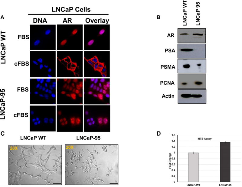
Figure 1.

Differential subcellular localization of the AR in prostate cancer LNCaP cells and the androgen resistant LNCaP-95 cells in normal or charcoal stripped containing media (A). Western blot showing expression of the AR and upregulation of the PCNA proteins in the LNCaP-95 cells and down regulation of PSA and PSMA compared to the wildtype LNCaP cells (B). A bright field (20×) microscopy image of the two cell lines showing distinct changes in the size and morphology of the androgen resistant LNCaP-95 cells (C). MTS cell proliferation assay in charcoal stripped FBS containing media showing a statistical difference between the wild type and LNCaP-95 cells at 48h (D).
