Abstract
Objective
In adults, obesity has been associated with several health outcomes including increased bone density. Our objective was to evaluate the association between percent body fat and fat mass with bone mineral density (BMD) in a nationally representative population of children and adolescents.
Study design
A total of 8,348 participants 8–18 years of age from the National Health and Nutrition Examination Survey (NHANES) 1999–2006 had whole body DXA scans performed. We conducted linear regressions to examine the relationship between percent body fat and fat mass with outcome variables of total body, pelvic and lumbar spine areal BMD (aBMD), controlling for lean body mass and assessing for gender and race/ethnicity interactions.
Results
We found evidence of gender and race/ethnicity interactions with percent body fat and total fat mass for the different BMD areas. Generally, there were decreases in total body aBMD (p<0.001) and lumbar spine aBMD (p<0.001) with increasing percent body fat and total fat mass, with less consistent patterns for pelvic aBMD.
Conclusion
Our findings of regional differences in the relationship of adiposity to aBMD in children and adolescents with significant interactions by gender and race/ethnicity emphasizes the need for further investigations to understand the impact of adiposity on bone health outcomes.
Introduction
Obesity and its related medical diseases continue to be a significant problem in both adults and children. Currently 31.8 percent of children aged 2 to 19 years in the U.S. are categorized as overweight or obese[1] and there are many consequences of this excess weight in children, including hypertension [2, 3], type 2 diabetes [4], sleep apnea, hyperlipidemia, and increased risk for immune diseases [5]. The effects of excess weight on bone health are less well studied, but since childhood and adolescence are critical stages for skeletal mineralization, it is important to understand how body composition during this period may influence bone mineralization and may affect bone health.
The majority of clinical studies evaluating the relationship between weight status and bone mineral density (BMD) have been performed in adult populations, which have shown a positive association of body mass index (BMI) with total body BMD [6]. Pediatric studies have reported contradictory results, with some reporting a negative association of visceral adipose tissue and BMD [7], and percent body fat with BMD and/or bone mineral content (BMC) [8], while others report a positive association of BMI on BMD [9] and BMC [8]. This same association has been suggested to not be the case for children who have a BMI greater than the 95th percentile [10]. Limitations of these studies include small sample sizes, and the use of BMI as a measure of body fat, which may not accurately categorize children with increased lean mass [11]. BMI also does not correlate with body fat similarly across ethnic and racial groups [11–13], or genders [14].
Few studies have looked at the association of fat mass on bone density in children and adolescents while adjusting for lean body mass [15]. Given that lean mass is a major contributor to BMI/weight status and has a direct mechanical impact on the bone that contributes to an increase in BMD [16], it is important to control for lean mass when investigating the relationship between fat mass and bone mineral density. The objective of our study was to examine associations between percent body fat and total fat mass on total body, pelvic and lumbar spine aBMD in a nationally representative sample of U.S. children and adolescents, and to examine gender and race/ethnicity interactions in these associations. We hypothesized that higher percent body fat would be associated with lower aBMD across all regions.
Materials and methods
We used data from National Health and Nutrition Examination Survey (NHANES) 1999–2006, a nationally representative cross-sectional survey of adults and children in the US civilian non-institutionalized population which oversamples individuals 12–19 years of age and minority populations and collects survey information about health, anthropometric measures, and laboratory data [17].
NHANES uses a complex, multistage, probability design in order to select representative participants [18]. It provides weight, height, gender, age, and race/ethnicity for each participant. As the survey reports, we used race/ethnicity classification as following: 1) White/non-Hispanic, 2) African American/non-Hispanic, 3) Hispanic, 4) Other races (which includes all non-Hispanic persons reporting a race other than White or African American, all participants who reported multiple races and the non-Hispanic Asians participants as well). NHANES performed whole body DXA scans in a subset of individuals 8 years and older using a Hologic QDR 4500 fan-beam densitometer (Hologic, Inc., Bedford, Massachusetts). Scans were conducted in the fast mode by trained radiology technologists and were analyzed using Hologic DOS software version 8.26:a3. Detailed documentation of the procedures for the DXA scans are available online [17, 19]. The NHANES DXA scan data provides bone and soft tissue measurements of total body, arms, legs, trunk and head; bone measurements from whole body DXA scans (total body, pelvic, left and right ribs, thoracic spine, lumbar spine); and values for the total body and bone regions including total mass (grams), Bone Mineral Content (BMC) (grams), bone area (cm2), areal bone mineral density (aBMD) (gm/cm2), fat mass (gm), lean mass excluding BMC (gm), lean mass including BMD (gm) and percent body fat [19]. NHANES provides the DXA data as multiply imputed datasets because of nonrandom missing data [17, 19].
Similar to our previous study [20], we calculated sex- and age-specific percentiles of body fat based on the literature [11, 20]. We excluded study participants with sex- and age-specific percentiles less than the 5th percentile, because these individuals could have other underlying disease or illness.
Our dependent variables were total body, pelvis and lumbar spine aBMD (g/cm2), which were chosen based on previous studies that have evaluated bone density in these areas [8, 15, 21]. Our primary independent variables were percent body fat and fat mass (in kg). Covariates included lean mass (kg), age, gender and race/ethnicity.
Statistical analysis
We performed separate multiple linear regressions to examine the relationship between percent body fat and total body, pelvic, and lumbar spine aBMD, adjusting for lean mass, age, gender and race/ethnicity. In an additional set of models, we performed separate multiple linear regressions to examine the relationship between total fat mass and total body aBMD, pelvic aBMD and lumbar spine aBMD, adjusting for lean mass, age, gender and race/ethnicity. We first examined 3-way interactions effects of fat variables x gender x race/ethnicity. All 3-way interactions were not significant except for the interaction of total fat mass predicting lumbar spine aBMD. We then examined 2-way interactions of percent body fat (percent body fat x gender; percent body fat x race/ethnicity) with each aBMD region, and total fat mass (total fat mass x gender; total fat mass x race/ethnicity) with each aBMD region, and only show results for models with significant interactions.
To illustrate the associations and interactions, we generated predicted outcome values for total body, pelvic and lumbar spine aBMD using selected percent body fat (or total fat mass) values for each gender and race/ethnicity while fixing all other predictor values at their mean. All statistical analyses account for the complex sampling design in NHANES using sample weights and sample design variables provided by NHANES[22]. We used STATA 13 statistical software for all analysis, which incorporates appropriate sampling weights to adjust for the complex sample design. A p-value threshold of 0.05 was considered to be significant.
Results
Of 9,797 individuals aged 8–18 years, we excluded children with incomplete DXA scans (n = 911), missing BMI (n = 35), missing age-, and with sex-adjusted percentile body fat less than the 5th percentile (n = 503), which left a sample size of 8,348 (Table 1).
Table 1. Demographic characteristics of the included population.
| Male | Female | ||||||||||||
|---|---|---|---|---|---|---|---|---|---|---|---|---|---|
| weighted% (unweighted n) | Overall Population | White | African American | Hispanic | Other * | White | African American | Hispanic | Other | White | African American | Hispanic | Other * |
| Overall | 8348 | 62.2% (2271) | 13.6% (2548) | 17.8% (3183) | 6.5% (346) | 57.2% (1270) | 55.9% (1436) | 57.5% (1848) | 59.5% (191) | 42.8% (1001) | 44.1% (1112) | 42.5% (1335) | 40.1% (155) |
| Male | 57.3% (4745) | ||||||||||||
| Body Fat Percentile | |||||||||||||
| 5–70 | 69.3% (5583) | 71.3% (1619) | 71.0% (1823) | 59.6% (1898) | 73.4% (243) | 70.4% (897) | 73.9% (1070) | 60.2% (1093) | 71.6% (131) | 72.6% (722) | 67.3% (753) | 58.9% (805) | 75.9% (112) |
| 70–85 | 15.2% (1338) | 14.3% (324) | 13.3% (329) | 20.0% (631) | 15.0% (54) | 15.7% (196) | 12.2% (168) | 18.8% (365) | 15.2% (31) | 12.4% (128) | 14.6% (161) | 21.6% (266) | 14.7% (23) |
| 85–90 | 5.8% (509) | 5.7% (127) | 5.2% (135) | 6.9% (228) | 4.7% (18) | 5.3% (67) | 4.6% (66) | 8.1% (139) | 4.7% (10) | 6.2% (60) | 6.1% (69) | 5.2% (89) | 4.7% (8) |
| > 90 | 9.7% (918) | 8.7% (201) | 10.5% (261) | 13.5% (426) | 7% (31) | 8.6% (110) | 9.4% (132) | 12.9% (251) | 8.4% (19) | 8.8% (91) | 12.0% (129) | 14.3% (175) | 4.8% (12) |
| Mean (SD) | |||||||||||||
| Age | 13.0 (4.4) | 13.1 (3.8) | 12.9 (4.6) | 12.8 (4.3) | 12.9 (4.3) | 13.1 (3.7) | 12.9 (4.3) | 12.8 (4.3) | 13.0 (4.0) | 13.2 (3.9) | 13.0 (4.8) | 12.9 (4.4) | 12.7 (4.6) |
| Height (cm) | 157.0 (21.1) | 158.0 (19.2) | 157.7 (21.9) | 154.2 (20.5) | 153.5 (20.3) | 160.2 (21.1) | 159.7 (23.4) | 156.4 (22.6) | 155.8 (21.1) | 155.1 (15.5) | 155.2 (18.2) | 151.3 (16.5) | 150.1 (17.9) |
| Weight (kg) | 55.4 (27.1) | 55.4 (23.9) | 58.8 (31.4) | 54.6 (27.2) | 50.8 (24.5) | 56.8 (25.5) | 58.5 (30.9) | 56.1 (29.1) | 53.1 (26.3) | 53.4 (21.2) | 59.1 (31.4) | 52.6 (24.4) | 47.5 (20.5) |
| DXA variables | |||||||||||||
| Total body aBMD | 0.98 (0.20) | 0.98 (0.18) | 1.03 (0.23) | 0.96 (0.20) | 0.96 (0.19) | 0.99 (0.19) | 1.03 (0.23) | 0.97 (0.21) | 0.96 (0.19) | 0.97 (0.17) | 1.03 (0.22) | 0.96 (0.19) | 0.95 (0.19) |
| Pelvic aBMD | 1.10 (0.32) | 1.10 (0.28) | 1.17 (0.37) | 1.08 (0.31) | 1.05 (0.31) | 1.09 (0.29) | 1.15 (0.37) | 1.06 (0.33) | 1.05 (0.32) | 1.12 (0.26) | 1.19 (0.35) | 1.10 (0.29) | 1.06 (0.29) |
| Lumbar spine aBMD | 0.86 (0.25) | 0.86 (0.22) | 0.91 (0.28) | 0.84 (0.24) | 0.84 (0.23) | 0.84 (0.22) | 0.88 (0.27) | 0.81 (0.24) | 0.82 (0.22) | 0.90 (0.22) | 0.96 (0.29) | 0.88 (0.23) | 0.88 (0.24) |
| Total percent fat | 29.2 (11.0) | 28.9 (9.7) | 28.6 (12.8) | 30.9 (11.4) | 28.4 (9.6) | 25.8 (9.1) | 24.8 (11.3) | 27.8 (11.3) | 25.9 (9.4) | 33.0 (8.0) | 33.5 (10.8) | 34.9 (8.8) | 32.2 (7.5) |
| Total fat mass (kg) | 16.8 (12.7) | 16.6 (11.0) | 17.8 (15.9) | 17.6 (13.2) | 14.9 (10.1) | 15.2 (10.3) | 15.2 (13.6) | 16.3 (13.0) | 14.3 (10.3) | 18.6 (11.4) | 21.1 (17.4) | 19.3 (13.1) | 15.9 (9.6) |
| Total lean mass (kg) | 37.3 (17.3) | 37.5 (15.5) | 39.6 (19.1) | 35.9 (16.9) | 34.8 (16.4) | 40.3 (17.4) | 41.9 (20.6) | 38.6 (18.9) | 37.7 (17.8) | 33.7 (10.7) | 36.7 (15.1) | 32.2 (12.0) | 30.5 (11.5) |
*The “Other race” category includes: all non-Hispanic participants who reported a race other than White or African American, all who reported multiple races and the non-Hispanic Asians.
When we compared included and excluded participants in the sample, individuals included in our sample were younger (13.0 vs. 13.4 years, p = 0.002), heavier (underweight 2.7% vs. 7.2%, normal weight 61.5% vs. 59.1%, overweight 17% vs. 10.0%, and obesity 18.8% vs. 13.7%; p = 0.001), and had a lower proportion of minority children (White 62.2% vs. 54.7%, African American 13.6% vs. 23.1%, Hispanic 17.8% vs. 15.1%, and other 6.5% vs. 7.1%; p = 0.001).
The demographic characteristics of the sample are shown in Table 1. The average age was 13 years, and 17% and 18.8% of subjects had overweight and obesity, respectively. A higher proportion of African American and Hispanic children had overweight or obesity compared with White children. Mean aBMD was higher for African American children compared with White and Hispanic children.
Total body aBMD
For total body aBMD, we found significant 2-way interactions for percent body fat (percent body fat x gender; percent body fat x race/ethnicity) (Table 2) and total fat mass (total fat mass (kg) x gender; total fat mass (kg) x race/ethnicity) (Table 3).
Table 2. Models of percent body fat and bone mineral density (aBMD) by region.
2-way interaction models of percent body fat with control variables of lean mass, gender, and race on outcomes of total, pelvic and lumbar spine aBMD.
| Total body aBMD | Pelvic aBMD | Lumbar spine aBMD | |||||||
|---|---|---|---|---|---|---|---|---|---|
| Unstandardized coefficients (B) | SE | p-value | Unstandardized coefficients (B). | SE | p-value | Unstandardized coefficients (B) | SE | p-value | |
| Model 1 | |||||||||
| % body fat | -0.0030 | 0.0002 | <0.0001 | 0.0003 | 0.0003 | 0.3960 | -0.0046 | 0.0004 | < 0.0001 |
| Lean mass (in kg) | 0.0068 | 0.0001 | <0.0001 | 0.0136 | 0.0003 | <0.0001 | 0.0071 | 0.0002 | <0.0001 |
| White | Ref | ||||||||
| African American | 0.0310 | 0.0083 | <0.0001 | 0.0611 | 0.0118 | <0.0001 | 0.0343 | 0.0115 | 0.0040 |
| Hispanic | -0.0042 | 0.0089 | 0.6390 | -0.0045 | 0.0133 | 0.7340 | -0.0044 | 0.0119 | 0.7150 |
| Other | -0.0490 | 0.0136 | 0.0010 | -0.0826 | 0.0247 | 0.0010 | -0.0523 | 0.0221 | 0.0220 |
| AA * %body fat | 0.0001 | 0.0002 | 0.0002 | -0.0008 | 0.0004 | 0.0220 | 0.0001 | 0.0004 | 0.8270 |
| Hispanic * %body fat | 0.0002 | 0.0003 | 0.0003 | 0.0002 | 0.0004 | 0.7140 | 0.0003 | 0.0004 | 0.4970 |
| Other * %body fat | 0.0017 | 0.0005 | <0.0001 | 0.0027 | 0.0008 | 0.0020 | 0.0021 | 0.0008 | 0.0090 |
| Male | Ref | ||||||||
| Female | 0.0128 | 0.0081 | 0.1180 | 0.0902 | 0.0140 | <0.0001 | 0.0816 | 0.0148 | <0.0001 |
| Female* %body fat | 0.0012 | 0.0003 | <0.0001 | 0.0007 | 0.0004 | 0.1080 | 0.0018 | 0.0004 | <0.0001 |
| Age | 0.0172 | 0.0005 | <0.0001 | 0.0146 | 0.0009 | <0.0001 | 0.0215 | 0.0008 | <0.0001 |
Table 3. Models of total fat mass and bone mineral density (aBMD) by region.
2-way interaction models of total fat mass (in kg) with control variables of lean mass, gender, and race/ethnicity on outcomes of total body and pelvic aBMD.
| Total body aBMD | Pelvic aBMD | |||||
|---|---|---|---|---|---|---|
| Unstandardized coefficients (B) | SE | p-value | Unstandardized coefficients (B) | SE | p-value | |
| Model 2 | ||||||
| Fat mass kg | -0.0044 | 0.0003 | <0.0001 | 0.0143 | 0.0003 | < 0.0001 |
| Lean mass kg | 0.0085 | 0.0002 | <0.0001 | -0.0019 | 0.0004 | <0.0001 |
| White | Ref | |||||
| African American | 0.0307 | 0.0049 | <0.0001 | 0.0359 | 0.0069 | <0.0001 |
| Hispanic | -0.0042 | 0.0056 | 0.458 | -0.0051 | 0.0086 | 0.5590 |
| Other | -0.0162 | 0.0084 | 0.0600 | -0.0523 | 0.0148 | 0.0010 |
| AA* Fat mass (kg) | 0.0002 | 0.0002 | 0.3870 | -0.0001 | 0.0003 | 0.8020 |
| Hispanic * Fat mass (kg) | 0.0004 | 0.0002 | 0.0002 | 0.0004 | 0.0004 | 0.3140 |
| Other* Fat mass (kg) | 0.0012 | 0.0005 | 0.0180 | 0.0032 | 0.0010 | 0.0010 |
| Male | Ref | |||||
| Female | 0.0030 | 0.0037 | 0.4310 | 0.0706 | 0.0068 | <0.0001 |
| Female* Fat mass (kg) | 0.0029 | 0.0002 | <0.0001 | 0.0030 | 0.0004 | <0.0001 |
| Age | 0.0158 | 0.0005 | <0.0001 | 0.0125 | 0.0009 | <0.0001 |
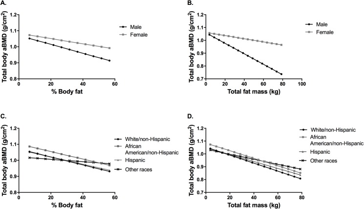
Fig 1 displays predicted values of total body aBMD according to gender and race. Overall there is a trend of lower total body aBMD with increasing percent body fat (Fig 1A) and increasing total fat mass (Fig 1B), with different slopes for males versus females and for the different racial groups (Fig 1C and Fig 1D). Males have a steeper rate of decrease in total body aBMD than females as percent body fat and total fat mass increase.
Fig 1. Models of adiposity on total body bone mineral density (aBMD).
Graphical display of effect of sex interaction (A and B) and race/ethnicity interaction (C and D) fixing all other predictors at their mean.
Pelvic aBMD
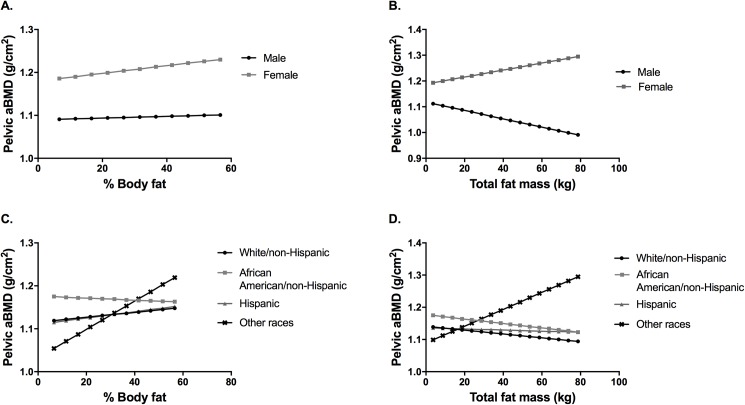
For pelvic aBMD, we found significant 2-way interactions for percent body fat (percent body fat x gender; percent body fat x race/ethnicity) (Table 2) and total fat mass (total fat mass (kg) x gender; total fat mass (kg) x race/ethnicity) (Table 3). Fig 2 displays predicted values of pelvic aBMD according to gender and race. With increasing percent body fat, there was a trend of higher pelvic aBMD for males and females (Fig 2A). With increasing total fat mass, there was a trend of higher pelvic aBMD for females, but lower pelvic aBMD for males (Fig 2B). Relationships were also mixed for the different races. With increasing percent body fat, White, Hispanic and Other races showed an increasing trend of pelvic aBMD, while it decreased in African Americans (Fig 2C). With increasing total fat mass, the pelvic aBMD decreased across races, except for the other race group (Fig 2D).
Fig 2. Models of adiposity on pelvic bone mineral density (aBMD).
Graphical display of effect of sex interaction (A and B) and race/ethnicity interaction (C and D) fixing all other predictors at their mean.
Lumbar spine aBMD
For lumbar spine aBMD, we only found significant 2-way interactions for the percent body fat model (percent body fat x gender; percent body fat x race/ethnicity) (Table 2) and the 3-way interaction model for total fat mass (total fat mass (kg) x gender x race/ethnicity) (Table 4).
Table 4. Model of total fat mass and lumbar spine bone mineral density (aBMD).
3-way interaction model of total fat mass (kg) with control variables of lean mass, gender, and race/ethnicity on outcomes of lumbar spine aBMD.
| Lumbar spine aBMD | |||
|---|---|---|---|
| Unstandardized coefficients (B) | SE | P-value | |
| Model 2 | |||
| Fat mass kg | -0.0066 | 0.0004 | <0.0001 |
| Lean mass kg | 0.0096 | 0.0003 | <0.0001 |
| White | Ref. | ||
| African American | 0.0180 | 0.0079 | 0.0250 |
| Hispanic | -0.0102 | 0.0077 | 0.1920 |
| Other | -0.0027 | 0.0176 | 0.8770 |
| African-American * Female | 0.0349 | 0.0144 | 0.0190 |
| Hispanic * Female | 0.0252 | 0.0147 | 0.0930 |
| Other * Female | -0.0194 | 0.0244 | 0.4290 |
| Male | Ref. | ||
| Female | 0.0560 | 0.0090 | <0.0001 |
| Female * Fat mass (kg) | 0.0049 | 0.0006 | <0.0001 |
| AA * Fat mass (kg) | 0.0010 | 0.0005 | 0.0570 |
| Hispanic * Fat mass (kg) | 0.0009 | 0.0004 | 0.0600 |
| Other * Fat mass (kg) | 0.0005 | 0.0010 | 0.6660 |
| Female * AA * Fat mass (kg) | -0.0017 | 0.0008 | 0.0400 |
| Female * Hispanic * Fat mass (kg) | -0.0014 | 0.0007 | 0.0620 |
| Female * Other * Fat mass (kg) | 0.0019 | 0.0016 | 0.2470 |
| Age | 0.0196 | 0.0008 | <0.0001 |
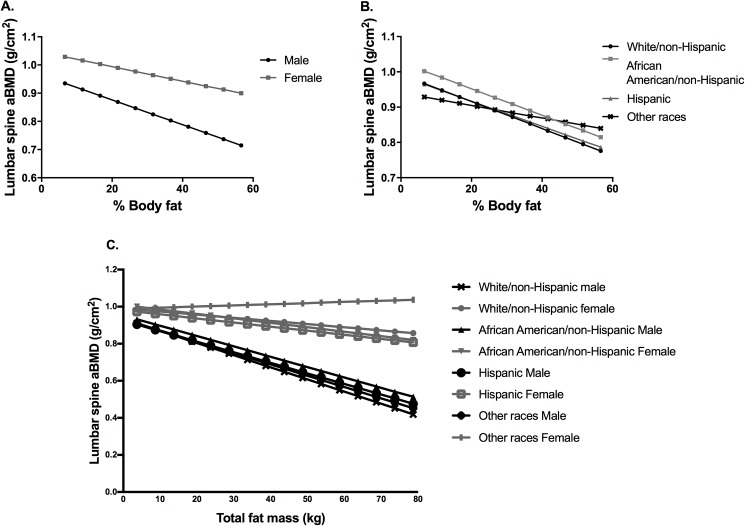
Fig 3 displays predicted values of lumbar spine aBMD according to gender and race/ethnicity. With increasing percent body fat, lumbar spine aBMD decreased in both genders (Fig 3A) and races (Fig 3B). Females have a higher average lumbar spine aBMD than males. African-Americans have the highest predicted lumbar spine aBMD followed by Whites and Hispanics. With increasing total fat mass, lumbar spine aBMD decreased for all categories except for females of other race/ethnicity (Fig 3C).
Fig 3. Models of adiposity on lumbar spine bone mineral density (aBMD).
Graphical display of effect of sex interaction (A) and race/ethnicity interaction (B) 3-way interaction model for total fat mass (kg) with lumbar spine aBMD (C).
Discussion
In our study, we found significant and novel interactions by gender and race/ethnicity in the associations between percent body fat and total fat mass (kg) with total body, pelvic and lumbar spine aBMD. Both total body aBMD and lumbar spine aBMD decreased with increasing percent body fat and total fat mass in both genders and all races. We found less consistent patterns for pelvic aBMD, which increased in both genders as percent body fat increased, but showed this positive trend with increasing total fat mass in females and the opposite in males.
Overall, females had higher total body, pelvic and lumbar spine aBMD than males in our models. These gender differences could be multifactorial: different pubertal status of participants, gender differences in body composition (with females having more fat mass than males, and males having higher lean mass), hormonal environment, and the effect of childhood obesity on puberty onset. While this may be dependent on pubertal status one of the limitations in this study is the lack of pubertal status in the database. However, another factor that might be leading to gender differences in our results is a difference in adipose tissue distribution. It has been well described that adipose tissue distribution is predominantly subcutaneous in females and more visceral central adiposity in males. This central adiposity is directly correlated with obesity-associated comorbidities, such as metabolic syndrome, dyslipidemia, insulin resistance and a low-grade inflammation which may affect the relationship between bone and adipose tissue in different ways [23]. A higher percent body fat has been associated with increased bone mineral density in girls during puberty and may be explained by these differences in adiposity during puberty [24]
Race differences in bone density have also been previously described with studies reporting that African American and Hispanic children have higher BMDs compared to Caucasian children [10], while others report a greater aBMD for African Americans compared to Caucasians, and Caucasians having a greater aBMD than Asian and Hispanics. [25] The genetic factors that determine bone size and mineralization are responsible for the majority of these racial differences [25]. As with gender, differences in lean mass by race may also explain the variable relationship of race on bone density [15] which is a reason these models adjusting for lean mass and still demonstrating a significant race difference are important.
The relationship between adiposity and bone mineral density (BMD) in the pediatric population is not completely understood, and the literature is quite mixed. Some studies have found negative associations between percent body fat and lumbar spine BMD [26] and whole body BMC [27] in females between 9 to 24 years of age (n = 521). Other studies have reported positive associations between fat mass and lean mass with total, lumbar spine and pelvic BMD in young premenopausal women aged 20 to 25 (n = 921) [28]. The race and sex interactions may account for some of the inconsistent findings of associations reported in the literature. A recent systematic review and meta-analysis found: 1) moderate quality of evidence for a higher total BMD in children with obesity when compared with those who were overweight, 2) moderate quality of evidence for a significant mean difference of lumbar spine BMD in the normal–weight group compared to the overweight or obese groups, and 3) no significant difference in femoral neck BMD in children with normal-weight when compared with those who were overweight. [29] They report significant statistical heterogeneity in all the results described above.[29]
Given the mixed results in the current literature our study using direct measures of adiposity (both percent body fat and total fat mass), lean mass, and areal bone mineral density (aBMD) using DXA across multiple regions of the body clarifies many of the results in prior studies. In addition, our study uses a nationally representative sample with a large number of participants and ethnic diversity with oversampling of minority children. Limitations of our study include the cross-sectional study design, which does not prove causality, the lack of information about long-term clinical outcomes like fractures, and a lack of information on clinical parameters such as pubertal status, hormone measurements, vitamin D levels, or physical activity. We also acknowledge that the children from our study population were younger, heavier, and had a lower proportion of minority children. Additional limitations due to what was included in this survey are the regions of spine and pelvis that are taken from whole body scans and hence may not be as accurate as focused DXA scans or volumetric BMD measured by Quantitative CT (QCT) scans. As recommended by the International Society For Clinical Densitometry, the use of DXA in children (≥3 years of age) and adolescents is the preferred method and it is well accepted due to the accurate and precise BMC and aBMD provided [30] and the low radiation exposure. [31] DXA is limited because it measures areal BMD instead of true volumetric BMD which may be important when the skeleton is still growing, and bone size might be influencing BMD measures [31]. Quantitative computerized tomography (QCT) measures bone size, shape and true volumetric BMD (vBMD) but are not yet widely available. [30, 32] Both negative [33, 34] and positive [35] associations between fat mass and cortical bone of the tibia, and a negative association between fat mass and the radius[36] have been reported with this technique. In addition, with the use of DXA, analysis can be performed in two regions along with the use of whole body BMD. It is possible given the lower ages of some of the participants that the total body less head (TBLH) variable could have yielded different results but analysis showed that there was a strong correlations between these two factors.
While the findings in our study overall suggest that obesity has a negative impact on total and lumbar aBMD, weight bearing regions such as the pelvis are possibly spared from the impact of adiposity on bone health. Future prospective studies are needed to clarify the impact of adiposity on bone density and quality in the pediatric population, using both DXA and QCT methods. Gender, race/ethnicity and pubertal status can also influence this association and it is crucial to understand this differences when interpreting DXA scans. While this retrospective clinical trial cannot directly evaluate mechanisms for the findings in the current study, pre-clinical investigations suggest that adiposity may directly correlate with marrow adiposity which is associated with a decline in bone density and secondary bone fragility. [37]
If childhood obesity is limiting peak bone accrual and increasing the risk for osteoporosis and/or fractures later in life, pediatricians can evaluate bone mineral density in children who have expanded adipose tissue. If lower BMD than expected is detected, some interventions like increasing physical activity, treating vitamin D and nutritional deficiencies can improve bone health during this critical period significantly. A close follow-up and the report of clinical outcomes of these children can also help close the gap existing in the literature.
Abbreviations
- NHANES
National Health Examination Survey
- BMD
Bone Mineral density
- aBMD
areal Bone Mineral Density
- BMC
Bone mineral content
- DEXA
Dual Energy X-ray Absorptiometry
Data Availability
Data are publicly available from the CDC website and the following link: https://wwwn.cdc.gov/nchs/nhanes/dxa/Dxa.aspx.
Funding Statement
Dr. Kanakadurga Singer receives support through a National Institute of Diabetes and Digestive and Kidney Diseases / National Institutes of Health Grant (K08DK101755), from the Department of Pediatrics Woodson Biostatistics Major Award and the Edith Briskin/SKS Foundation Taubman Emerging Scholars Program from the University of Michigan. The funders had no role in study design, data collection and analysis, decision to publish, or preparation of the manuscript.
References
- 1.Ogden CL, Carroll MD, Kit BK, Flegal KM. Prevalence of childhood and adult obesity in the United States, 2011–2012. JAMA. 2014;311(8):806–14. Epub 2014/02/27. doi: 10.1001/jama.2014.732 . [DOI] [PMC free article] [PubMed] [Google Scholar]
- 2.Friedemann C, Heneghan C, Mahtani K, Thompson M, Perera R, Ward AM. Cardiovascular disease risk in healthy children and its association with body mass index: systematic review and meta-analysis. BMJ. 2012;345:e4759 doi: 10.1136/bmj.e4759 ; PubMed Central PMCID: PMCPMC3458230. [DOI] [PMC free article] [PubMed] [Google Scholar]
- 3.Sorof J, Daniels S. Obesity hypertension in children: a problem of epidemic proportions. Hypertension. 2002;40(4):441–7. . [DOI] [PubMed] [Google Scholar]
- 4.Must A, Strauss RS. Risks and consequences of childhood and adolescent obesity. Int J Obes Relat Metab Disord. 1999;23 Suppl 2:S2–11. . [DOI] [PubMed] [Google Scholar]
- 5.Kuperman EF, Showalter JW, Lehman EB, Leib AE, Kraschnewski JL. The impact of obesity on sepsis mortality: a retrospective review. BMC Infect Dis. 2013;13(1):377 Epub 2013/08/21. doi: 10.1186/1471-2334-13-377 ; PubMed Central PMCID: PMC3765286. [DOI] [PMC free article] [PubMed] [Google Scholar]
- 6.Lloyd JT, Alley DE, Hawkes WG, Hochberg MC, Waldstein SR, Orwig DL. Body mass index is positively associated with bone mineral density in US older adults. Arch Osteoporos. 2014;9:175 doi: 10.1007/s11657-014-0175-2 . [DOI] [PubMed] [Google Scholar]
- 7.Junior IF, Cardoso JR, Christofaro DG, Codogno JS, de Moraes AC, Fernandes RA. The relationship between visceral fat thickness and bone mineral density in sedentary obese children and adolescents. BMC pediatrics. 2013;13:37 Epub 2013/03/21. doi: 10.1186/1471-2431-13-37 ; PubMed Central PMCID: PMC3606829. [DOI] [PMC free article] [PubMed] [Google Scholar]
- 8.Mosca LN, Goldberg TB, da Silva VN, da Silva CC, Kurokawa CS, Bisi Rizzo AC, et al. Excess body fat negatively affects bone mass in adolescents. Nutrition. 2014;30(7–8):847–52. doi: 10.1016/j.nut.2013.12.003 . [DOI] [PubMed] [Google Scholar]
- 9.Leonard MB, Shults J, Wilson BA, Tershakovec AM, Zemel BS. Obesity during childhood and adolescence augments bone mass and bone dimensions. Am J Clin Nutr. 2004;80(2):514–23. . [DOI] [PubMed] [Google Scholar]
- 10.McCormick DP, Ponder SW, Fawcett HD, Palmer JL. Spinal bone mineral density in 335 normal and obese children and adolescents: evidence for ethnic and sex differences. J Bone Miner Res. 1991;6(5):507–13. doi: 10.1002/jbmr.5650060513 . [DOI] [PubMed] [Google Scholar]
- 11.Freedman DS, Wang J, Thornton JC, Mei Z, Sopher AB, Pierson RN Jr., et al. Classification of body fatness by body mass index-for-age categories among children. Archives of pediatrics & adolescent medicine. 2009;163(9):805–11. Epub 2009/09/09. doi: 10.1001/archpediatrics.2009.104 ; PubMed Central PMCID: PMC2846460. [DOI] [PMC free article] [PubMed] [Google Scholar]
- 12.Freedman DS, Wang J, Thornton JC, Mei Z, Pierson RN Jr., Dietz WH, et al. Racial/ethnic differences in body fatness among children and adolescents. Obesity (Silver Spring). 2008;16(5):1105–11. Epub 2008/03/01. doi: 10.1038/oby.2008.30 . [DOI] [PubMed] [Google Scholar]
- 13.Dugas LR, Cao G, Luke AH, Durazo-Arvizu RA. Adiposity is not equal in a multi-race/ethnic adolescent population: NHANES 1999–2004. Obesity (Silver Spring). 2011;19(10):2099–101. Epub 2011/03/26. doi: 10.1038/oby.2011.52 . [DOI] [PubMed] [Google Scholar]
- 14.Goran MI, Allison DB, Poehlman ET. Issues relating to normalization of body fat content in men and women. Int J Obes Relat Metab Disord. 1995;19(9):638–43. Epub 1995/09/01. . [PubMed] [Google Scholar]
- 15.Gracia-Marco L, Ortega FB, Jimenez-Pavon D, Rodriguez G, Castillo MJ, Vicente-Rodriguez G, et al. Adiposity and bone health in Spanish adolescents. The HELENA study. Osteoporos Int. 2012;23(3):937–47. Epub 2011/05/13. doi: 10.1007/s00198-011-1649-3 . [DOI] [PubMed] [Google Scholar]
- 16.Kohrt WM, Barry DW, Schwartz RS. Muscle forces or gravity: what predominates mechanical loading on bone? Med Sci Sports Exerc. 2009;41(11):2050–5. doi: 10.1249/MSS.0b013e3181a8c717 ; PubMed Central PMCID: PMC3037021. [DOI] [PMC free article] [PubMed] [Google Scholar]
- 17.National Center for Health Statistics, National Health Examination Survey. Available from: http://www.cdc.gov/nchs/products/elec_prods/subject/nhes3.htm-description1.
- 18.Centers for Disease Control and Prevention—NHANES Survey Design Factors. Available from: https://www.cdc.gov/nchs/tutorials/nhanes/surveydesign/SampleDesign/intro.htm.
- 19.National Center for Health Statistics, National Health Examination Survey. [Internet]. Available from: Dexa reports https://wwwn.cdc.gov/nchs/nhanes/dxa/Dxa.aspx.
- 20.Singer K, Eng DS, Lumeng CN, Gebremariam A, Lee JM. The relationship between body fat mass percentiles and inflammation in children. Obesity (Silver Spring). 2014;22(5):1332–6. Epub 2014/01/28. doi: 10.1002/oby.20710 . [DOI] [PMC free article] [PubMed] [Google Scholar]
- 21.Patsch JM, Kiefer FW, Varga P, Pail P, Rauner M, Stupphann D, et al. Increased bone resorption and impaired bone microarchitecture in short-term and extended high-fat diet-induced obesity. Metabolism. 2011;60(2):243–9. doi: 10.1016/j.metabol.2009.11.023 . [DOI] [PMC free article] [PubMed] [Google Scholar]
- 22.Johnson CL, Paulose-Ram R, Ogden CL, Carroll MD, Kruszon-Moran D, Dohrmann SM, et al. National health and nutrition examination survey: analytic guidelines, 1999–2010. Vital Health Stat 2 2013;(161):1–24. . [PubMed] [Google Scholar]
- 23.Gower BA, Casazza K. Divergent effects of obesity on bone health. J Clin Densitom. 2013;16(4):450–4. doi: 10.1016/j.jocd.2013.08.010 ; PubMed Central PMCID: PMCPMC5321047. [DOI] [PMC free article] [PubMed] [Google Scholar]
- 24.Streeter AJ, Hosking J, Metcalf BS, Jeffery AN, Voss LD, Wilkin TJ. Body fat in children does not adversely influence bone development: a 7-year longitudinal study (EarlyBird 18). Pediatr Obes. 2013;8(6):418–27. doi: 10.1111/j.2047-6310.2012.00126.x . [DOI] [PubMed] [Google Scholar]
- 25.Bachrach LK. Acquisition of optimal bone mass in childhood and adolescence. Trends Endocrinol Metab. 2001;12(1):22–8. . [DOI] [PubMed] [Google Scholar]
- 26.Lazcano-Ponce E, Tamayo J, Cruz-Valdez A, Diaz R, Hernandez B, Del Cueto R, et al. Peak bone mineral area density and determinants among females aged 9 to 24 years in Mexico. Osteoporos Int. 2003;14(7):539–47. doi: 10.1007/s00198-002-1363-2 . [DOI] [PubMed] [Google Scholar]
- 27.Weiler HA, Janzen L, Green K, Grabowski J, Seshia MM, Yuen KC. Percent body fat and bone mass in healthy Canadian females 10 to 19 years of age. Bone. 2000;27(2):203–7. . [DOI] [PubMed] [Google Scholar]
- 28.Wang MC, Bachrach LK, Van Loan M, Hudes M, Flegal KM, Crawford PB. The relative contributions of lean tissue mass and fat mass to bone density in young women. Bone. 2005;37(4):474–81. doi: 10.1016/j.bone.2005.04.038 . [DOI] [PubMed] [Google Scholar]
- 29.Van Leeuwen J, Koes BW, Paulis WD, Van Middelkoop M. Differences in bone mineral density between normal-weight children and children with overweight and obesity: a systematic review and meta-analysis. Obes Rev. 2017. doi: 10.1111/obr.12515 . [DOI] [PubMed] [Google Scholar]
- 30.Gordon CM, Leonard MB, Zemel BS, International Society for Clinical D. 2013 Pediatric Position Development Conference: executive summary and reflections. J Clin Densitom. 2014;17(2):219–24. doi: 10.1016/j.jocd.2014.01.007 . [DOI] [PubMed] [Google Scholar]
- 31.Binkovitz LA, Henwood MJ. Pediatric DXA: technique and interpretation. Pediatr Radiol. 2007;37(1):21–31. doi: 10.1007/s00247-006-0153-y ; PubMed Central PMCID: PMCPMC1764599. [DOI] [PMC free article] [PubMed] [Google Scholar]
- 32.Leonard MB, Zemel BS, Wrotniak BH, Klieger SB, Shults J, Stallings VA, et al. Tibia and radius bone geometry and volumetric density in obese compared to non-obese adolescents. Bone. 2015;73:69–76. doi: 10.1016/j.bone.2014.12.002 ; PubMed Central PMCID: PMCPMC4540475. [DOI] [PMC free article] [PubMed] [Google Scholar]
- 33.Pollock NK, Laing EM, Baile CA, Hamrick MW, Hall DB, Lewis RD. Is adiposity advantageous for bone strength? A peripheral quantitative computed tomography study in late adolescent females. Am J Clin Nutr. 2007;86(5):1530–8. . [DOI] [PubMed] [Google Scholar]
- 34.Taes YE, Lapauw B, Vanbillemont G, Bogaert V, De Bacquer D, Zmierczak H, et al. Fat mass is negatively associated with cortical bone size in young healthy male siblings. J Clin Endocrinol Metab. 2009;94(7):2325–31. doi: 10.1210/jc.2008-2501 . [DOI] [PubMed] [Google Scholar]
- 35.Sayers A, Tobias JH. Fat mass exerts a greater effect on cortical bone mass in girls than boys. J Clin Endocrinol Metab. 2010;95(2):699–706. doi: 10.1210/jc.2009-1907 ; PubMed Central PMCID: PMCPMC2817095. [DOI] [PMC free article] [PubMed] [Google Scholar]
- 36.Wey HE, Binkley TL, Beare TM, Wey CL, Specker BL. Cross-sectional versus longitudinal associations of lean and fat mass with pQCT bone outcomes in children. J Clin Endocrinol Metab. 2011;96(1):106–14. doi: 10.1210/jc.2010-0889 ; PubMed Central PMCID: PMCPMC3038473. [DOI] [PMC free article] [PubMed] [Google Scholar]
- 37.Scheller EL, Khoury B, Moller KL, Wee NK, Khandaker S, Kozloff KM, et al. Changes in Skeletal Integrity and Marrow Adiposity during High-Fat Diet and after Weight Loss. Front Endocrinol (Lausanne). 2016;7:102 doi: 10.3389/fendo.2016.00102 ; PubMed Central PMCID: PMCPMC4961699. [DOI] [PMC free article] [PubMed] [Google Scholar]
Associated Data
This section collects any data citations, data availability statements, or supplementary materials included in this article.
Data Availability Statement
Data are publicly available from the CDC website and the following link: https://wwwn.cdc.gov/nchs/nhanes/dxa/Dxa.aspx.


