Abstract
Touch perception on the fingers and hand is essential for fine motor control, contributes to our sense of self, allows for effective communication, and aids in our fundamental perception of the world. Despite increasingly sophisticated mechatronics, prosthetic devices still do not directly convey sensation back to their wearers. We show that implanted peripheral nerve interfaces in two human subjects with upper limb amputation provided stable, natural touch sensation in their hands for more than 1 year. Electrical stimulation using implanted peripheral nerve cuff electrodes that did not penetrate the nerve produced touch perceptions at many locations on the phantom hand with repeatable, stable responses in the two subjects for 16 and 24 months. Patterned stimulation intensity produced a sensation that the subjects described as natural and without “tingling,” or paresthesia. Different patterns produced different types of sensory perception at the same location on the phantom hand. The two subjects reported tactile perceptions they described as natural tapping, constant pressure, light moving touch, and vibration. Changing average stimulation intensity controlled the size of the percept area; changing stimulation frequency controlled sensation strength. Artificial touch sensation improved the subjects’ ability to control grasping strength of the prosthesis and enabled them to better manipulate delicate objects. Thus, electrical stimulation through peripheral nerve electrodes produced long-term sensory restoration after limb loss.
INTRODUCTION
The sense of touch is essential to experience and manipulate the world around us (1, 2). In addition to loss of function, loss of sensation is a devastating consequence of upper limb amputation. Sensory perception is important for control of the prosthetic limb, a sense of embodiment (3), and in the reduction of phantom limb pain (4, 5). Those with limb loss rely primarily on visual and auditory feedback from the device motors to control the prosthetic limb (6). The prosthesis is perceived by the user as a foreign tool extending beyond, but not as part of, their body (7). Sensory substitution, such as vibration on a residual limb with intensity proportional to pressure at the prosthetic fingertip, improves prosthetic control in limited situations (8, 9), but has not been widely adopted as the vibration is often described as distracting (8). Single-channel nerve cuff electrodes were used to produce sensation in the perceived hand nearly four decades ago (10). Each subject, however, reported different sensory perceptions such as general fist clenching, vibration, and paresthesia. The sensation was over large regions of the hand. More recently, intrafascicular electrodes have produced tactile sensation but were only implanted for 4 weeks or less, and some exhibited a continuous threshold increase and full loss of sensory stimulation capability as early as 10 days (11, 12). Paresthesia was associated with 30 to 50% of the stimulating channels. However, the restored sensation allowed a subject to correctly identify three different objects, illustrating the value of sensory feedback on functional control (12). Electrodes inserted into the sensory cortex of nonhuman primates (13) have demonstrated localized sensory feedback.
Here, nonpenetrating peripheral nerve cuff electrodes (14, 15) were shown to provide multiple tactile perceptions at multiple locations on the phantom hand in two upper limb amputees with stable responses for up to 24 months.
RESULTS
The cuff electrode peripheral interface was selective and stable
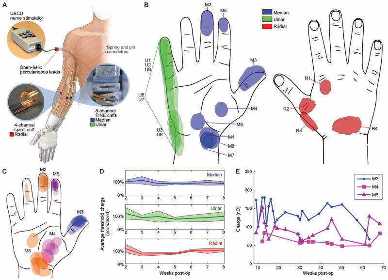
Cuffs contained either four or eight independent stimulus channels. Each cuff was an electrically insulating silicone sheath with multiple electrical contacts spaced evenly around the outside of the peripheral nerve. The first subject, subject 1, was a 49-year-old male who suffered a wrist disarticulation in a 2010 industrial accident. We implanted cuffs on his median, radial, and ulnar nerves in his mid-forearm in May 2012, providing a total of 20 stimulation channels: 8 each on the median and ulnar nerves and 4 on the radial nerve (Fig. 1A). The second subject, subject 2, was a 47-year-old male with a below-elbow amputation resulting from a 2004 industrial accident. We implanted cuffs on his median and radial nerves in his mid-upper arm in January 2013, providing eight stimulation channels on each nerve. After surgery, leads from the cuffs protruded from the subjects’ upper arm for connection to stimulation equipment during laboratory visits.
Fig. 1. Stability and selectivity of implanted cuff electrodes.
(A) We implanted three cuffs with a total of 20 channels in the forearm of subject 1: a four-contact spiral cuff on the radial nerve of the forearm and an eight-contact FINE on the median and ulnar nerves. The electrode leads ran subcutaneously to the upper arm and connected to open-helix percutaneous leads via spring-and-pin connectors (27–29). A Universal External Control Unit (UECU, Ardiem Medical) supplied single-channel, charge-balanced, monopolar nerve stimulation. (B) Sensation locations after threshold stimulation at week 3 post-op. Cuff electrodes were highly selective, with each contact producing either a unique location or unique sensation. Here, the letter represents the nerve and the number represents the stimulus channel within the nerve cuff around that nerve. Thus, M3 is the third stimulus channel within the median nerve cuff. Ulnar (U) locations presented the most overlap at threshold, but differentiated in area expansion at suprathreshold responses. The subjects drew the borders around areas of perception. Areas outside the template, for example, M3, represent a small wrap-around of sensation on the digit. (C) Repeated weekly overlapping threshold locations of channels M2, M3, M4, M5, and M8 for weeks 3 through 10 post-op indicated consistent location perception. Locations remained stable for all stimulation waveforms used. (D) Mean normalized threshold charge density for all channels on the median (blue), ulnar (green), and radial (red) cuffs of subject 1 shown as a solid line. Shaded areas indicate the 95% confidence interval. An unbiased, stepwise search determined the threshold. Frequency was a constant 20 Hz. During weeks 2 to 8, percept thresholds for subject 1 were 95.5 ± 42.5 nC (n = 59), 70.7 ± 59.2 nC (n = 50), and 40.7 ± 12.4 nC (n = 24) for the median, ulnar, and radial nerves, respectively. Linear regression of the threshold stimulation intensity for perception over 8 weeks for every channel was unchanging [18/19, analysis of variance (ANOVA) test, P ≥ 0.067] or decreasing (1/19, ANOVA, P = 0.044). Subject 2 was also stable (P ≥ 0.087) with thresholds of 141 ± 46 nC and 95 ± 47 nC for the median and radial nerves, respectively. (E) Threshold tracking of median channels M3, M4, and M5 to 68 weeks and thereafter showed no significant change in threshold over time (P = 0.053, 0.587, and 0.773, respectively).
During monthly visits by the subjects to the Cleveland Veterans Affairs Medical Center, lasting between 4 and 6 hours, the subjects described perceived sensations in response to trains of electrical pulses on each channel (Fig. 1A). We identified the threshold for sensory perception by slowly increasing the intensity of the stimulation pulse until the subject indicated feeling a sensation. The subject was blind to pulse intensity. Random insertion of null pulses ensured the subject was not anticipating stimulation. For each change in stimulus intensity, the subjects verbally described the perceived sensation and traced its location on a schematic of a hand. Of the 20 available channels implanted in subject 1, stimulation produced sensation from 19 channels at 15 unique locations on the perceived hand (Fig. 1B). Of the 16 available channels implanted in subject 2, stimulation produced sensation from 14 channels at 9 unique locations (fig. S1). Over the duration of the study, the locations of percepts were repeatable and stable (Fig. 1C). In both subjects, percepts were produced at multiple, independent, small, and well-defined locations on the hand, including the thumb and fingertips. All perceived locations were consistent with innervation patterns of the median, ulnar, and radial nerves on which the electrodes were implanted. The peripheral sensory pathways and the subject’s sensory perception did not appear to have reorganized after injury, which confirmed earlier studies (16). The sensory stimulation threshold remained stable the first 8 weeks after implant (Fig. 1D), ranging from 40.7 to 95.5 nC for subject 1 and 95 to 141 nC for subject 2. The thresholds were stable for up to 68 weeks (Fig. 1E). Electrode impedances remained stable around 3 kilohms (fig. S2).
Stimulation produced sensory perception before muscle activity that would interfere with myoelectric control. In subject 1, the cuffs were implanted distal to the motor branches of residual muscles. In subject 2, the electrodes were located on mixed motor and sensory nerves. Stimulation did not interfere with standard myoelectrical prosthesis control, even when the motor and sensory fibers were in the same nerve. Both subjects participating in this study were active individuals. Their normal daily activity did not affect the peripheral nerve electrode performance or the sensory restoration provided by the electrode, nor did the implants affect their daily activities.
Constant stimulation intensity produced paresthesia
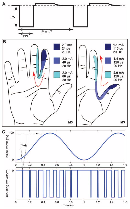
The standard nerve stimulation paradigm is a train of identical, charge-balanced, square electrical pulses characterized by pulse amplitude (PA), pulse width (PW), and either pulse repetition frequency (f) or interpulse interval (IPI = 1/f) (Fig. 2A). Traditionally, these three parameters are time invariant and fixed in value: PA0, PW0, and IPI0 = 1/f0. With constant stimulation intensity pulse trains with stimulation frequency ranging from 1 to 1000 Hz, the subjects reported an unnatural sensation of paresthesia, described as “electrical,” in 96% of 151 trials over a 10-month period. We systematically mapped the sensory perceptions produced for many different constant values of PW0, PA0, and f0. Increasing PA0 or PW0 increased the intensity and the size of the percept area (Fig. 2B). The increasing area of sensation was somatotopically organized, suggesting a somatotopic organization of sensory fibers within the peripheral nerve. During stimulation trains up to 60 s, paresthesia did not resolve into a natural sensation, as was previously reported with intrafascicular electrodes (11).
Fig. 2. Waveform patterns.
(A) Square, charge-balanced, cathodic-first stimulation pulsing pattern. Prior neural stimulation maintained constant parameters, such as pulse amplitude (PA), pulse width (PW), and interpulse interval (IPI) or frequency (f). (B) In general, constant PA and PW modulate the area of perception. M5 showed a channel-specific recruitment pattern as PW was increased from 24 to 60 μs. M3 showed that the percept area increases as PA increases from 1.1 to 2.0 mA. These recruitment patterns matched the sensory nerve innervation patterns of the digital nerve. (C) An example of full-scale modulation, using a sinusoidal (1 Hz) PW envelope that produced a natural sensation of pulsing pressure (top plot). The schematic resulting stimulation waveform where the IPI is 0.1 s (10 Hz) is shown in the bottom plot. Our stimulation trials typically used an IPI of 0.01 s (100 Hz).
Stimulation intensity modulation with a time-variant pulse width resulted in “natural” pressure perception
Full-scale modulation feels like a pressure pulse
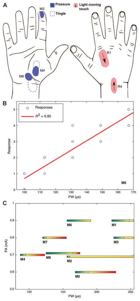
Modulation of stimulation intensity resulted in a description by subject 1 of the perception changing from “tingly” to “as natural as can be.” He variously described the sensation as pulsing pressure, constant pressure, vibration, tapping, and rubbing on a texture. In the full-scale modulation pattern, the width of the pulses in the pulse train (f0 = 100 Hz) followed a slow (fmod = 1 Hz) sinusoidal envelope. The pulse width varied between 0 μs and a maximum of B μs (Fig. 2C, see Methods). In response to this stimulation, subject 1 reported a 1-Hz pulsing pressure sensation and described it “as if I was feeling my own pulse or heartbeat—just like putting my fingers here,” as he demonstrated his fingers against his carotid artery pulse in his neck. When the peak pulse width was set to a first noticeable level, B = Bth, the sensation was described as the back of a pen repeatedly pushing “very lightly” on a small, localized area of the skin (Fig. 3A). The sensory transformation occurred in both subjects. Both subjects could tap synchronously with the perceived pressure pulse using the intact limb for visual confirmation of the pulse. The tapping matched the frequency of the intensity modulation, fmod, as fmod varied between 0.5 and 4 Hz. As the maximum pulse width, B, increased, the intensity of the pressure pulse increased and the pulsing frequency remained matched to fmod.
Fig. 3. Full-scale modulation sinusoidal PW envelope.
(A) At threshold (Bth), a pulsing pressure was felt at the bold circle area (M3, M4, M8; blue). Increasing the PW to a secondary threshold (Btingle) introduced an additional pulsing paresthesia, which typically covered a larger area that overlapped the pressure location. Increasing the PW further caused the area of par-esthesia to increase but the area of constant pressure did not increase. Light moving touch was described as someone lightly brushing the skin with a finger. It consistently moved in the same direction for a given stimulus (R1, R4; pink). (B) Psychometric rating of sensation intensity as a function of PWmax showing a relationship between PW and the strength of the perceived intensity. The subject was provided five PWmax stimuli (100, 114, 131, 150, and 167 μs), and each level was presented three to six times in random order. (C) Threshold windows for natural sensation were measured on every channel of the median cuff. Pressure occurred at Bth (green), was accompanied by paresthesia at Btingle (black line, yellow), and was overwhelmed by paresthesia at BMask (red). The largest PW windows for a particular channel were found when PA was lowest. Higher levels of stimulation were avoided for M6 because of pain response.
There was a psychometric correlation between perceived intensity of the pressure pulse and the maximum pulse width. The subjects rated their perception of five different values of peak pulse width, B. Each stimulation level was repeated multiple times and all presented in random order. The subject was blind to the stimulation level. The verbal rating of perceived intensity correlated significantly with B (P < 0.05, R2 = 0.85, n = 22) (Fig. 3B).
At the fingertips, the subjects described the pulsing sensation to be like pressing on the tip of a ball-point pen. The perceived sensory modalities across all 19 active channels and all trials in subject 1 included pulsing pressure (86%), light moving touch (7%), or tapping (7%) (Tables 1 and 2). Subject 2 reported perceiving pulsing pressure during stimulation with contacts on the median nerve (75%) and vibration during stimulation with contacts on the radial nerve (75%). The range of modulation producing natural sensation is defined by the threshold pulse width, Bth, and maximum pulse width, Btingle, with Bth < B < Btingle. When B increased above Btingle, the subject reported a light tingle sensation in addition to and surrounding the region of natural perception (Fig. 3A). When B was increased past an upper limit, BMask, where B > BMask > Btingle, paresthesia dominated and overwhelmed the natural sensory perception (Fig. 3C).
Table 1. Sensation qualities reported during a single experimental session.
M6 transitioned to a sensation of a needle within a vein at higher stimulation.
| Median | Ulnar | Radial | |||
|---|---|---|---|---|---|
|
|
|
|
|||
| Ch. | Modality | Ch. | Modality | Ch. | Modality |
| M1 | Light moving touch | U1 | Pressure | R1 | Pressure/light moving touch |
| M2 | Pressure | U2 | Pressure on bone | R2 | Pressure |
| M3 | Pressure of ball-point pen | U3 | Pressure on bone | R3 | Pressure/tingling |
| M4 | Pressure | U4 | Pressure of ball-point pen | R4 | Pressure/light moving touch/foam texture |
| M5 | Pressure of ball-point pen | U5 | Tingling pulse | ||
| M6 | Uncomfortable, deep, dull vibration | U6 | Light moving touch, tickle | ||
| M7 | Light moving touch | U7 | Pressure | ||
| M8 | Pressure | U8 | Pressure on bone | ||
Table 2. Average channel response for each cuff with full-scale modulation, where n is the number of unique, natural responses per nerve, per experimental visit.
Natural, non-tingling sensation was achieved on every channel with sinusoidal varying PW stimulation. Columns may not sum to 100% because some channels have multiple sensations depending on stimulation.
| Median (n = 45) | Ulnar (n = 24) | Radial (n = 13) | |
|---|---|---|---|
| Pressure (pulsing or tapping at 1 Hz) | 85.0% | 86.5% | 87.5% |
| Moving touch | 8.8% | 3.1% | 12.5% |
| Vibration or tapping (>1 Hz) | 7.3% | ||
| Discomfort or pain | 6.8% | 3.1% |
Small-scale, offset modulation feels like constant pressure
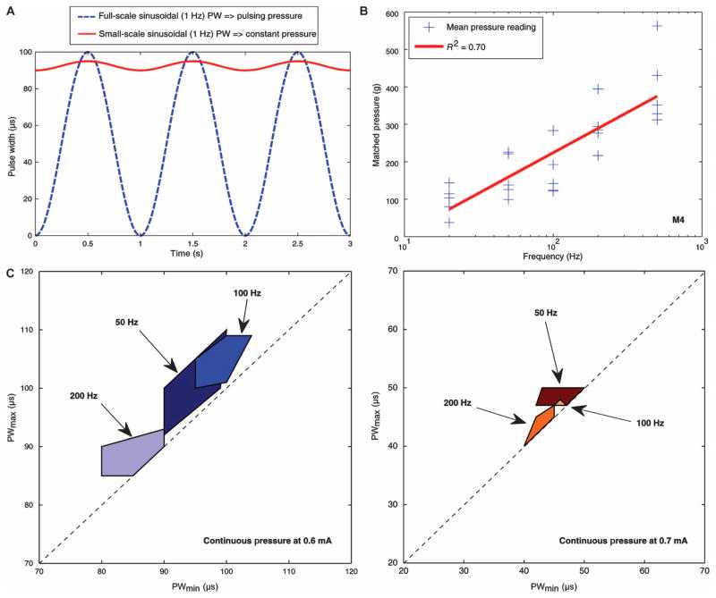
Increasing the minimum pulse width reduced the pulsing quality of the sensation. The frequency of the train of stimulation pulses, f0, remained 100 Hz and the modulation envelope frequency, fmod, remained 1 Hz. As the minimum pulse width was raised above 0 μs, the subject felt smaller variation in the pulsing and described a “lingering pressure” during the sinusoidal pulse width modulation. When the modulation was sufficiently small, the subject reported a continuous pressure described as though “someone just laid a finger on my hand.” The typical size of pulse width modulation resulting in constant sensation, PWpk-pk, was small, on the order of 5 μs. The best results were achieved when the average pulse width, PWoffset, was about 90% of the Bth required to produce the natural pulsing sensation with full-scale modulation (Fig. 4A, see Methods).
Fig. 4. Small-scale, offset modulation.
(A) Typical example of a small-scale, offset (SSO) modulation using sinusoidal (1 Hz) PW with offset stimulation on M4 (solid, red line). PWpk-pk = 90 to 95 μs was the lowest stimulation level that produced constant pressure sensation. For comparison, the threshold for pulsing pressure from full-scale modulation is shown (dotted blue line). (B) Contralateral pressure matching indicated that frequency can control the intensity of constant pressure sensation. The subject was provided SSO modulation with the IPI set to 50, 20, 10, 5, or 2 ms (20, 50, 100, 200, or 500 Hz) on channel M4 and asked to match the perceived pressure with the contralateral hand. Pressure intensity was proportional to the stimulus frequency. (C) The PWmin-max window that produced a sensation of constant pressure was influenced by the PA (left figure: 0.6 mA, right figure: 0.7 mA), which altered both the size and the location of the window. We found that frequency had a weaker effect on the window but affected the intensity. At PA of 0.5 mA, there was no response. For PA 0.8 mA and above, the data suggested that the window for continuous pressure sensation decreased.
The quality of the sensation varied depending on the type of skin in which the sensation was perceived. Small-scale pulse width modulation elicited constant pressure sensation when the perception corresponded to glabrous skin only (6 of 6 sites in subject 1 and 4 of 5 sites in subject 2). On sites with mixed glabrous and hairy skin or hairy skin only (5 of 11 sites in subject 1), small-scale modulation resulted in a sensation of constant pressure. Of the other 6 of 11 mixed or hairy skin sites, 4 remained as paresthesia, 1 was constant vibration, and 1 was described as a “cotton ball” lightly rubbed on skin (Table 3). The sensations in hairy skin were described as pulsing “natural” pressure during full-scale modulation, but as the modulation envelope was decreased, both subjects reported that these sensations resolved to a constant vibration, itch, or tingling sensation.
Table 3. Perceived sensations elicited from small-scale pulse width modulation on all available channels.
Constant pressure sensation was commonly associated with glabrous skin location on the perceived hand.
| Median | Ulnar | Radial | ||||||
|---|---|---|---|---|---|---|---|---|
|
|
|
|
||||||
| Ch. | Perceived location skin type | Small-scale PW modulation sensation | Ch. | Perceived location skin type | Small-scale PW modulation sensation | Ch. | Perceived location skin type | Small-scale PW modulation sensation |
| M1 | Hairy | Itch, tingle, pulse | U1 | Hairy | Constant vibration | R1 | Hairy | Tingling |
| M2 | Glabrous | Constant pressure | U2 | — | No response | R2 | Hairy | Constant pressure |
| M3 | Glabrous | Constant pressure | U3 | Glabrous, hairy | Tingling | R3 | Hairy | Tingling |
| M4 | Glabrous | Constant pressure | U4 | Hairy | Tingling | R4 | Hairy | Hair movement, cotton ball |
| M5 | Glabrous | Constant pressure | U5 | Glabrous | Constant pressure, Itch | |||
| M6 | Deep | N.T.* | U6 | Glabrous, hairy | Constant pressure | |||
| M7 | Hairy | Constant pressure | U7 | Glabrous | Constant pressure | |||
| M8 | Glabrous, hairy | Constant pressure | U8 | — | N.T.† | |||
Not tested: usually stimulation on M6 resulted in uncomfortable sensations.
Not tested: connector needed replacement. External connector lasted 1.6 years before requiring simple replacement.
To confirm that the natural sensation was a consequence of the modulation of the simulation and not of other effects, such as sensory accommodation, we repeatedly decreased PWpk-pk modulation to zero and back to a value that produced constant pressure. The percept always transformed to paresthesia when PWpk-pk was zero and returned to continuous pressure when modulated. As PWoffset increased, the subject reported an increase in sensation intensity. When the maximum pulse width exceeded Btingle, pressure was accompanied by paresthesia. The size and range of the PWpk-pk window needed to produce constant pressure depended on the stimulation channel, pulse amplitude (PA0), offset of the modulation (PWoffset), and pulse train frequency (IPI0) (Fig. 4C).
Quality of a sensory perception is a consequence of higher-order processing of multiple sensory inputs. Time-varying or patterned intensity of the stimulation changed the quality of sensory perception related to higher-order processing. Variation in stimulation intensity varied the population of activated sensory fibers, which we defined as population coding. Population coding is different from the traditional temporal coding resulting from patterned changes of pulse frequency only and was more effective in controlling sensory perception.
Stimulation frequency controls intensity of sensation
Microneurography studies of sensation (17) show that axonal firing frequency encodes intensity of pressure sensation. The perception of pressure intensity during small-scale modulation was a function of the pulse repetition frequency (f = 1/IPI). The subject was presented with an initial stimulation pulse rate with the IPI = 0.02 s and instructed that the perceived intensity was defined as “5.” He then scored subsequent sensations relative to the initial sensation and the upper end of the scale was unbound. The lightest intensity (score 1) occurred with the longest IPI = 0.2 s and the subject described the sensation as if “a finger was just resting on the surface of the skin.” The greatest intensity (score 13) was at the shortest IPI = 0.002 s and was reported as “white knuckle” forceful pressure. To relate the perception of pressure to a physical value, the subject pressed the intact hand on a force sensor having a shape and position that matched the perceived shape and position of the phantom sensation. There was a direct relationship between the log of frequency and matched pressure sensation in subject 1 (ANOVA, P < 0.05, R2 = 0.70, n = 25) (Fig. 4B). Data from subject 2 are shown in fig. S3.
Sensory feedback improved functional performance and user confidence
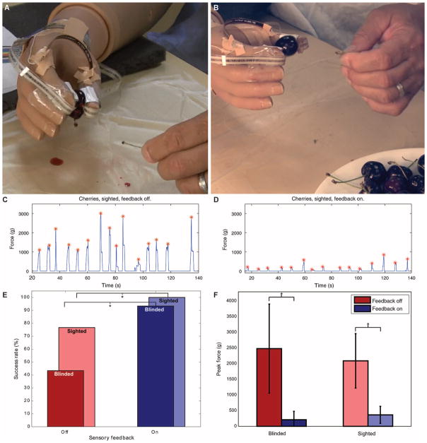
Pulling the stem from a cherry requires control of the grasping pressure on the fruit. If the pressure is too light, the cherry slips; if it is too heavy, the cherry is damaged or crushed (Fig. 5, A and B). We performed this test under four conditions combining sensory (S) and audiovisual (AV) feedback: S−/AV+, S−/AV−, S+/AV−, and S+/AV+. Sensory feedback provided to the thumb and index finger matched the physical pressure measured at matched positions on the prosthesis. Stimulation frequency to each location was proportional to force measured by force-sensitive-resistor sensors mounted on the corresponding fingertips of the prosthetic hand. Stimulation frequency to the contact that produced pressure sensation on the thenar eminence of the phantom limb was proportional to the opening span of the prosthesis.
Fig. 5. Functional tasks with sensory feedback.
(A) Without the sensory feedback system enabled, the subject was often unable to adequately control the grip force in a delicate task such as holding a cherry while removing the stem. (B) With the sensory feedback enabled, the subject felt contact with the cherry and the force applied. He successfully gripped the cherry and removed the stem without damaging the fruit. (C) Total force from thumb and index tip sensors when subject 1 had audiovisual feedback (sighted) but not sensory feedback (feedback off). Peak force during the trial is denoted with a red asterisk. (D) Total force from thumb and index tip sensors when subject 1 had both audiovisual feedback (sighted) and sensory feedback (feedback on). Peak forces (red asterisks) were significantly reduced. (E) Sighted and blinded performance with the sensory feedback on or off during the cherry task showed an improvement in success rate (test of proportions, P < 0.005, n = 15 per condition). (F) Peak forces were significantly lower in the feedback enabled condition under both blinded and sighted conditions (Welch’s t test, P < 0.001, n = 15 per condition).
When sensory feedback was not provided, the subject successfully plucked 43 and 77% of the cherries without (S−/AV−) or with (S−/AV+) audiovisual feedback, respectively. When sensory feedback was provided, the subject successfully plucked 93 and 100% of the cherries without (S+/AV−) or with (S+/AV+) audiovisual feedback, respectively (Fig. 5E). Sensory feedback significantly improved performance (43 to 93% success) without audiovisual feedback (test of two proportions, P < 0.001, n = 15 per condition) (Fig. 5E). With audiovisual feedback, sensation still significantly improved performance with 77 to 100% success (P < 0.005) (Fig. 5E). Fingertip grip forces were significantly reduced with sensory feedback (Fig. 5, C, D, and F). The subject’s self-reported confidence in performing the functional trials was significantly higher with sensory feedback compared to without sensory feedback (one-tailed t test, P = 0.0305).
Without sensation, prosthesis users typically use the prosthesis for gross tasks such as bracing and holding. The improved control and confidence resulting from sensory feedback may lead to greater use of the prosthesis for fine activity and improve balanced bilateral activities and, hence, a more normal appearance and integration of the prosthesis into daily activity.
Subjects reported that sensory feedback eliminated pain in the phantom hand
Subject 1 reported that before the study, his phantom hand felt like it was always clenched in a fist. Furthermore, multiple times per week, he would experience pain that he described as his fist being squeezed in a vice. After sensory stimulation began, he reported that it felt like his phantom hand was opening again, and that, over time, the phantom pain episodes diminished and eventually disappeared. Subject 2 reported that before sensory stimulation, he experienced pain described as “a nail being driven through [his] thumb” about twice per month. Since beginning sensory stimulation, he has not had another episode of phantom pain. The results of the pain survey from the Trinity Amputation and Prosthesis Experience Scales (TAPES) given throughout the study are shown in table S1. These are similar to findings from other single-subject case studies (5, 18) and warrant more rigorous investigation into the benefits of sensory feedback and neural interfaces in the management of phantom pain.
DISCUSSION
Peripheral nerve cuff electrode interfaces provided more than a year of multi-location, multi-perception sensory feedback in two human subjects. Patterned stimulation intensity controlled the quality of sensory perception. Our hypothesis was that the patterned intensity introduced information in the peripheral nerve by population coding that had influence on higher-order processes to produce complex sensory perceptions. Independent control of pulse width, pulse amplitude, stimulation frequency, and the patterns by which these parameters were varied controlled the spatial extent, intensity, and quality of perception. This level of control was possible at multiple locations innervated by a single peripheral nerve. There was independent control of 19 different locations on the hand with only three implanted cuffs having a total of 20 electrical contacts on mostly sensory nerves in subject 1, and 9 different locations in the hand and 3 in the arm from two cuffs having a total of 16 contacts on mixed motor and sensory nerves in subject 2. Higher density contact arrays may further improve coverage on the hand. The implanted electrodes were stable for up to 2 years in the two individuals who had active lifestyles. A few of the activities that each of the subjects reported included chopping wood, home renovations, and camping. The performance of the electrodes remained stable during the external forces associated with the activity and during muscle activity and motion.
Tasks requiring fine grasp control were difficult without sensation even with audiovisual feedback when they were able to see the prosthesis. With sensory feedback, functional performance improved, especially when audiovisual feedback was withheld by putting a blindfold and noise-canceling headphones on the subjects. The addition of sensory feedback alleviated the visual and attentional demands typically required to use a myoelectric prosthesis and enhanced the user’s confidence in task performance. The subjects reported feeling like they were grabbing the object, not just using a tool to grab the object.
The complex perceptions described in response to patterned stimulation intensity paradigms suggest that sensory perception is a pattern recognition activity (19). Paresthesia results from ectopic or unnatural patterns of fiber activation (20, 21). Whereas perception can arise from activation of a single fiber, it is more natural for many fibers to be active during a sensory task (22). This activity is then integrated centrally to produce meaningful perception. Nerve electrodes activate populations of axons on the basis of size and location relative to the stimulating contact (23). Because the fibers associated with slowly adapting type 1 (SA1), slowly adapting type 2 (SA2), rapidly adapting type 1 (RA1), and Pacinian corpuscle (PC) mechanoreceptors all have similar diameters and stimulation threshold characteristics, it is not possible to selectively activate a population consisting of a single fiber type. With a constant pulse intensity, there is synchronous activity of a mixed population of axons. This is the abnormal firing pattern observed when paresthesia is induced from ischemic block (20) and is typical in traditional electrical stimulation.
The patterned stimulus intensity recruited varying populations of axons at each pulse, thereby creating a pattern in the population activation. At the lowest pulse width of the SSO modulation paradigm, a small population of axons was suprathreshold and actively firing. Because the pulse width never decreased below this level, this population of the neurons will be active at the frequency of the pulse train. This is similar to a constant, slowly adapting pattern of activity in response to a constant pressure. At higher pulse widths, the stimulation is sufficient to activate other slightly more distant or smaller diameter neurons. That population will have a transient or bursting activation pattern that mimics the pattern more typical of rapidly adapting fibers. The higher-order processing of this population pattern resulted in the perceived quality of the sensation. Sinusoidal pulse amplitude modulation would likely produce similar population recruitment patterns and sensations. With modulation patterns more complex than a sinusoidal pattern reported here, it may be possible to introduce more complex population codes.
The sensation was described by the subjects as “natural.” It is unlikely that the population activation was a perfect mimic of the patterns observed in slowly and rapidly adapting fibers in response to touch. Thus, the resulting activation pattern was not strictly natural at the axonal level. However, processing in the thalamic relays and columns of the primary sensory cortex appeared robust to errors in the pattern, resulting in the reported natural sensory perception. Normal processing in the brain is highly tolerant of abnormal patterns and classifies patterns according to best matching prior sensory experiences (19).
The subjects’ responses to restored sensation demonstrate its value to quality of life. The subjects strongly preferred having sensory feedback. They described the sensation as natural and not requiring additional interpretation, as is required by sensory substitution techniques. When asked about performing object grasping tasks with sensory feedback enabled, subject 1 stated that, “I knew that I had it,” referring to whatever object was part of the test. Both subjects desired a fully implanted system that would provide them with permanent sensation. Whether sensation was natural or paresthetic, subject 1 stated that, “I’d rather have it in a heartbeat,” and “I miss it when I leave.”
When sensation was active, both subjects perceived the hand and prosthetic hand to be nearly perfectly colocated in space. When sensation was not active, the prosthesis was viewed by the subjects as a tool that extended beyond their hands. Subject 1 described his normal perception of the prosthesis as though “[he] is holding the prosthetic hand [at the base of the prosthesis] with [his] phantom hand.”
The extraneural electrode selectivity was effective. Every stimulating contact provided sensation at well-defined and unique locations on a predominantly sensory nerve (subject 1). The subject could identify sensation at multiple sites independently. There is mounting evidence that peripheral nerves are somatotopically organized (24–26), and hence, multi-channel cuff electrodes are able to produce somatotopically selective results similar to those reported in acute trials with intrafascicular electrodes (5, 12, 16). The peripheral interface in this trial produced punctate sensations in the hand with 19 of 20 channels (95%) or 9 of 16 channels (56%) from a proximal implant location. Natural tactile sensation has been reported in prior work but has not been explicitly quantified or otherwise detailed, making direct comparison with the current study difficult (5, 12). Prior work only examined the response to a pulse train stimulation paradigm. By introducing patterned stimulation intensity, we are able to control the quality of the sensation. The results were stable for more than a year after implant in subject 2, and for more than 2 years in subject 1. On mixed motor and sensory nerves (subject 2), the results were similar in that isolated and distinct locations were perceived. Even more encouraging, sensation could be produced without motor activation or interference with myoelectric control.
Sensory perceptions were elicited corresponding to several distinct physical locations across the missing limb. However, only three locations of sensation were implemented during the functional tasks in this study. This was limited by the capabilities of the users’ prostheses and our stimulation system. It has yet to be shown that information, including relative intensity and timing, from multiple locations is integrated by the user for stereognosis and functional improvement. The limited function of the two degrees-of-freedom prostheses used by both of our subjects limited the value of multiple channels of sensory feedback. The impact of multiple sensations on the acceptance of an anthropomorphic prosthesis with more degrees of freedom and better control options will be important to determine. We also expect performance to improve with continuous usage as the user learns how to incorporate sensory feedback. The plasticity of perception, especially with neural stimulation, is poorly understood and will not be elucidated until the sensory feedback system is implemented continuously outside of the laboratory setting. Continuous sensory feedback may become an unconscious part of the control of the myoelectric prosthesis, hence moving closer to normal human function. Preliminary trials with more sophisticated modulation patterns show that better perception of more complex textures is possible. The exploration of effects of the stimulation dimensions defined in Eq. 1 is ongoing. Last, grasp position feedback was important but rudimentary in the functional trials reported. We did not fully map or explore proprioception or kinesthetic perceptions. Controllable proprioception and kinesthetic sensations were only elicited when the electrodes were implanted proximal to the motor branches, that is, in subject 2. This would indicate that although skin stretch has been implicated in proprioception, the intrafusal motor fibers are important for proprioception in our system.
Peripheral nerve cuff electrodes are stable and produced sensory feedback in human subjects with multiple modes of sensation at multiple points on the hand. Sensory feedback significantly improved grasping performance using a prosthesis. Patterned stimulation intensity controlled perceived sensory quality and has potential application in other areas of somatosensory neuromodulation, such as pain, autonomic function, and deep brain stimulation. Modulation of the frequency of stimulation produced natural, graded intensity of sensation. The human experiments have provided data not readily available in nonhuman studies, specifically in a verbal description of perception. Closing the loop by providing natural sensory feedback significantly improved success rates on a functional task requiring variable grip strength.
MATERIALS AND METHODS
Study design
Our central hypothesis was that direct nerve stimulation with selective, nonpenetrating peripheral nerve cuff electrodes on the residual upper limb nerves can elicit graded sensation at multiple locations perceived in the missing hand of human subjects with upper limb loss. The inclusion criteria included unilateral, upper limb loss amputees, age 21 or older, who are current users of a myoelectric prosthesis or prescribed to use one. Potential subjects were excluded because of poor health (uncontrolled diabetes, chronic skin ulceration, history of uncontrolled infection, active infection) or the presence of significant, uncontrolled persistent pain in the residual or phantom limb.
This was a first-in-man, self-controlled, nonrandomized case study of two subjects to demonstrate feasibility of cuff electrodes to generate sensory perception. Subject 1 had a wrist disarticulation and was 19 months post-loss at the time of the implant in May 2012. Subject 2 had a below-elbow amputation and was 93 months (7.75 years) post-amputation at the time of implant in January 2013.
Methods
In subject 1, surgeons implanted three electrodes in the residual limb of a then 46-year-old male who had a unilateral wrist disarticulation from work-related trauma in 2010. The surgery was an outpatient procedure. At the time of implant, the subject had been a regular user of a myoelectric prosthesis for 7 months. Eight-contact flat interface nerve electrodes (FINEs) (14) were implanted on the median and ulnar nerves and a four-contact CWRU (Case Western Reserve University) spiral electrode (15) was implanted on the radial nerve. FINE opening size for the nerve was 10 mm wide by 1.5 mm high for both the median and ulnar nerves. The internal diameter of the spiral electrode was 4 mm for the radial nerve. Peripheral nerve histology from human cadavers guided specifications of electrode sizes, and surgeons confirmed proper electrode fit during the procedure. All electrodes were implanted in the mid-forearm (Fig. 1A) and connected to percutaneous leads (27–29) that exited through the upper arm.
In subject 2, surgeons implanted two multi-contact nerve cuff electrodes of a then 46-year-old male who had a below-elbow amputation from work-related trauma in 2004. At the time of implant, the subject had been a regular user of a myoelectric prosthesis for 7 years. Eight-contact FINEs were implanted on the median and radial nerves. FINE opening size for the nerve was 10 mm wide by 1.5 mm high for both the median and radial nerves. All electrodes were implanted in the mid-upper arm. The subject was discharged from the hospital the day after surgery. All other surgical details were identical to subject 1. Ardiem Medical supplied implanted components (cuff electrodes, percutaneous leads, connectors) and MOOG sterilized the components with ethylene oxide.
Stimulation experiments began after 3 weeks to allow the electrodes and tissue response to stabilize. Subjects did not report any adverse sensation in the implanted locations or changes in their phantom sensations. In weekly sessions beginning after the stabilization period, we applied stimulation through each contact for up to 10 s. For all trials, the subject was blind to the stimulation strength. After stimulation, the subject would describe any perceived sensation and sketch its location on a blank hand diagram. We randomly intermixed null trials with no stimulation to ensure the subject was not anticipating sensation. The Cleveland Department of Veterans Affairs Medical Center Institutional Review Board (IRB) approved all procedures, and the study was conducted under a Food and Drug Administration (FDA) Investigational Device Exemption.
Experimental setup
The stimulation system consists of a computer that controls stimulation parameters and sends the commands to a single board computer running xPC Target (MathWorks Inc.). Ardiem Medical fabricated a custom-designed stimulator (Cleveland FES Center). An isolator provides optical isolation between devices plugged into the wall and the subject. To prevent overstimulation, we limit charge density to less than 50 μC/cm2 and stimulation to less than a 50% duty cycle during all stimulation protocols.
The stimulator has 24 channels of controlled-current stimulation outputs, with a maximum stimulation amplitude of 5.6 mA, a maximum stimulation pulse width of 255 μs, and a compliance voltage of 50 V. The stimuli are monopolar, biphasic, charge-balanced, cathodic-first pulses with return to a common anode, which is a 5.08 cm × 10.16 cm surface electrode on the dorsal surface of the upper arm immediately proximal to elbow. There is a minimum delay of 0.6 ms between each channel of stimulus; thus, the stimulator does not output truly simultaneous stimulation when multiple channels are active.
Generic framework of electrical stimulation
The generic stimulation waveform, Λ, is a train of pulses, Ψ, separated by an interpulse interval, IPI. For each pulse shape, the pulse parameters, Δ, are selected to activate a population of neurons. To elicit a sensory perception from touch, the pulse parameters, Δ, and IPI are a function of measured external inputs over time, ϕ(t), and time, t. Patterns in the pulse parameters will vary the population of axons excited and affect qualities of sensory perception. Hence, Δ and IPI are defined as a function of the desired tactile perception and time, that is, Δ(ϕ(t),t) and IPI(ϕ(t),t) (Eq. 1).
| (1) |
The psychometric relationships between modulation of stimulation parameters and sensory perception were assessed for ψ as a charge-balanced, biphasic, square pulse, having parameters Δ = {PA(ϕ,t), PW(ϕ,t)} with IPI(t), where PA is pulse amplitude and PW is pulse width. Perception is controlled by the time-dependent change of the parameters, referred to as patterned stimulation intensity. We systematically examined each parameter of stimulation according to each of the following conditions.
Stimulating with time invariant parameters
In a typical stimulation paradigm, the pulse intensity parameters are set to a fixed values, that is, PA(ϕi) = PA0 and PW(ϕi) = PW0, and the pulse train frequency is constant, IPI(ϕi) = IPI0 = 1/f0.
Stimulating with a time-variant pulse width, PW(ϕi,t)
Full-scale modulation
For this set of trials, the following equation (Eq. 2) defines the specific version of Eq. 1 for the full-scale modulation pattern of stimulation intensity:
| (2) |
a and b are parameters that control the size of the pulse width modulation. In these trials, the interpulse interval was held constant at IPI0 = 0.01 s. The pulse width was modulated in a slow (fmod = 1 Hz) sinusoidal envelope with b = a = B/2, where B is the peak of the varying pulse width. The pulse width, therefore, ranged from 0 μs to B μs (Fig. 2D).
SSO modulation
Small modulation is defined by Eq. 2 with a = (PWmax − PWmin)/2 = PWpk-pk/2 and b = PWoffset. The typical value of PWpk-pk was 5 μs, IPI0 was 0.01 s, fmod = 1 Hz, and PWoffset was set to about 90% of the Bth required to produce the natural pulsing sensation (Fig. 4A).
Threshold detection method
See the Supplementary Materials.
Contralateral pressure matching
We tested pressure matching with SSO modulation on channel M4 (corresponding to the thenar eminence) of subject 1. For each trial, the subject received 5 s of stimulation and matched the pressure sensation in the contralateral hand by pressing on a manipulator for 5 s. The last 2 s of the matched pressure data were averaged per trial. The manipulator shape resembled the perceived sensation, and the user pressed with the same palmar location on the contralateral hand as the perceived sensation. The manipulator shape was an about 1.27 cm diameter circle flat tip with rounded edges and made of balsa wood. We placed this manipulator on top of a FlexiForce sensors model A201 (Tekscan Inc.) with 0 to 454 g range. The sensor was calibrated before each trial and sampled at 10 Hz.
Functional testing
The subject used his intact hand to pluck the stem off of a cherry, with the fruit grasped by the prosthetic hand. The subject used his standard prosthetic hand, which was the velocity-controlled, SensorHand Speed (Otto Bock HealthCare) with automatic slip detection/correction turned off. Control of the hand was driven by subjects using standard surface electromyography signals. Thin FlexiForce sensors (Tekscan model A201) mounted on the end pads of the thumb and index finger measured the pressure applied to the cherry by the prosthetic. A bend sensor mounted between index finger and thumb provided measurement of the opening span. The instantaneous stimulation frequency applied to the nerve was linearly proportional to the real-time force sensor output. The frequency range was 10 to 125 Hz. The sensory feedback stimulation system has a measured mean delay of 112 ± 35 ms between measurement and applied sensation. The subject reports no noticeable delay in sensation during sighted tasks. We administered the test 60 times with sensory feedback off and 30 times with sensory feedback on. For half the trials in each set, designated AV−, the subject wore a sleeping mask to remove visual feedback and noise protection ear muffs over ear buds playing white noise to remove audio feedback. Failures were defined as any production of juice or visible cracks in the fruit skin during the task, and determined by a single evaluator for all trials. After each trial with the subject blinded, we asked the subject to rate his confidence in his overall performance before the mask and headphones were removed.
Statistical analysis
Unless otherwise stated, we used ANOVA statistical testing and set significance as α = 0.05.
Supplementary Material
Acknowledgments
We thank M. Schmitt for clinical support and management of the IRB compliance. We thank J. Wall for regulatory support and managing FDA compliance. We thank L. Miller and S. Bensmaia for review and editing early drafts of the manuscript.
Funding: This material is based on work supported by the Department of Veterans Affairs, Veterans Health Administration, Office of Research Development, Rehabilitation Research and Development under Merit Review #A6156R and Career Development Award 1IK1RX000724-01A1.
Footnotes
www.sciencetranslationalmedicine.org/cgi/content/full/6/257/257ra138/DC1
Materials and Methods
Fig. S1. Subject 2 perceptual locations at near-threshold stimulation levels.
Fig. S2. Stable electrode impedances across study duration.
Fig. S3. Subject 2 contralateral pressure matching.
Table S1. TAPES Pain Survey data.
Competing interests: D.J.T. is Founder and President of Bear Software, LLC. D.J.T. is an inventor on the patent entitled “The flat interface nerve electrode and a method for use” (US6456866 B1); Case Western Reserve University holds the patents on this technology. D.W.T., M.A.S., and D.J.T. are named inventors on a provisional patent on the stimulation paradigm described in the paper, which is jointly held by Case Western Reserve University and the Louis-Stokes Cleveland VA Medical Center.
Author contributions: D.W.T. and M.A.S. contributed equally to experiment design, data collection, analysis, and documentation. M.W.K. and J.R.A. conducted the implantation surgeries. J.T. provided occupational therapy support. D.J.T. was the principal investigator and contributed significantly to the experimental work.
REFERENCES AND NOTES
- 1.Robles-De-La-Torre G. The importance of the sense of touch in virtual and real environments. IEEE Multimed. 2006;13:24–30. [Google Scholar]
- 2.Augurelle AS, Smith AM, Lejeune T, Thonnard JL. Importance of cutaneous feedback in maintaining a secure grip during manipulation of hand-held objects. J Neurophysiol. 2003;89:665–671. doi: 10.1152/jn.00249.2002. [DOI] [PubMed] [Google Scholar]
- 3.Marasco PD, Kim K, Colgate JE, Peshkin MA, Kuiken TA. Robotic touch shifts perception of embodiment to a prosthesis in targeted reinnervation amputees. Brain. 2011;134:747–758. doi: 10.1093/brain/awq361. [DOI] [PMC free article] [PubMed] [Google Scholar]
- 4.Ramachandran VS, Rogers-Ramachandran D. Synaesthesia in phantom limbs induced with mirrors. Proc Biol Sci. 1996;263:377–386. doi: 10.1098/rspb.1996.0058. [DOI] [PubMed] [Google Scholar]
- 5.Rossini PM, Micera S, Benvenuto A, Carpaneto J, Cavallo G, Citi L, Cipriani C, Denaro L, Denaro V, Di Pino G, Ferreri F, Guglielmelli E, Hoffmann KP, Raspopovic S, Rigosa J, Rossini L, Tombini M, Dario P. Double nerve intraneural interface implant on a human amputee for robotic hand control. Clin Neurophysiol. 2010;121:777–783. doi: 10.1016/j.clinph.2010.01.001. [DOI] [PubMed] [Google Scholar]
- 6.Childress DS. Closed-loop control in prosthetic systems: Historical perspective. Ann Biomed Eng. 1980;8:293–303. doi: 10.1007/BF02363433. [DOI] [PubMed] [Google Scholar]
- 7.Murray CD. In: Psychoprosthetics. Gallagher P, Desmond D, MacLachlan M, editors. Springer; London: 2008. pp. 119–129. [Google Scholar]
- 8.Pylatiuk C, Kargov A, Schulz S. Design and evaluation of a low-cost force feedback system for myoelectric prosthetic hands. J Prosthet Orthot. 2006;18:57–61. [Google Scholar]
- 9.Witteveen H, Droog E, Rietman J, Veltink P. Vibro- and electrotactile user feedback on hand opening for myoelectric forearm prostheses. IEEE Trans Biomed Eng. 2012;59:2219–2226. doi: 10.1109/TBME.2012.2200678. [DOI] [PubMed] [Google Scholar]
- 10.Clippinger FW, Avery R, Titus BR. A sensory feedback system for an upper-limb amputation prosthesis. Bull Prosthet Res. 1974 Fall;:247–258. [PubMed] [Google Scholar]
- 11.Dhillon GS, Krüger TB, Sandhu JS, Horch KW. Effects of short-term training on sensory and motor function in severed nerves of long-term human amputees. J Neurophysiol. 2005;93:2625–2633. doi: 10.1152/jn.00937.2004. [DOI] [PubMed] [Google Scholar]
- 12.Raspopovic S, Capogrosso M, Petrini FM, Bonizzato M, Rigosa J, Di Pino G, Carpaneto J, Controzzi M, Boretius T, Fernandez E, Granata G, Oddo CM, Citi L, Ciancio AL, Cipriani C, Carrozza MC, Jensen W, Guglielmelli E, Stieglitz T, Rossini PM, Micera S. Restoring natural sensory feedback in real-time bidirectional hand prostheses. Sci Transl Med. 2014;6:222ra19. doi: 10.1126/scitranslmed.3006820. [DOI] [PubMed] [Google Scholar]
- 13.Berg JA, Dammann JF, III, Tenore FV, Tabot GA, Boback JL, Manfredi LR, Peterson ML, Katyal KD, Johannes MS, Makhlin A, Wilcox R, Franklin RK, Vogelstein RJ, Hatsopoulos NG, Bensmaia SJ. Behavioral demonstration of a somatosensory neuroprosthesis. IEEE Trans Neural Syst Rehabil Eng. 2013;21:500–507. doi: 10.1109/TNSRE.2013.2244616. [DOI] [PubMed] [Google Scholar]
- 14.Tyler DJ, Durand DM. Chronic response of the rat sciatic nerve to the flat interface nerve electrode. Ann Biomed Eng. 2003;31:633–642. doi: 10.1114/1.1569263. [DOI] [PubMed] [Google Scholar]
- 15.Naples GG, Mortimer JT, Scheiner A, Sweeney JD. A spiral nerve cuff electrode for peripheral nerve stimulation. IEEE Trans Biomed Eng. 1988;35:905–916. doi: 10.1109/10.8670. [DOI] [PubMed] [Google Scholar]
- 16.Dhillon GS, Lawrence SM, Hutchinson DT, Horch KW. Residual function in peripheral nerve stumps of amputees: Implications for neural control of artificial limbs. J Hand Surg Am. 2004;29:605–615. doi: 10.1016/j.jhsa.2004.02.006. [DOI] [PubMed] [Google Scholar]
- 17.Knibestöl BYM, Vallbo AB. Intensity of sensation related to activity of slowly adapting mechanoreceptive units in the human hand. J Physiol. 1980;300:251–267. doi: 10.1113/jphysiol.1980.sp013160. [DOI] [PMC free article] [PubMed] [Google Scholar]
- 18.Horch K, Meek S, Taylor TG, Hutchinson DT. Object discrimination with an artificial hand using electrical stimulation of peripheral tactile and proprioceptive pathways with intrafascicular electrodes. IEEE Trans Neural Syst Rehabil Eng. 2011;19:483–489. doi: 10.1109/TNSRE.2011.2162635. [DOI] [PubMed] [Google Scholar]
- 19.Makin JG, Fellows MR, Sabes PN. Learning multisensory integration and coordinate transformation via density estimation. PLOS Comput Biol. 2013;9:e1003035. doi: 10.1371/journal.pcbi.1003035. [DOI] [PMC free article] [PubMed] [Google Scholar]
- 20.Mogyoros I, Bostock H, Burke D. Mechanisms of paresthesias arising from healthy axons. Muscle Nerve. 2000;23:310–320. doi: 10.1002/(sici)1097-4598(200003)23:3<310::aid-mus2>3.0.co;2-a. [DOI] [PubMed] [Google Scholar]
- 21.Ochoa JL, Torebjörk HE. Paraesthesiae from ectopic impulse generation in human sensory nerves. Brain. 1980;103:835–853. doi: 10.1093/brain/103.4.835. [DOI] [PubMed] [Google Scholar]
- 22.Dimitriou M, Edin BB. Discharges in human muscle spindle afferents during a key-pressing task. J Physiol. 2008;586:5455–5470. doi: 10.1113/jphysiol.2008.160036. [DOI] [PMC free article] [PubMed] [Google Scholar]
- 23.Schiefer MA, Tyler DJ, Triolo RJ. Probabilistic modeling of selective stimulation of the human sciatic nerve with a flat interface nerve electrode. J Comput Neurosci. 2012;33:179–190. doi: 10.1007/s10827-011-0381-5. [DOI] [PMC free article] [PubMed] [Google Scholar]
- 24.Badia J, Pascual-Font A, Vivó M, Udina E, Navarro X. Topographical distribution of motor fascicles in the sciatic-tibial nerve of the rat. Muscle Nerve. 2010;42:192–201. doi: 10.1002/mus.21652. [DOI] [PubMed] [Google Scholar]
- 25.Watchmaker GP, Gumucio Ca, Crandall RE, Vannier Ma, Weeks PM. Fascicular topography of the median nerve: A computer based study to identify branching patterns. J Hand Surg Am. 1991;16:53–59. doi: 10.1016/s0363-5023(10)80013-9. [DOI] [PubMed] [Google Scholar]
- 26.Prodanov D, Nagelkerke N, Marani E. Spatial clustering analysis in neuroanatomy: Applications of different approaches to motor nerve fiber distribution. J Neurosci Methods. 2007;160:93–108. doi: 10.1016/j.jneumeth.2006.08.017. [DOI] [PubMed] [Google Scholar]
- 27.Knutson JS, Naples GG, Peckham PH, Keith MW. Electrode fracture rates and occurrences of infection and granuloma associated with percutaneous intramuscular electrodes in upper-limb functional electrical stimulation applications. J Rehabil Res Dev. 2002;39:671–683. [PubMed] [Google Scholar]
- 28.Letechipia JE, Peckham PH, Gazdik M, Smith B. In-line lead connector for use with implanted neuroprosthesis. IEEE Trans Biomed Eng. 1991;38:707–709. doi: 10.1109/10.83572. [DOI] [PubMed] [Google Scholar]
- 29.Polasek KH, Hoyen HA, Keith MW, Kirsch RF, Tyler DJ. Stimulation stability and selectivity of chronically implanted multicontact nerve cuff electrodes in the human upper extremity. IEEE Trans Neural Syst Rehabil Eng. 2009;17:428–437. doi: 10.1109/TNSRE.2009.2032603. [DOI] [PMC free article] [PubMed] [Google Scholar]
Associated Data
This section collects any data citations, data availability statements, or supplementary materials included in this article.


