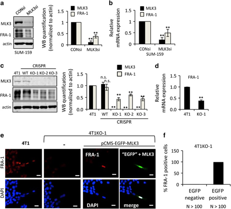
Figure 2.
MLK3 is required for FRA-1 expression in TNBC cells. Cellular lysates and/or mRNA samples were collected from (a and b) SUM-159 cells treated with 50 nm control siRNA or MLK3 siRNA for 24 h, (c) parental 4T1, WT clone and three 4T1 CRISPR MLK3-knockout clones (KO-1, KO-2 and KO-3) and (d) parental 4T1 or 4T1KO-1 cells. Cellular lysates were subjected to immunoblotting with indicated antibodies. Western blot quantification of the indicated protein normalized to actin is expressed as mean±s.d. from at least three independent experiments. The mRNAs were subjected to qRT–PCR with primers for the indicated genes. Relative mRNA expression is displayed as mean±s.d. from at least three independent experiments performed in triplicate. (e and f) Parental 4T1 cells or 4T1KO-1 cells were transfected with bi-cistronic vector expressing EGFP and MLK3 (pCMS-EGFP-MLK3) for 24–48 h and were subjected to immunofluorescence staining using a FRA-1 antibody. FRA-1 staining is shown in red and GFP, which indicates co-expression of MLK3, is shown in green. Nuclei were counterstained with DAPI (blue); Scale bar, 25 μm; NS, not statistically significant; **P<0.01.

