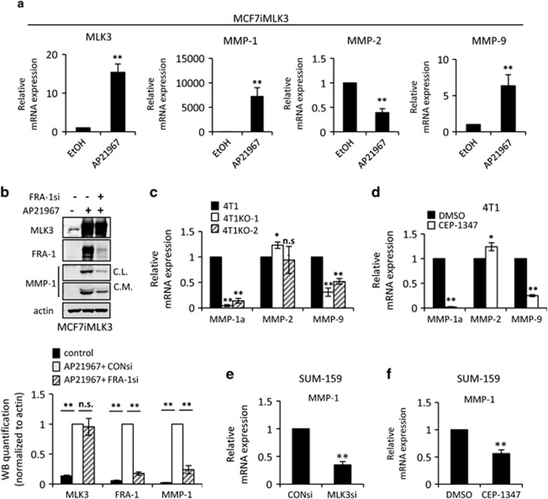
Figure 5.
MLK3 induces MMP-1 and MMP-9 expression. Cellular lysates or mRNAs, as indicated, were isolated from (a) MCF7iMLK3 cells treated with vehicle or 50 nm AP21967 for 24 h, (b) MCF7iMLK3 cells treated with vehicle or with 50 nm AP21967 plus either 50 nm control or FRA-1 siRNA, as indicated, for 24 h, (c) parental 4T1 cells and two 4T1 MLK3-knockout clones (4T1KO-1 and 4T1KO-2), (d) 4T1 cells treated with vehicle or 400 nm CEP-1347 for 24 h, and SUM-159 cells treated with (e) 50 nm control or MLK3 siRNA for 24 h, or (f) vehicle or 400 nm CEP-1347 for 24 h. The mRNAs were subjected to qRT–PCR with primers to the indicated genes. Relative mRNA expression is displayed as the mean±s.d. from at least three independent experiments performed in triplicate. Cellular lysates were subjected to immunoblotting with indicated antibodies. Western blot quantification of the indicated protein normalized to actin is expressed as mean±s.d. from at least three independent experiments. Con, Control; CL, cellular lysate; CM, concentrated conditioned medium; NS, not statistically significant; *P<0.05; **P<0.01.

