Figure 2.
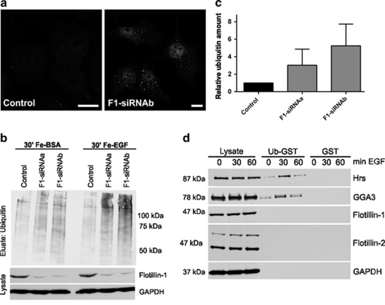
Increased amount of ubiquitinated cargo in endosomes of EGF-stimulated flotillin-1-knockdown cells.(a) Control siRNA and flotillin-1 siRNA-transfected cells were stimulated with 100 ng/ml EGF for 30 min, fixed and immunostained for ubiquitin. An enhanced ubiquitin staining was observed in punctate perinuclear structures in flotillin-1-knockdown cells. Scale bar: 10 μm. (b) Endosomes were isolated with magnetic columns from control siRNA and flotillin-1-depleted cells treated with Ferrofluid-coupled BSA or Ferro-EGF for 30 min. Samples from homogenate and eluate were analysed for ubiquitin by western blot. (c) Increased ubiquitin signal in endosomes of flotillin-1-knockdown cells. Densitometric analysis of ubiquitin signals shown in b from three independent experiments±s.d. Data were normalised to GAPDH in homogenates. (d) GST pulldown with ubiquitin-GST or GST from HeLa cells treated for 0, 30 or 60 min with EGF shows an increased binding of Hrs and GGA3 to ubiquitin upon EGF stimulation, whereas flotillins do not bind to ubiquitin-GST. Ubiquitin interactors were detected by western blot using specific antibodies, GAPDH was used as an input control.

