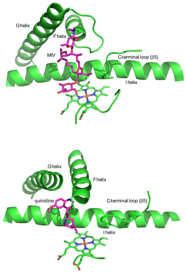
Figure 10.
Comparison of crystallographic M-IV binding orientation in MycG (top, PDB entry 2Y98) with that of inhibitor quinidine in CYP2D6 (bottom, PDB entry 4WNU, ref. (27)). Molecules are oriented so that the heme plane and orientation is in approximately the same tilt and direction in both structures.

