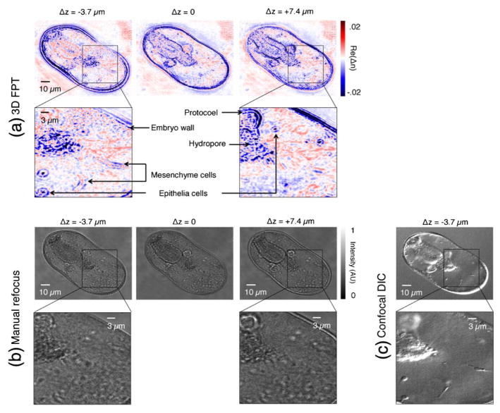
Fig. 7.
3D reconstruction of a starfish embryo at larval stage. (a) Three different axial planes of the FPT tomogram show significant feature variation (e.g., protocol is completely missing from Δz = −3.7 μm plane, expected developing mesenchyme cells are only visible in Δz = −3.7 μm plane). (b) Such axial information, and even certain structures (e.g., mesenchyme cells and various epithelia cells, marked in (a)) are completely missing from standard microscope images after manual refocusing. (c) A high-resolution DIC confocal scan of the Δz = −3.7 μm plane confirms presence of structures of interest. See Visualization 2.

