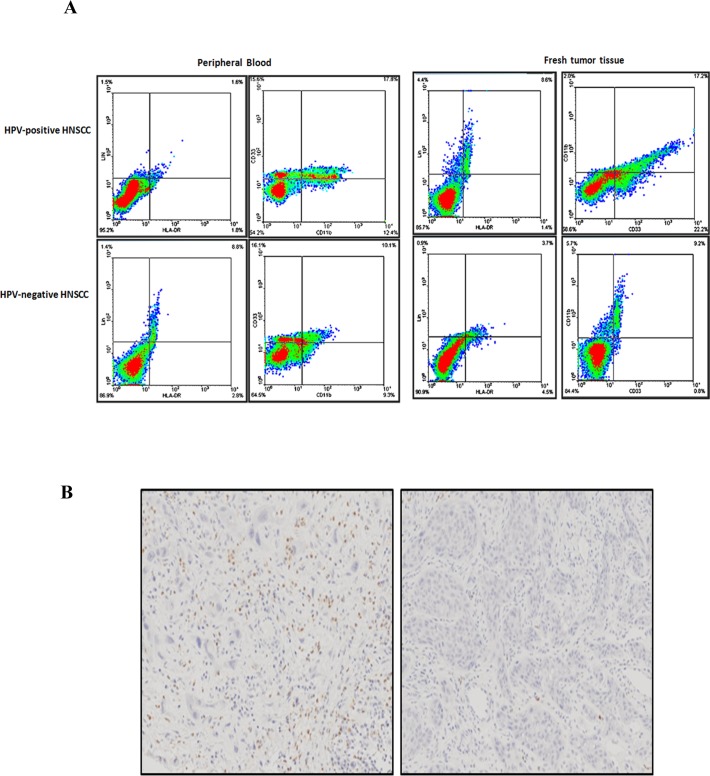
Figure 2. MDSCs expression in peripheral blood and fresh tumor tissue samples of HNSCC.
A. Representative flow cytometry image of CD11b+ LIN- HLA-DR- CD33+ MDSCs in peripheral blood and fresh tumor tissues of HPV-positive HNSCC and HPV-negative HNSCC. We first examined the percentage of LIN- HLA-DR- cells, and then screened the percentage of CD11b+ CD33+ cells in LIN- HLA-DR- cells. The peripheral blood sample and the fresh tumor tissue were from the same patients. The percentage of CD11b+ LIN- HLA-DR- CD33+ MDSCs in blood and tissue was almost the same. B. Representative immunohistochemical image of MPO in paraffin sections of HNSCC patients. Right was MPO in strong positive staining. Left was MPO in negative staining.

