Abstract
Background
18F-fluorodeoxyglucose (18F-FDG) positron emission tomography (PET) and PET/CT have become two of the most powerful tools for malignant lymphoma exploration, but their diagnostic role in primary central nervous system lymphoma (PCNSL) is still disputed. The purpose of our study is to identify the usefulness of 18F-FDG PET and PET/CT for detecting PCNSL.
Results
A total of 129 patients, obtained from eight eligible studies, were included for this systematic review and meta-analysis. The performance of 18F-FDG PET and PET/CT for diagnosing PCNSL were as follows: the pooled sensitivity was 0.88 (95% CI: 0.80–0.94), specificity was 0.86 (95% CI: 0.73–0.94), positive likelihood ratio (PLR) was 3.99 (95% CI: 2.31–6.90), negative likelihood ratio (NLR) was 0.11 (95% CI: 0.04-0.32), and diagnostic odds ratio (DOR) was 33.40 (95% CI: 10.40–107.3). In addition, the area under the curve (AUC) and Q index were 0.9192 and 0.8525, respectively.
Materials and Methods
PubMed/MEDLINE, Embase and Cochrane Library were systematically searched for potential publications (last updated on July 16th, 2016). Reference lists of included articles were also checked. Original articles that reported data on patients who were suspected of having PCNSL were considered suitable for inclusion. The sensitivities and specificities of 18F-FDG PET and PET/CT in each study were evaluated. The Stata software and Meta-Disc software were employed in the process of data analysis.
Conclusions
18F-FDG PET and PET/CT showed considerable accuracy in identifying PCNSL in immunocompetent patients and could be a valuable radiological diagnostic tool for PCNSL.
Keywords: 18F-fluorodeoxyglucose, positron emission tomography, positron emission tomography/computed tomography, primary central nervous system lymphoma, diagnosis
INTRODUCTION
Primary central nervous system lymphomas (PCNSL) are extranodal malignant lymphomas that arise within the brain, eyes, leptomeninges, or spinal cord in the absence of systemic lymphoma at the time of diagnosis. The annual incidence of PCNSL in developed countries is 0.5 cases per 100,000 persons, accounting for 3–5% of primary brain tumors [1]. However, epidemiological data have shown a striking increase in the immunocompetent population over the past decades, while the incidence seems to be decreasing in patients with acquired immunodeficiency syndrome (AIDS), owning to the development of highly active antiretroviral therapy (HAART) [2–5]. Recent clinical studies have demonstrated an increase in the overall survival rates by a large margin due to the combined treatment of high-dose methotrexate-(MTX-) based chemotherapy and whole-brain radiotherapy [6]. Moreover, younger age and higher Karnofsky performance score (KPS) at the time of diagnosis are believed to be associated with prolonged survival time [7]. Accordingly, it is essential to make an early diagnosis of PCNSL and initiate early treatment correspondingly, prior to a decline of the patient's physical condition. Thus far, the diagnosis of PCNSL still relies on invasive stereotactic brain biopsy, which will inevitably be linked with a heavy expense and risk of injury. Under this circumstance, it is imperative to determine an accurate, reliable and cost-effective method for PCNSL screening.
Contrast-enhanced magnetic resonance imaging (MRI) is the standard diagnostic imaging technique when PCNSL is suspected and often shows characteristic radio-morphological features such as a lesion location adjacent to the cerebrospinal fluid (CSF) space, strong and homogenous contrast-enhancement, moderate edema and an absence of necrosis [8]. If MRI cannot be performed, then another option is contrast-enhanced computed tomography (CT), which is diagnostically equivalent in most cases [9]. The radiologic findings of MRI and CT, however, are not pathognomonic for PCNSL. Similar findings can be seen in malignant gliomas, brain metastases, and inflammatory diseases [10]. Consequently, in diagnosing PCNSL, another credible and effective radiological modality is desirable.
As a modern metabolic imaging modality, 18F-fluorodeoxyglucose (18F-FDG) positron emission tomography (PET) has shown remarkable sensitivity and specificity in the detection of systemic non-Hodgkin's lymphoma (NHL). More importantly, it has been shown to provide high accuracy in the differentiation between cerebral lymphomas and either high-grade gliomas or infectious lesions in AIDS patients [8, 11, 12]. This method alone or combined with CT (18F-FDG PET/CT) has been proposed as a non-invasive and accurate tool to assess disease progression in cancer patients [13, 14]. Since the tumor tissue usually has a high cellular density with an accelerated glucose metabolism, lesions of PCNSL often show a high FDG concentration. Thus, 18F-FDG PET and PET/CT are good at distinguishing PCNSL with hypermetabolic lesions from infection with hypometabolic lesions [15]. Furthermore, preliminary data suggest that 18F-FDG PET and PET/CT may be excellent in making a distinction between PCNSL and other brain tumors [16, 17]. A retrospective study by Makino et al. reported that, when it came to PCNSL and GBM with similar MRI findings, the addition of 18F-FDG PET could improve diagnostic accuracy compared to that with conventional MRI [18]. However, Kawai and colleagues see things differently. They found little benefit of PET to discriminate PCNSL from other neoplastic and benign diseases compared with MRI, especially for atypical lesions [19]. Unfortunately, similar studies are relatively rare, so it is difficult to draw a firm conclusion.
With the development of 18F-FDG PET and PET/CT, a number of studies have reported varying results about their detection ability of PCNSL. Nevertheless, the present evidence is mainly from some small sample clinical studies. Consequently, a systematic review aimed at clarifying the potential effects of 18F-FDG PET and PET/CT in PCNSL radiological diagnosis was essential. For this reason, we conducted a meta-analysis to evaluate the role of 18F-FDG PET and PET/CT in the diagnosis of immunocompetent patients with PCNSL.
RESULTS
Search strategy and study selection
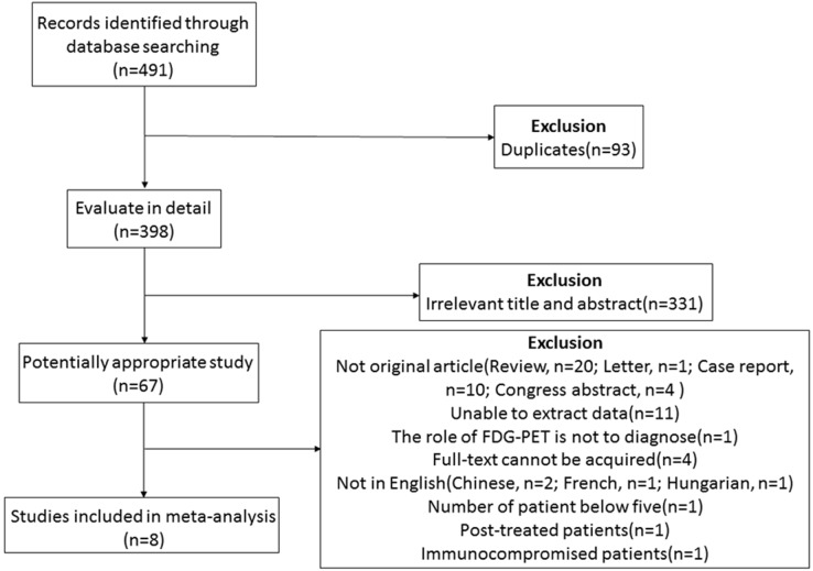
As previously mentioned, we searched up to July 16th, 2016, which yielded a total of 491 papers: 352 in Embase, 111 in PubMed/MEDLINE, 11 in Cochrane Library, and 17 by a manual search. Among them, 93 duplicate publications were excluded. After screening for titles and abstracts, we reviewed 67 articles in detail. Of these, 59 were excluded. The exclusion criteria were as follows: (1) Articles that could not reveal the performance of 18F-FDG PET and PET/CT in the diagnosis of PCNSL (n = 1). (2) Publications without sufficient data to acquire or calculate the true-positive (TP), false-positive (FP), true-negative (TN), and false-negative (FN) (n = 11). (3) Publications without primary data, such as comments, letters, case reports, conference proceedings, guidelines, and reviews (n = 35). (4) Articles with less than five PCNSL patients enrolled (n = 1). (5) Articles with patients who have been treated before (n = 1). (6) Articles with patients who were in a situation of immunodeficiency or immunosuppression (n = 1). (7) Articles in which the full-text versions could not be acquired or articles published in a non-English language (n = 8). Finally, eight studies with a total of 129 patients were eligible for inclusion (see Figure 1).
Figure 1. Studies evaluated for inclusion in this meta-analysis.
Data extraction and quality assessment
Independently, two investigators reviewed and extracted data from the articles. Any disagreements were resolved by discussion and a consensus. The main characteristics of the eight eligible studies are exhibited in Table 1. Most of these studies were assessed as having a low-risk of bias, according to Quality Assessment of Diagnostic Accuracy Studies-2 (QUADAS-2). Regarding the study design, all of the included studies were retrospective studies. Patients were entirely immunocompetent, and PCNSL was at least confirmed by histopathology. The patient-based diagnostic parameters of 18F-FDG PET and PET/CT in PCNSL from these studies are shown in Table 2. Among them, four studies used only 18F-FDG PET, three studies used PET/CT, and only one study used both techniques. Additionally, the lesion-based diagnostic parameters from only three studies are shown in Table 3. The sensitivity for diagnosing ranged from 87.5% to 100%. Furthermore, the highest specificity was 96.30%.
Table 1. Main characteristics of the included studies.
| Study | Country | Year | Number of patients | Sex (M/F) | Mean age | Imaging | Immune system | Study design |
|---|---|---|---|---|---|---|---|---|
| Palmedo et al [17] | Germany | 2005 | 7 | 4/3 | 66.4 ± 4.9 | FDG-PET | Immunocompetent | retrospective |
| Karantanis et al [36] | America | 2007 | 14 | 10/4 | 58.4 ± 12.2 | FDG-PET/CT | Immunocompetent | retrospective |
| Kosaka et al [16] | Japan | 2008 | 34 | 17/17 | 64.2 | FDG-PET, FDG-PET/CT | Immunocompetent | retrospective |
| Kawai et al* [19, 32] | Japan | 2010 | 17 | 9/8 | 65.1 ± 8.7 | FDG-PET | Immunocompetent | retrospective |
| Kawase et al* [27] | Japan | 2010 | 6 | 3/3 | 71.8 ± 8.9 | FDG-PET | Immunocompetent | retrospective |
| Makino et al [18] | Japan | 2011 | 21 | 13/8 | 67 | FDG-PET/CT | Immunocompetent | retrospective |
| Okada et al [30] | Japan | 2012 | 18 | 10/8 | 59.3 ± 14.9 | FDG-PET | Immunocompetent | retrospective |
| Mercadal et al [28] | Spain | 2015 | 12 | 6/6 | 61.4 ± 12.1 | FDG-PET/CT | Immunocompetent | retrospective |
*Seven patients were overlapped between these two studies, therefore, we removed the data of the seven patients from the study of Kawase et al.
Table 2. PET in PCNSL: Patient-based data.
| Study | N(M/F) | TP | FP | FN | TN | Median SUVmax | Confirmation |
|---|---|---|---|---|---|---|---|
| Palmedo et al [17] | 7 (4/3) | 6 | 0 | 1 | 0 | 6.6 (0–10.7) | MRI, Histopathology |
| Karantanis et al [36] | 14 (10/4) | 13 | 0 | 1 | 0 | - | MRI, Histopathology |
| Kosaka et al [16] | 34 (17/17) | 7 | 1 | 0 | 26 | 22.17 ± 5.03a | Histopathology, Clinical and radiologic follow-up |
| Kawai et al [19, 32] | 17 (9/8) | 13 | 0 | 4 | 0 | 12.4 (6.3–23.3) | MRI, Histopathology |
| Kawase et al* [27] | 6 (3/3) | 6 | 0 | 0 | 0 | 10.89 (8.59–20.33) | Histopathology |
| Makino et al [18] | 21 (13/8) | 14 | 2 | 0 | 5 | –(7.9–30.5) | Histopathology |
| Okada et al [30] | 18 (10/8) | 6 | 1 | 1 | 10 | 11 (4.8–33.9) | Histopathology |
| Mercadal et al [28] | 12 (6/6) | 12 | 0 | 0 | 0 | 25 (6–39) | Histopathology |
a: Mean ± SD.
*Seven patients were overlapped between the study of Kawai et al. and Kawase et al. therefore, we removed the data of the seven patients from the study of Kawase et al.
Table 3. PET in PCNSL: Lesion-based data.
| Study | Total | TP | FP | FN | TN | Median SUVmax | Confirmation |
|---|---|---|---|---|---|---|---|
| Palmedo et al [17] | 9 | 8 | 0 | 1 | 0 | 6.6 (0–10.7) | MRI, Histopathology |
| Karantanis et al [36] | 16 | 14 | 0 | 2 | 0 | - | MRI, Histopathology |
| Kosaka et al [16] | 34 | 7 | 1 | 0 | 26 | 22.17 ± 5.03a | Histopathology, clinical and radiologic follow-up |
a: Mean ± SD.
Heterogeneity and threshold effect assessment
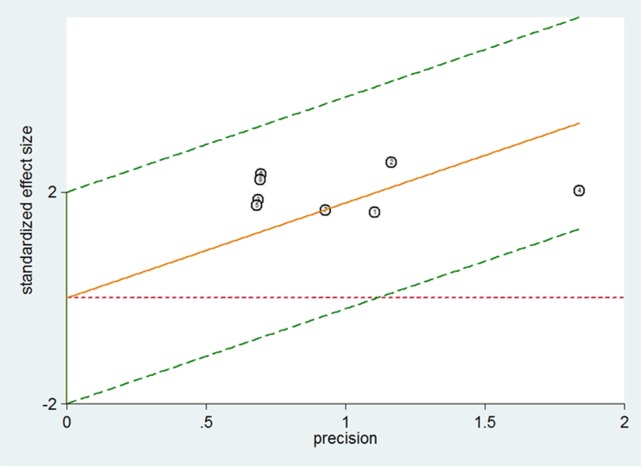
The heterogeneity among the included studies were checked with the Chi-square test. There was no significant heterogeneity in the sensitivity (χ2 = 5.66, p = 0.5796) and specificity (χ2 = 7.64, p = 0.3656) of 18F-FDG PET and PET/CT in the patient-based data (see Figure 2). The Spearman correlation coefficient was –0.327 (p = 0.429), among the eight studies with patient-based data. This result indicated that no threshold effect existed. Thus, the Mantel–Haenszel method (fixed effects model) was adopted to estimate the pooled data.
Figure 2. Galbraith plots of studies for the sensitivities of 18F-FDG PET and PET/CT in the diagnosis of PCNSL.
Diagnostic performance
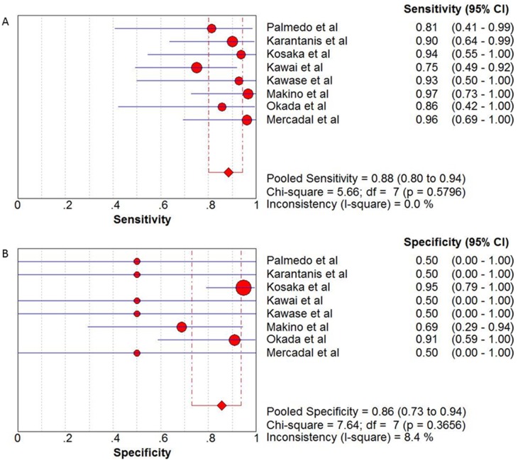
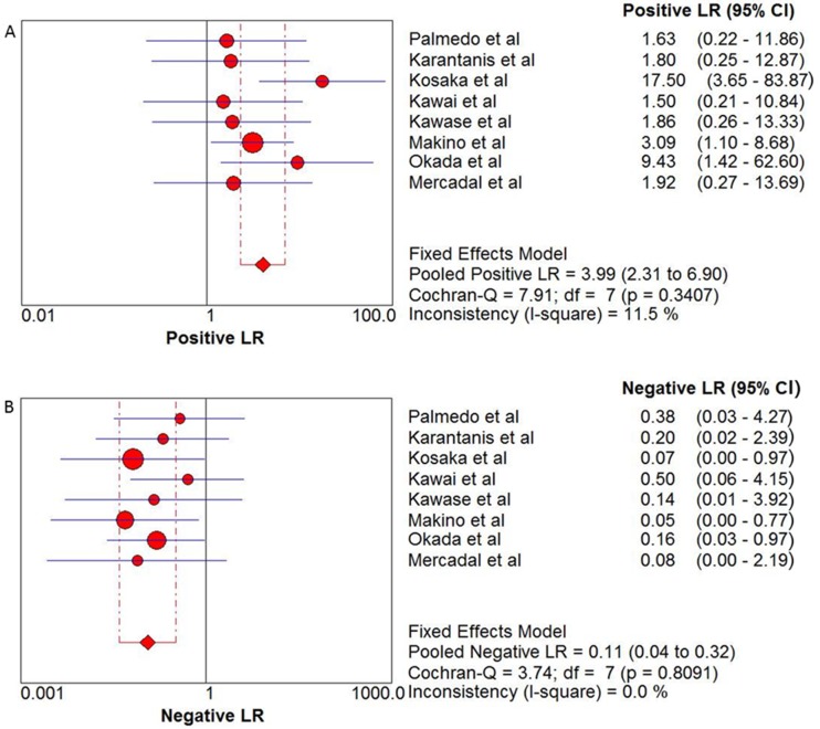
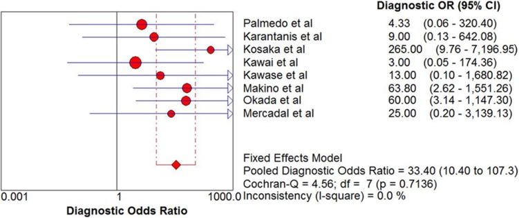
The pooled sensitivity and specificity of 18F-FDG PET and PET/CT in the diagnosis of PCNSL were 0.88 (95% CI: 0.80–0.94) and 0.86 (95% CI: 0.73–0.94), respectively (see Figure 3). In addition, the pooled positive likelihood ratio (PLR) was 3.99 (95% CI: 2.31–6.90) and negative likelihood ratio (NLR) was 0.11 (95% CI: 0.04–0.32) (see Figure 4). The pooled diagnostic odds ratio (DOR) was 33.40 (95% CI: 10.40–107.3) (see Figure 5).
Figure 3.
(A) Sensitivity and 95% confidence intervals for studies assessing the diagnostic value of 18F-FDG PET and PET/CT in patients with PCNSL. (B) Specificity and 95% confidence intervals for studies assessing the diagnostic value of 18F-FDG PET and PET/CT in patients with PCNSL. *The diamond represents the 95% CI of the pooled estimate.
Figure 4.
(A) Positive LR and 95% confidence intervals for studies assessing the diagnostic value of 18F-FDG PET and PET/CT in patients with PCNSL. (B) Negative LR and 95% confidence intervals for studies assessing the diagnostic value of 18F-FDG PET and PET/CT in patients with PCNSL. *The diamond represents the 95% CI of the pooled estimate.
Figure 5. DOR and 95% confidence intervals for studies assessing the diagnostic value of 18F-FDG PET and PET/CT in patients with PCNSL.
*The diamond represents the 95% CI of the pooled estimate.
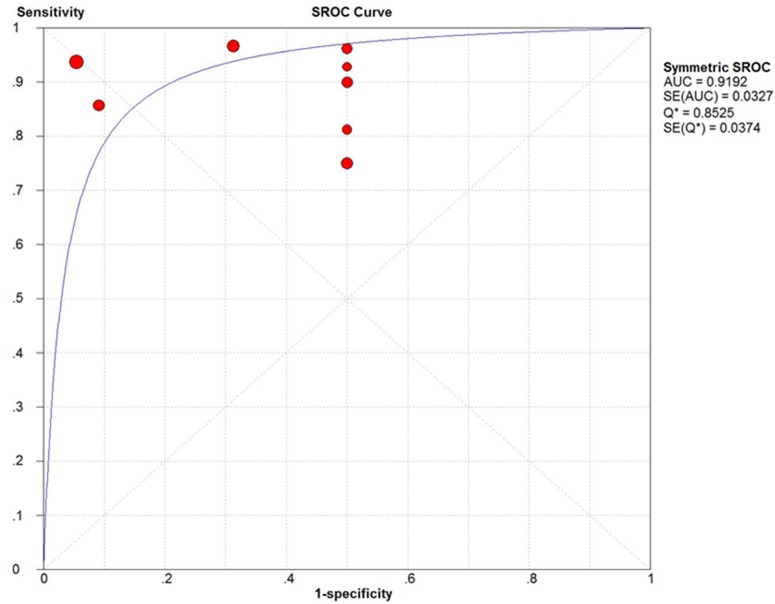
The summary receiver operating characteristic curve (SROC) and the Q index for 18F-FDG PET and PET/CT in the diagnosis of PCNSL are shown in Figure 6. The area under the curve (AUC) was 0.9192 and the Q index was 0.8525.
Figure 6. The SROC curves of 18F-FDG PET and PET/CT in the patient-based data of PCNSL.
DISCUSSION
PCNSL, an uncommon variant of extranodal non-Hodgkin lymphoma (NHL), can affect any part of the neuraxis, including the eyes, brain, leptomeninges, or spinal cord [20]. So far, PCNSL has been challenging to study, owing to the rarity of the disease. As a result, it was a tough task that to establish an effective diagnosis and treatment standard. Nevertheless, because PCNSL has highly aggressive tumors, both the successful treatment and improvement of prognosis would benefit from an early diagnosis.
Currently, MRI and CT are still the first-line imaging examinations used in the detection of brain lesions. Neuroimaging with cranial MRI using fluid-attenuated inversion recovery and T1-weighted sequences before and after contrast injection is the preferred method of choice for diagnosis and follow-up [21]. Studies have shown that typical findings in immunocompetent patients are homogenously enhancing single lesions (60–70% of cases) or multiple lesions (30–40% of cases) without necrosis and with a relatively small amount of edema [22]. In most cases, the possibility of PCNSL could be recognized by MRI based on these imaging signs. Still, glioblastoma multiforme (GBM), brain metastasis, and some non-neoplasm lesions with enhancement are the most likely misdiagnosed diseases [23–25]. In a dilemma, an accurate initial diagnosis is of vital importance because the management and prognosis of these diseases are quite at odds with each other. For instance, if the patient is highly suspected of having GBM, a craniotomy would be recommended. In contrast, for PCNSL, stereotactic biopsy is usually performed to confirm the diagnosis. Moreover, the subsequent chemotherapy standards vary for these two kinds of tumors.
In such a difficult situation, a powerful molecular imaging tool called 18F-FDG PET, which is expected to characterize the lesion on a metabolic and molecular level, is attractive. It is well known that FDG-uptake on diffuse large B-cell lymphoma is higher than that on other types of lymphoma, and this characteristic feature could be helpful in differentiating between B-cell lymphoma and other histological tumors [26]. Similarly, PET can identify degenerative diseases, multiple sclerosis, infectious diseases and cerebral infarction [27]. In recent years, the use of 18F-FDG PET and PET/CT has increased in routine practice at the time of diagnosis. Taking several studies together, this technique has a diagnostic sensitivity in PCNSL of 76%–100% [4, 28]. Maximum standardized uptake value (SUVmax) is higher in PCNSL than in gliomas [29, 30].
The first results of 18F-FDG PET in PCNSL were revealed by Rosenfeld et al. They found FDG-uptake in PCNSL lesions that were similar to lesions of anaplastic gliomas [31]. A few years afterwards, Palmedo et al. revealed that PCNSL lesions usually showed high FDG-uptake, which could be detected by 18F-FDG PET, with high sensitivity in immunocompetent patients [17]. Similarly, Kosaka et al. suggested that 18F-FDG PET had a reliable ability to differentiate between PCNSL and GBM when the cutoff value of SUVmax was 15 [16]. In agreement with the results of Kosaka et al., Makino et al. demonstrated a cutoff point of 12 [18]. However, Kawai et al. found the obvious advantage of PET to discriminate PCNSL from other tumorous and non-tumorous diseases in lesions with atypical MRI findings [19]. Although the sensitivity in the research of Kawai et al. was lower than that of the others, they noted that pretreatment FDG values could predict treatment response and tumor progression in patients with newly diagnosed PCNSL [32]. Overall, those studies were short of conviction, since the low incidence of PCNSL led to small sample size of study. The value of 18F-FDG PET and PET/CT in the diagnosis of PCNSL is still uncertain.
In this review, four studies used PET alone and three studies used PET/CT alone. Uniquely, in the study of Kosaka et al., both techniques were performed. Indeed, it should be noted that there are differences between PET and PET/CT. PET is a functional diagnostic imaging technique using compounds labeled with positron-emitting radioisotopes to measure cell metabolism [33]. In combination with CT, termed PET/CT, this method could detect lesion location more conspicuously. However, in a meta-analysis on the detection rate of 18F-FDG PET in marginal zone lymphoma of the mucosa-associated lymphoid tissue (MALT), there was no significant difference between the detection rate of PET/CT and PET alone [14]. Furthermore, we examined the heterogeneity between the eight studies that we included and no heterogeneity was found in the sensitivity and specificity of 18F-FDG PET and PET/CT in the patient-based data. Therefore, we put together two techniques in our study aiming to reveal the ability of 18F-FDG PET and PET/CT in finding disease, rather than finding the location of lesions.
In patients with PCNSL, compared with the immunocompetent population, the immunocompromised population often showed significant differences in clinical presentation, imaging feature, treatment and prognosis evaluation. Of note, in immunocompetent patients, necrosis and the resulting ring-enhancing lesions are rare. Conversely, imaging features are more variable in immunocompromised patients [34]. Additionally, in account of the better prognosis of the immunocompetent patients, our study mainly focused on assessing the value of 18F-FDG PET and PET/CT in the diagnosis of immunocompetent patients with PCNSL.
To the best of our knowledge, this is the first systematic review and meta-analysis aimed at revealing the diagnostic value of 18F-FDG PET and PET/CT in immunocompetent PCNSL patients. Overall, eight studies with 129 immunocompetent patients of PCNSL were included in the meta-analysis. In general, the results of our meta-analysis showed that 18F-FDG PET and PET/CT have a high diagnostic accuracy in patients with PCNSL. In regard to the patient-based analysis, the pooled sensitivity was 0.88 and the pooled specificity was 0.86. Moreover, the pooled PLR and NLR were 3.99 and 0.11, respectively. AUC presents the area under SROC, ranging from 0.5 to 1, which is used to evaluate the overall diagnosis effect. The larger the area is, the more powerful the detection ability of PET and PET/CT. In our meta-analysis, the AUC value was 0.9192. Q index is the point on the SROC curve at which the sensitivity and specificity are equal. Q index could assess the comprehensive diagnostic accuracy. In our meta-analysis, the Q index was 0.8525. Concerning these two parameters, our study indicated that 18F-FDG PET and PET/CT were of great value in the detecting of PCNSL in the immunocompetent population.
Indeed, it should be underlined that we included in this review the studies with patients who were suspected as PCNSL. 18F-FDG PET or PET/CT was applied to these patients as a radiological diagnostic tool. Based on the results of histopathology or clinical and radiologic follow-up, the TP, TN, FN and FP could be acquired or calculated. Unlike the ideal situation, 18F-FDG PET or PET/CT was used for PCNSL screening in the entire population. It could be attributed to that all of the included studies had adopt this criterion. In fact, the expense of 18F-FDG PET and PET/CT is great, as they are not suitable for cancer screening in the asymptomatic population. In this condition, although the specificity for diagnosing of PCNSL may be underestimated, the sensitivity would not be influenced. Still, we should have a clear understanding that PET cannot replace a surgical biopsy, which is essential for definitive diagnosis. Stereotactic biopsy is usually performed in a clinic to confirm the PCNSL diagnosis, which could compensate the lack of specificity in the initial diagnosis of 18F-FDG PET and PET/CT.
It is worth mentioning that, in regard to the pooled analysis, we only calculated the pooled sensitivity, specificity, PLR, NLR and DOR of the patient-based data (instead of the lesion-based data). This is mainly because most of the researchers of the included studies have adopted this method for data collection. Only in three studies would it have been possible to acquire the lesion-based data. The number of the lesions were 9, 16 and 34 for those three studies. The sample size is too small to conduct a pooled analysis. However, we should not ignore the potential diagnostic capability of 18F-FDG PET and PET/CT from the perspective of lesion-based analysis. It is hoped that future studies will focus on this issue.
Nevertheless, this systematic review and meta-analysis had some limitations. First, it is limited by the current existing literature. Although the diagnostic capability of 18F-FDG PET and PET/CT was well discussed in this review, it was not possible to assess the value of other aspects of disease management, such as staging, follow-up, and prognosis. Therefore, the clinical application value of 18F-FDG PET and PET/CT on PCNSL was not fully examined. Second, all of the studies were retrospective and the sample size was relatively small, which would weaken the confidence in making statistical conclusions. Further investigations were required to remedy this defect. In addition, since there were differences in observers’ experiences and the performance of instruments among the studies, potential measurement biases could exist.
Overall, based on current investigations, the findings of our meta-analysis demonstrate that 18F-FDG PET and PET/CT are valuable radiological diagnostic tools in immunocompetent PCNSL patients. They are conductive to narrowing the differential diagnosis for patients who were suspected as having PCNSL. Furthermore, they may provide useful information in addition to that obtained by MRI. We recommend 18F-FDG PET and PET/CT as appropriate choices for the routine diagnostic imaging method in PCNSL.
MATERIALS AND METHODS
Search strategy
Embase, PubMed/MEDLINE and Cochrane Library databases were searched based on the following strategy: (‘‘PET’’ OR ‘‘positron emission tomography’’) AND (‘‘primary central nervous system lymphoma’’ OR ‘‘primary CNS lymphoma’’ OR ‘‘PCNSL’’). The search was last updated on July 16th, 2016. There was no beginning date limit used. Reference lists of the included studies were also manually screened in before-mentioned databases in order to find the relevant additional study.
Study selection
The inclusion criteria were: (1) Original articles revealing the performance of 18F-FDG PET or PET/CT in the initial diagnosis of PCNSL. (2) Studies in which the patients were diagnosed as having PCNSL, which was then confirmed by histopathology or clinical and radiologic follow-up. (3) Studies in which PET or PET/CT was used as the single reference standard for PCNSL diagnosis. (4) The most recent article or article with the most comprehensive information was included if the data from same patient were used in more than one article. (5) Articles with sufficient data to acquire or calculate the TP, FP, TN and FN. (6) Articles in which the full-text version could be acquired and articles that were published in the English language.
The exclusion criteria were: (1) Articles that cannot reveal the performance of 18F-FDG PET and PET/CT in the initial diagnosis of PCNSL. (2) Publications without primary data, such as comments, letters, case reports, conference proceedings, guidelines, and reviews. (3) In vitro studies and animal experiments. (4) Articles with patients who were in a situation of immunodeficiency or immunosuppression. (5) Articles with patients with diabetes mellitus. (6) Articles with less than five PCNSL patients enrolled. (7) Articles with patients who have been treated before (including steroid, surgical resection, chemotherapy, or radiotherapy). (8) Articles in which a full-text version could not be acquired or articles published in a non-English language.
Data extraction and quality assessment
Independently, two investigators (Y.Z and J.T) reviewed the titles, abstracts and full-text articles, and extracted the data from the eligible studies. For each included study, basic information was collected concerning the study (name of the first authors, country of origin, year of publication, and study design), population characteristics of participants (number of subjects, sex and age distributions, immune states) and technical aspects of the study (imaging method used, SUVmax). In addition, the numbers of TP, FP, TN and FN findings for PET or PET/CT were recorded, as well as the method of PCNSL determination. Any disagreements would be resolved by discussion and a consensus.
The QUADAS-2 was used to estimate the quality of the included studies. This tool consists of four domains: patient selection, index test, reference standard and flow, and timing. Each domain involves the assessment of risk of bias (“low,” “high,” or “unclear”) and the applicability of diagnostic accuracy studies [35].
Statistical analysis
Based on patient or lesion, the data of SUVmax, TP, FP, TN and FN were acquired or calculated from the primary data of each included study. For patient-based data, pool estimates of sensitivity, specificity, PLR, NLR and DOR were analyzed. Owing to no threshold effect existing, the Mantel–Haenszel method was performed to estimate the pooled data, which were presented as 95% confidence intervals. The SROC analysis was performed and plotted. The related AUC value and Q index were also estimated. Statistical analyses were executed using Stata software (version 13.0, StataCorp, College Station, Texas, USA). Meta-Disc software (version 1.4, Clinical Biostatistics Ramón y Cajal Hospital, Madrid, Spain) was adopted for drawing the plots and curve. The results of the statistical analysis were considered significant when the p value was < 0.05.
Acknowledgments
This study was supported in part by grants from the National Natural Science Foundation of China (Grant No. 81670195 to Zi Chen, Grant No. 81673064 and 81472875 to Meng Pan).
Authors’ contributions
Y.Z. and J.T. performed the literature review, conceptualized and designed the data analysis, and wrote the first draft of the article. H.L. and J.J. contributed to the acquisition, analysis and interpretation of the data. M.P. and Z.C. were responsible for the study design, and they also reviewed and revised the manuscript. All authors commented on drafts of the article and have approved the final draft of the manuscript.
CONFLICTS OF INTEREST
The authors declare no conflicts of interest.
REFERENCES
- 1.Sierra del Rio M, Rousseau A, Soussain C, Ricard D, Hoang-Xuan K. Primary CNS lymphoma in immunocompetent patients. Oncologist. 2009;14:526–39. doi: 10.1634/theoncologist.2008-0236. [DOI] [PubMed] [Google Scholar]
- 2.Olson JE, Janney CA, Rao RD, Cerhan JR, Kurtin PJ, Schiff D, Kaplan RS, O’Neill BP. The continuing increase in the incidence of primary central nervous system non-Hodgkin lymphoma: a surveillance, epidemiology, and end results analysis. Cancer. 2002;95:1504–10. doi: 10.1002/cncr.10851. [DOI] [PubMed] [Google Scholar]
- 3.Kadan-Lottick NS, Maria CS, Gurney JG. Decreasing incidence rates of primary central nervous system lymphoma. Cancer. 2002;95:193–202. doi: 10.1002/cncr.10643. [DOI] [PubMed] [Google Scholar]
- 4.Kawai N, Miyake K, Yamamoto Y, Nishiyama Y, Tamiya T. 18F-FDG PET in the diagnosis and treatment of primary central nervous system lymphoma. Biomed Research International. 2013;2013:247152. doi: 10.1155/2013/247152. [DOI] [PMC free article] [PubMed] [Google Scholar]
- 5.Wöhrer A. Epidemiology and Brain Tumours: Practical Usefulness. European Association of Neurooncology Magazine. 2013:3. [Google Scholar]
- 6.Mohile P. Advances in Primary Central Nervous System Lymphoma. Current Oncology Reports. 2016;17:60. doi: 10.1007/s11912-015-0483-8. [DOI] [PubMed] [Google Scholar]
- 7.Abrey LE, Ben-Porat L, Panageas KS, Yahalom J, Berkey B, Curran W, Schultz C, Leibel S, Nelson D, Mehta M. Primary central nervous system lymphoma: the Memorial Sloan-Kettering Cancer Center prognostic model. Journal of Clinical Oncology. 2006;24:5711–5. doi: 10.1200/JCO.2006.08.2941. [DOI] [PubMed] [Google Scholar]
- 8.Maza S, Buchert R, Brenner W, Munz DL, Thiel E, Korfel A, Kiewe P. Brain and whole-body FDG-PET in diagnosis, treatment monitoring and long-term follow-up of primary CNS lymphoma. Radiology & Oncology. 2013;47:103–10. doi: 10.2478/raon-2013-0016. [DOI] [PMC free article] [PubMed] [Google Scholar]
- 9.Batchelor T, Loeffler JS. Primary CNS lymphoma. Journal of Clinical Oncology. 2006;24:1281–8. doi: 10.1200/JCO.2005.04.8819. [DOI] [PubMed] [Google Scholar]
- 10.Wilhelm K, Thomas N, Agnieska K, Stefan H, Eckhard T, Michael B, Michael W, Ulrich H. Primary central nervous system lymphomas (PCNSL): MRI features at presentation in 100 patients. Journal of neuro-oncology. 2005;72:169–77. doi: 10.1007/s11060-004-3390-7. [DOI] [PubMed] [Google Scholar]
- 11.Kimura N, Yamamoto Y, Kameyama R, Hatakeyama T, Kawai N, Nishiyama Y. Diagnostic value of kinetic analysis using dynamic 18F-FDG-PET in patients with malignant primary brain tumor. Nuclear Medicine Communications. 2009;30:602–9. doi: 10.1097/MNM.0b013e32832e1c7d. [DOI] [PubMed] [Google Scholar]
- 12.Heald AE, Hoffman JM, Bartlett JA, Waskin HA. Differentiation of central nervous system lesions in AIDS patients using positron emission tomography (PET) International Journal of Std & Aids. 1996;7:337–46. doi: 10.1258/0956462961918239. [DOI] [PubMed] [Google Scholar]
- 13.Treglia G, Cason E, Fagioli G. Recent applications of nuclear medicine in diagnostics (I part) Italian Journal of Medicine. 2013;4:159–166. doi: 10.4081/itjm.2010.84. [DOI] [Google Scholar]
- 14.Treglia G, Zucca E, Sadeghi R, Cavalli F, Giovanella L, Ceriani L. Detection rate of fluorine-18-fluorodeoxyglucose positron emission tomography in patients with marginal zone lymphoma of MALT type: a meta-analysis. Hematological Oncology. 2014;33:113–24. doi: 10.1002/hon.2152. [DOI] [PubMed] [Google Scholar]
- 15.Baraniskin A, Deckert M, Schulte-Altedorneburg G, Schlegel U, Schroers R. Current strategies in the diagnosis of diffuse large B-cell lymphoma of the central nervous system. British Journal of Haematology. 2012;156:421–32. doi: 10.1111/j.1365-2141.2011.08928.x. [DOI] [PubMed] [Google Scholar]
- 16.Kosaka N, Tsuchida T, Uematsu H, Kimura H, Okazawa H, Itoh H. 18F-FDG PET of common enhancing malignant brain tumors. Ajr American Journal of Roentgenology. 2008;190:365–9. doi: 10.2214/AJR.07.2660. [DOI] [PubMed] [Google Scholar]
- 17.Palmedo H, Urbach H, Bender H, Schlegel U, Schmidt-Wolf IGH, Matthies A, Linnebank M, Joe A, Bucerius J, Biersack HJ. FDG-PET in immunocompetent patients with primary central nervous system lymphoma: correlation with MRI and clinical follow-up. European Journal of Nuclear Medicine & Molecular Imaging. 2006;33:164–8. doi: 10.1007/s00259-005-1917-6. [DOI] [PubMed] [Google Scholar]
- 18.Makino K, Hirai T, Nakamura H, Murakami R, Kitajima M, Shigematsu Y, Nakashima R, Shiraishi S, Uetani H, Iwashita K. Does adding FDG-PET to MRI improve the differentiation between primary cerebral lymphoma and glioblastoma? Observer performance study. Annals of Nuclear Medicine. 2011;25:432–8. doi: 10.1007/s12149-011-0483-1. [DOI] [PubMed] [Google Scholar]
- 19.Kawai N, Okubo S, Miyake K, Maeda Y, Yamamoto Y, Nishiyama Y, Tamiya T. Use of PET in the diagnosis of primary CNS lymphoma in patients with atypical MR findings. Annals of Nuclear Medicine. 2010;24:335–43. doi: 10.1007/s12149-010-0356-z. [DOI] [PubMed] [Google Scholar]
- 20.Gerstner ER, Batchelor TT. Primary central nervous system lymphoma. Arch Neurol. 2010;67:291–7. doi: 10.1001/archneurol.2010.3. [DOI] [PubMed] [Google Scholar]
- 21.Hoang-Xuan K, Bessell E, Bromberg J, Hottinger AF, Preusser M, Ruda R, Schlegel U, Siegal T, Soussain C, Abacioglu U, Cassoux N, Deckert M, Dirven CM, et al. Diagnosis and treatment of primary CNS lymphoma in immunocompetent patients: guidelines from the European Association for Neuro-Oncology. Lancet Oncol. 2015;16:e322–32. doi: 10.1016/s1470-2045(15)00076-5. [DOI] [PubMed] [Google Scholar]
- 22.Korfel A, Schlegel U. Diagnosis and treatment of primary CNS lymphoma. Nat Rev Neurol. 2013;9:317–27. doi: 10.1038/nrneurol.2013.83. [DOI] [PubMed] [Google Scholar]
- 23.Kim DS, Na DG, Kim KH, Kim JH, Kim E, Yun BL, Chang KH. Distinguishing tumefactive demyelinating lesions from glioma or central nervous system lymphoma: added value of unenhanced CT compared with conventional contrast-enhanced MR imaging. Radiology. 2009;251:467–75. doi: 10.1148/radiol.2512072071. [DOI] [PubMed] [Google Scholar]
- 24.Wang S, Kim S, Chawla S, Wolf RL, Knipp DE, Vossough A, O’Rourke DM, Judy KD, Poptani H, Melhem ER. Differentiation between glioblastomas, solitary brain metastases, and primary cerebral lymphomas using diffusion tensor and dynamic susceptibility contrast-enhanced MR imaging. American Journal of Neuroradiology. 2011;32:507–14. doi: 10.3174/ajnr.A2333. [DOI] [PMC free article] [PubMed] [Google Scholar]
- 25.Ma JH, Kim HS, Rim NJ, Kim SH, Cho KG. Differentiation among glioblastoma multiforme, solitary metastatic tumor, and lymphoma using whole-tumor histogram analysis of the normalized cerebral blood volume in enhancing and perienhancing lesions. Ajnr American Journal of Neuroradiology. 2010;31:235–42. doi: 10.3174/ajnr.A2161. [DOI] [PMC free article] [PubMed] [Google Scholar]
- 26.Yamaguchi S, Hirata K, Kobayashi H, Shiga T, Manabe O, Kobayashi K, Motegi H, Terasaka S, Houkin K. The diagnostic role of (18)F-FDG PET for primary central nervous system lymphoma. Annals of Nuclear Medicine. 2014;28:603–9. doi: 10.1007/s12149-014-0851-8. [DOI] [PubMed] [Google Scholar]
- 27.Kawase Y, Yamamoto Y, Kameyama R, Kawai N, Kudomi N, Nishiyama Y. Comparison of 11C-methionine PET and 18F-FDG PET in patients with primary central nervous system lymphoma. Molecular Imaging & Biology. 2011;13:1284–9. doi: 10.1007/s11307-010-0447-1. [DOI] [PubMed] [Google Scholar]
- 28.Mercadal S, Cortésromera M, Vélez P, Climent F, Gámez C, Gonzálezbarca E. [Positron emission tomography combined with computed tomography in the initial evaluation and response assessment in primary central nervous system lymphoma]. [Article in Spanish] Medicina Clínica. 2015;144:503–6. doi: 10.1016/j.medcli.2014.09.010. [DOI] [PubMed] [Google Scholar]
- 29.Lewitschnig S, Gedela K, Toby M, Kulasegaram R, Nelson M, O’Doherty M, Cook GJ. 18F-FDG PET/CT in HIV-related central nervous system pathology. European Journal of Nuclear Medicine & Molecular Imaging. 2013;40:1420–7. doi: 10.1007/s00259-013-2448-1. [DOI] [PubMed] [Google Scholar]
- 30.Okada Y, Nihashi T, Fujii M, Kato K, Okochi Y, Ando Y, Yamashita M, Maesawa S, Takebayashi S, Wakabayashi T. Differentiation of newly diagnosed glioblastoma multiforme and intracranial diffuse large B-cell Lymphoma using (11)C-methionine and (18)F-FDG PET. Clinical Nuclear Medicine. 2012;37:843–9. doi: 10.1097/RLU.0b013e318262af48. [DOI] [PubMed] [Google Scholar]
- 31.Rosenfeld SS, Hoffman JM, Coleman RE, Glantz MJ, Hanson MW, Schold SC. Studies of primary central nervous system lymphoma with fluorine-18-fluorodeoxyglucose positron emission tomography. Journal of Nuclear Medicine. 1992;33:532–6. [PubMed] [Google Scholar]
- 32.Kawai N, Zhen HN, Miyake K, Yamamaoto Y, Nishiyama Y, Tamiya T. Prognostic value of pretreatment 18F-FDG PET in patients with primary central nervous system lymphoma: SUV-based assessment. Journal of neuro-oncology. 2010;100:225–32. doi: 10.1007/s11060-010-0182-0. [DOI] [PubMed] [Google Scholar]
- 33.Langer A. A systematic review of PET and PET/CT in oncology: a way to personalize cancer treatment in a cost-effective manner? BMC Health Serv Res. 2010;10:283. doi: 10.1186/1472-6963-10-283. [DOI] [PMC free article] [PubMed] [Google Scholar]
- 34.Tang YZ, Booth TC, Bhogal P, Malhotra A, Wilhelm T. Imaging of primary central nervous system lymphoma. Clinical Radiology. 2011;66:768–77. doi: 10.1016/j.crad.2011.03.006. [DOI] [PubMed] [Google Scholar]
- 35.Whiting PF, Rutjes AW, Westwood ME, Mallett S, Deeks JJ, Reitsma JB, Leeflang MM, Sterne JA, Bossuyt PM. QUADAS-2: a revised tool for the quality assessment of diagnostic accuracy studies. Annals of Internal Medicine. 2011;155:529–36. doi: 10.7326/0003-4819-155-8-201110180-00009. [DOI] [PubMed] [Google Scholar]
- 36.Karantanis D, O'eill BP, Subramaniam RM, Witte RJ, Mullan BP, Nathan MA, Lowe VJ, Peller PJ, Wiseman GA. 18F-FDG PET/CT in primary central nervous system lymphoma in HIV-negative patients. Nuclear Medicine Communications. 2007;28:834–41. doi: 10.1097/MNM.0b013e328264ae7f. [DOI] [PubMed] [Google Scholar]


