Abstract
β-lactam antibiotics target penicillin-binding proteins (PBPs) preventing peptidoglycan synthesis and this inhibition is circumvented in methicillin resistant Staphylococcus aureus (MRSA) strains through the expression of an additional PBP, named PBP2A. This enzyme is encoded by the mecA gene located within the Staphylococcal Chromosome Cassette mec (SCCmec) mobile genetic element, of which there are 12 types described to date. Previous investigations aimed at analysing the synergistic activity of two β-lactams, oxacillin and cefoxitin, found that SCCmec type IV community-acquired MRSA strains exhibited increased susceptibility to oxacillin in the presence of cefoxitin, while hospital-acquired MRSA strains were unaffected. However, it is not clear if these differences in β-lactam resistance are indeed a consequence of the presence of the different SCCmec types. To address this question, we have exchanged the SCCmec type I in COL (HA-MRSA) for the SCCmec type IV from MW2 (CA-MRSA). This exchange did not decrease the resistance of COL against oxacillin and cefoxitin, as observed in MW2, indicating that genetic features residing outside of the SCCmec element are likely to be responsible for the discrepancy in oxacillin and cefoxitin synergy against these MRSA strains.
Introduction
The human pathogen Staphylococcus aureus is a leading cause of infections ranging from superficial wound infections to more serious illnesses including bacteremia, endocarditis or toxic shock syndrome1. Patient treatment commonly involves the use of β-lactams, antibiotics that prevent cell wall synthesis by targetting the four S. aureus penicillin-binding proteins (PBPs) responsible for the transpeptidation of the peptidoglycan2. The use of methicillin, an early semisynthetic β-lactam, was soon followed by the emergence of methicillin resistant S. aureus (MRSA). Today, MRSA strains demonstrate resistance to multiple antibiotics and include not only hospital-acquired (HA-MRSA), but also the later emerging community-acquired (CA-MRSA) strains, which tend to be more virulent3.
The key determinant of β-lactam resistance is the expression of PBP2A, an additional PBP that has low affinity for β-lactams, thereby maintaining transpeptidation activity in the presence of otherwise lethal concentrations of these antibiotics4. PBP2A is encoded by mecA, a gene located within a mobile genetic element called the Staphylococcal Chromosome Cassette mec (SCCmec)5. To date, there are 12 SCCmec types described, varying greatly in size (~21 kb to 67 kb) and most commonly HA-MRSA strains carry SCCmec types I, II and III, while CA-MRSA strains have SCCmec types IV and V6–8. All MRSA strains possess a mec gene complex, a cassette chromosome recombinase (ccr) gene complex and junkyard or joining (J) regions8. The mec gene complex includes mecA and its regulatory genes mecI and mecR1, though depending on the SCCmec type, these regulatory genes may be disrupted by insertional inactivation sequences9. The ccr gene complex encodes site-specific recombinases responsible for the integration and excision of the SCCmec at the 3′ end of the orfX gene, referred to as the attB site10. While this site is well-defined, the mechanism of integration and excision and the acquisition of the genetic element itself are still not fully elucidated and many of its ORFs have not been well characterised. Homology to genetic regions identified in Staphylococcus sciuri, Staphylococcus fleuretti, Staphylococcus xylosus, Staphylococcus hominis, and Macrococcus caseolyticus makes them all possible SCCmec sources, and although the exact mechanism of SCCmec acquisition remains unknown, one possibility is via bacteriophage-mediated transduction11–15. S. aureus strains carry a huge array of bacteriophages, which are thought to play a key role in the transfer of DNA within the species16. In fact, it has been shown that the mec genes can be introduced into MSSA backgrounds by transduction17. However, no bacteriophages have been shown to transfer DNA between different staphylococcal species, supporting the idea of SCCmec acquisition via conjugation18.
The increase in MRSA incidence has led to a need for alternative therapies, the focus of which has been not only the identification of new antibiotics with novel killing mechanisms, but also the study of synergistic activity of currently available drugs. One such example is the use of two β–lactams, oxacillin and cefoxitin, which have highest affinity for different PBPs (PBP1/PBP2, and PBP4, respectively)19. Addition of cefoxitin reduces the minimum inhibitory concentration (MIC) of oxacillin in CA-MRSA SCCmec type IV strains MW2 and USA300, suggesting that PBP4 is required for β–lactam resistance in these strains19. Accordingly, genetic inactivation of pbpD encoding PBP4 was also found to decrease resistance to oxacillin19. Surprisingly, this effect is not observed in HA-MRSA SCCmec type I strain COL, and further blind testing of clinical isolates found that all tested type IV SCCmec strains demonstrated a synergistic oxacillin and cefoxitin inhibitory effect, while HA-MRSA strains did not19, 20. It was therefore posited that the differences in β-lactam resistance observed in CA-MRSA and HA-MRSA strains may be due to differences in the genetic composition of the SCCmec type.
In this work, we aimed to analyse the effects of exchanging the SCCmec type I of COL with type IV of MW2, in order to determine whether the resistance of HA-MRSA strain COL to the synergistic action of oxacillin and cefoxitin was specifically dependent on the type of SCCmec. The results shown here indicate that the genetic differences between SCCmec type I and type IV do not significantly alter the resistance level or the morphological response of COL to the challenge of these β-lactams, indicating that there are additional key genetic factors involved.
Materials and Methods
Bacterial strains and growth conditions
All strains used in this study are listed in Table 1. E. coli strains were grown at 37 °C in Luria-Bertani broth (LB, Difco) or on LB agar (LA, Difco) supplemented with 100 μg/ml of ampicillin (Apollo Scientific), where appropriate. S. aureus strains were grown at 37 °C or 30 °C where indicated, in tryptic soy broth (TSB, Difco) or on tryptic soy agar (TSA, Difco), with the addition of 10 μg/ml of erythromycin (Apollo Scientific) or 10 μg/ml of tetracycline (Sigma-Aldrich) when necessary.
Table 1.
Bacterial strains used in this study.
| Strain | Relevant Features | Reference |
|---|---|---|
| Plasmids | ||
| pSR | Multi-copy plasmid encoding ccrA2 and ccrB2 genes of N315; TetR | 10 |
| pMAD | E. coli/S. aureus allelic exchange vector; AmpR, EryR | 23 |
| pMAD-MW2Δspa | 600b of up- and downstream regions of the spa gene of MW2 cloned into pMAD; AmpR, EryR | This study |
| pMAD-SACOL0057 | SACOL0057 inserted between 650 bp of the up- and downstream regions of attB site of MW2 cloned into pMAD; AmpR, EryR | This study |
| Escherichia coli | ||
| DC10B | Δdcm in DH10B background; Dam methylation only; for cloning | 51 |
| Staphylococcus aureus | ||
| RN4220 | restriction-negative derivative of 8325-4 | 52 |
| COL | HA-MRSA; TetS; SCCmec type I, ST250, pbla− | 28 |
| COL-S | COL with SCCmec type I excised, pbla- | 28 |
| MW2 | CA-MRSA; SCCmec type IV, ST1, pbla+ | 53 |
| COL type IV | COL with SCCmec type IV, pbla- | This study |
| RN4220 pPBP4-J | pPBP4-J integrated into RN4220, insertionally inactivating pbpD encoding PBP4; EryR | 22 |
| COL pbpDmut | pPBP4-J integrated into COL, insertionally inactivating pbpD encoding PBP4; EryR; pbla- | This study |
| MW2 pbpDmut | pPBP4-J integrated into MW2, insertionally inactivating pbpD encoding PBP4; EryR; pbla+ | This study |
| COL type IV pbpDmut | pPBP4-J integrated into COL type IV, insertionally inactivating pbpD encoding PBP4; EryR; pbla- | This study |
| MW2-S | MW2 with SCCmec type IV excised; pbla+ | This study |
| MW2-SΔspa | MW2 with SCCmec type IV excised and deletion of spa gene; pbla+ | This study |
| MW2-SΔspa0057 | MW2 with SCCmec type IV excised, deletion of spa gene and insertion of SACOL0057 downstream of orfX; pbla+ | This study |
Strain construction
For construction of COL type IV, MW2 DNA was transferred to COL-S (lacking SCCmec) by transduction. Bacteriophage 80α was used to infect the donor strain, MW2, and the lysate was collected and sterilised by filtration. The lysate was next incubated with COL-S, the recipient strain, agitated for 20 min at 37 °C, and COL type IV was selected for on a plate containing 12 µg/ml oxacillin in the bottom 10 ml 0.3GL agar, followed by 20 ml 0.3GL agar without antibiotics (giving an average oxacillin concentration of 4 µg/ml)21. COL type IV was confirmed by next generation sequencing (NGS).
For excision of the SCCmec element of MW2, plasmid pSR was transduced from RN4220 into MW2 using bacteriophage 80α and selecting for tetracycline resistance, as previously described10. This strain was then grown in TSB and back-diluted 1:500 every 12 hrs for a total of 70 hrs, before plating on TSA. Colonies were patched on TSA as well as TSA supplemented with 4 µg/ml oxacillin to confirm the loss of SCCmec, and 10 µg/ml tetracycline to confirm the loss of plasmid pSR, before PCR confirmation (primers 11/12 for pSR; primers 13/8 for SCCmec excision) and sequencing of the SCCmec excision region.
For construction of PBP4 mutants, plasmid pPBP4-J22 was transduced from RN4220 into COL, MW2 and COL type IV using bacteriophage 80α and erythromycin selection, as previously described21, resulting in COL pbpDmut, MW2 pbpDmut and COL type IV pbpDmut, respectively.
Primers used in this study are listed in Supplementary Table S1. For construction of MW2-SΔspa, the 600 bp upstream and downstream of the spa gene were amplified from MW2 chromosomal DNA using primer pairs 1/2 and 3/4, respectively. The PCR fragments were then fused by overlap PCR using primers 1/4, resulting in a single 1.2 kb PCR fragment. Following digestion with EcoRI/BamHI, this PCR fragment was ligated to pMAD, which had been similarly digested. This plasmid, pMAD-MW2Δspa, was initially obtained in DC10B, confirmed by sequencing and electroporated into RN4220 at 30 °C (the permissive temperature), selecting for erythromycin resistance, as previously described21, 23. Bacteriophage 80α was used to transduce this plasmid into MW2-S, and integration and excision using erythromycin selection was performed as previously described21. Deletion of the spa gene was confirmed by sequencing and this strain was named MW2-SΔspa.
For construction of MW2-SΔspa0057, the 650 bp upstream and downstream of the attB site were amplified from MW2 chromosomal DNA using primer pairs 5/6 and 9/10, respectively. The 550 bp downstream of the attB site of COL (encoding SACOL0057) were amplified from COL chromosomal DNA using primer pair 7/8. The three PCR fragments were then fused by overlap PCR using primer pair 5/10. Both pMAD and the PCR product were digested with BglII/BamHI and ligated, before recovery in DC10B and confirmation by sequencing. The plasmid, named pMAD-SACOL0057, was next introduced into RN4220 at the permissive temperature (by erythromycin selection), transduced using bacteriophage 80α into MW2-SΔspa and following integration and excision, strain MW2-SΔspa0057 was obtained.
Next Generation Sequencing (NGS)
Chromosomal DNA was purified from COL and COL type IV using a standard phenol-chloroform extraction technique24. DNA was sequenced using the Illumina MiSeq system at Instituto Gulbenkian de Ciência, Oeiras, Portugal, producing 300 bp paired end reads with over 100x average coverage. The sequence reads of COL were assembled with SeqMan NGen 12 software (DNASTAR, Inc) using the COL genome (NCBI Accession NC_002951.2) as a reference. This sequence was then used as a reference for assembly of COL type IV, where single nucleotide polymorphisms (SNPs) with a consensus below 50% were excluded from SNP analysis, those with 50–85% consensus were confirmed by region specific sequencing by GATC Biotech and those above 85% were accepted.
Minimum Inhibitory Concentration (MIC) determination by Population Analysis Profiles (PAPs) and microdilution
PAPs were performed to assess the MIC of strains against oxacillin, cefoxitin and a combination of both. Overnight cultures of S. aureus strains were diluted tenfold from 10−1 to 10−7 and 10 µl of each dilution as well as the non-diluted culture was spread on a TSA plate containing twofold dilutions of 0.0625–1,024 µg/ml oxacillin (Sigma), or 1–1,024 µg/ml cefoxitin (Sigma). Plates were incubated at 37 °C for 48 hrs and colony forming units (CFU) were counted. The MIC was defined as the concentration at which growth of 99.9% of the population was inhibited, therefore causing a 3 log drop in CFU/ml. For testing of synergistic activity, twofold dilutions of oxacillin were tested in the presence of ¼ X MIC of cefoxitin (8 or 64 µg/ml), similar to previous experiments as described by Memmi et al. (2008).
MIC determination by microdilution was performed in 96-well plates, where overnight cultures were diluted to a final OD600 nm of 0.0025 (cell density of ~ 5×10⁵ CFU/ml) in wells containing twofold dilutions of the following antibiotics in TSB: 2–1,024 µg/ml cephradine (Sigma), 0.25–64 µg/ml vancomycin (Sigma), 2–1,024 µg/ml bacitracin (Sigma), 2–1,024 µg/ml D-cycloserine (Apollo Scientific), 0.25–64 µg/ml daptomycin (Cubist Pharmaceuticals) in the presence of 50 μg/ml of Ca++, 0.25–64 µg/ml chloramphenicol (Sigma), 2–1,024 µg/ml nalidixic acid (Sigma). Plates were incubated at 37 °C and assessed for growth after 48 hrs, whereby the MIC was defined as the lowest concentration of antibiotic at which growth was prevented.
Total protein extraction and western blot analysis
Extraction of proteins and western blotting were performed as previously described, with minor alterations25. In brief, S. aureus strains were grown in 50 ml TSB with and without 0.5 µg/ml oxacillin (and with 10 µg/ml erythromycin for pbpDmut strains) to an OD600 nm of 0.8. Cells were lysed using glass beads in a Fast Prep FP120 (Thermo Electro Corporation), and separated from the glass beads by centrifugation for 1 min at 4,200 rpm, before removal of unbroken cells by centrifugation for 15 min at 16,000 × g. Protein content was quantified using the BCA protein assay kit (Pierce) and 20 mg of protein extract were loaded in each well of a 10% SDS-PAGE gel and separated by 80 V for 4 hrs. Samples were transferred to a Hybond-P Polyvinylidene difluoride (PVDF) membrane (GE Healthcare) using a semidry transfer cell (Biorad), and cut along the 50 kDa marker to separate the PBP2A and MreC containing regions. Each membrane was blocked for 1 hr with 5% milk in PBST (0.5% Tween 20 in phosphate buffered saline) before incubation at 4 °C with anti-PBP2A (Slidex MRSA detection, Biomerieux) or anti-mreC antibody26 overnight. Washed membranes were then incubated for 1 h with HRP-conjugated goat anti-mouse and HRP-conjugated goat anti-rabbit secondary antibodies (GE Healthcare), respectively. Bands were visualised using the ECL Plus Western blotting detection kit (Amersham) and Chemidoc XRS + Imaging System (Biorad).
Structured Illumination Microscopy (SIM)
Overnight cultures of COL and COL type IV were back-diluted 1:200 in 20 ml TSB and grown to an OD600 nm of 0.2, before 5 ml aliquots were transferred to test tubes. Cultures were grown for a further 1 hr, agitated at 37 °C, in the presence or absence of oxacillin (256 µg/ml for COL; 512 µg/ml for COL type IV) and 1 ml of culture was then pelleted, washed in 1 ml PBS and incubated at 37 °C agitated for 5 min with 1 µg/ml Hoechst 33342 (Invitrogen), 10 µg/ml Nile Red (Invitrogen) and 0.8 µg/ml BODIPY FL conjugated vancomycin (Van-FL, Molecular Probes). Cells were then pelleted and washed with PBS before being mounted on a 1.2% PBS agarose pad and SIM imaging was performed, due to its improved resolution compared to conventional microscopy, using a Plan-Apochromat 63x/1.4 oil DIC M27 objective, in an Elyra PS.1 microscope (Zeiss) with a Pco.edge 5.5 camera. Images were acquired using five grid rotations, with grating periods of 34 µm period for 561 nm laser (100 mW), 28 µm period for 488 nm laser (100 mW) and 23 µm period for 405 nm laser (50 mW) and images were reconstructed using ZEN software (black edition, 2012, version 8.1.0.484) based on a structured illumination algorithm, using synthetic, channel specific optical transfer functions, as described previously27.
Data availability
NGS of COL type IV identified SNPs indicated in Table S2.
Results
Exchange of SCCmec Type I for Type IV in HA-MRSA strain COL
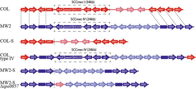
In order to investigate the specific differential roles of SCCmec types I and IV in susceptibility to the action of combined oxacillin and cefoxitin in S. aureus, it was necessary to analyse these mobile genetic elements in the same genetic background. Therefore we decided to attempt the exchange of the native SCCmec type I in COL (MIC of 512 µg/ml) for the SCCmec type IV from MW2. The SCCmec of COL had previously been excised28 and the COL-S strain (MIC of 2 µg/ml) served as the recipient strain while MW2 was the donor strain for transductions using bacteriophage 80α. Since the acquisition of type IV SCCmec could confer a low level of oxacillin resistance, selection was performed with 4 µg/ml oxacillin. We were able to obtain a COL type IV clone, which was identified by PCR and confirmed by Next Generation Sequencing (NGS). NGS showed that in addition to the exchange in SCCmec type, the immediate downstream region in strain COL type IV carried non-homologous genes mw0048-0053 and lacked sacol0057-0059 (Fig. 1). Compared to the sequenced COL genome, there were an extra 96 SNPs, all of which resided within 5 kb of the SCCmec element and were identical to the MW2 genome (see Supplementary Table S2). This suggests that the bacteriophage 80α took up 34 kb of MW2 chromosomal DNA, which encoded the SCCmec genetic element (24 kb) and approximately 10 kb of flanking regions, and that the insertion of the SCCmec sequence into COL-S may have taken place via a double crossover event (Figure 1).
Figure 1.

Schematic representation of S. aureus strains with SCCmec types I and IV. The SCCmec element and surrounding regions of strains COL, MW2, COL type IV, COL-S, MW2-S and MW2-SΔspa0057 are graphically represented here. Annotated ORFs are shown as arrows and non-annotated homologous regions are represented as boxes. The SACOL ORFs are colored in red, the MW ORFs are in blue and the SCCmec element is highlighted by a black box (not all SCCmec genes are shown). Homologous regions of COL and MW2 genomes are shown in dark colors and indicated by dotted lines, while non-homologous regions are in light colors. Next generation sequencing of COL type IV indicates a double crossover event occurring downstream of yycJ (mw0022 homologue) and upstream of sacol0063 (mw0056 homologue). Not to scale.
One additional genetic modification in the genome of COL type IV compared to COL was found to be a deletion within the map gene, encoding the Major Histocompatibility Complex Class II Analog protein (Map), also referred to as the extracellular adherence protein (Eap)29, 30. This protein is a member of the secretable expanded repertoire adhesive molecules (SERAM) family, which has been linked to roles in adherence to fibroblasts, activation of proinflammatory cytokines, and impaired wound healing in vivo 31–33. The protein shows size variability across S. aureus clinical strains, with four to six tandem repeats, and the mutation in COL type IV resulted in the deletion of one out of five tandem repeats. The map gene of COL-S was sequenced and found to also contain this deletion, indicating that this modification did not arise as a result of the acquisition of SCCmec type IV, but was already present in the recipient strain. Therefore, given that Map has not previously been associated with β-lactam resistance or SCCmec acquisition, and its natural size variation, it is highly unlikely that this deletion will affect the findings put forward in this paper.
Having constructed COL type IV, we wanted to assess the basal level of expression of PBP2A. Both type I and type IV SCCmec contain insertional inactivations within the mec inducer and repressor (mecIR) and in strain COL, this results in constitutive expression of mecA 9, 19. However, in MW2, the presence of the β-lactamase carrying plasmid, pbla, causes repression of mecA transcription in the absence of β-lactams, and the loss of this plasmid leads to constitutive mecA expression19, 34. Since COL type IV does not carry pbla (as confirmed by NGS), it was expected to have high basal levels of PBP2A expression. Western blot analysis confirmed that PBP2A levels were similar in COL type IV compared to COL and did not significantly increase following oxacillin challenge (Fig. 2). In contrast, detection of PBP2A in MW2 required the addition of oxacillin (Fig. 2), as previously shown19. These results confirmed that the type IV SCCmec does not carry mecA regulatory elements capable of repressing PBP2A expression.
Figure 2.

Western blot analysis of PBP2A expression levels. Total protein extracts of COL, COL type IV, MW2 and their PBP4 mutants grown in the presence and absence of 0.5 µg/ml oxacillin (Oxa) were separated by 10% SDS-PAGE gel and assessed for PBP2A expression using anti-PBP2A antibody (top panel). The same PVDF membrane was probed using anti-mreC antibody as a loading control (bottom panel).
Unaltered resistance of COL type IV versus COL to the synergistic effect of oxacillin and cefoxitin
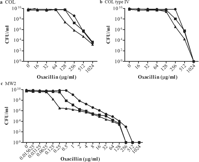
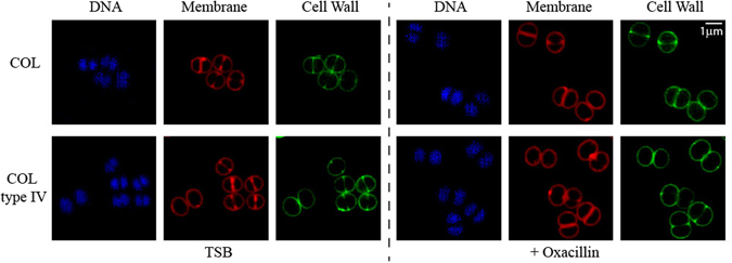
The question we aimed to answer was whether the requirement of PBP4 for β-lactam resistance observed in type IV SCCmec CA-MRSA strains was encoded in the SCCmec cassette. In other words, if the exchange of SCCmec in COL affected resistance to oxacillin in the presence of cefoxitin. To this end, the MICs of cefoxitin for MW2, COL, and COL type IV were first evaluated by a population analysis profile (PAP) method, and determined to be 32 µg/ml, 256 µg/ml and 256 µg/ml, respectively. The synergistic activity of oxacillin and cefoxitin was then determined by PAP using increasing concentrations of oxacillin in the presence and absence of ¼ X MIC of cefoxitin (Fig. 3). COL type IV resistance to oxacillin was marginally affected by the addition of cefoxitin (twofold drop), mimicking COL, while the oxacillin MIC for MW2 dropped 32-fold in the presence of cefoxitin. These results were reinforced by genetically inactivating the key target of cefoxitin, PBP4, encoded by pbpD, in strains COL, COL type IV and MW2, and only observing an appreciable drop in oxacillin resistance in the MW2 background (Fig. 3). The expression levels of PBP2A were found to be maintained in all strains irrespective of PBP4 presence (Fig. 2). We also questioned whether there was any difference in the morphological response of COL and COL type IV to antibiotic challenge. To this end, COL and COL type IV were grown to early exponential phase before exposure to oxacillin for 1 hr, and visualised by SIM, having been incubated with DNA, membrane and cell wall staining dyes. Under these conditions, no differences in the morphological response to oxacillin were observed (Fig. 4).
Figure 3.

Population Analysis Profiles of S. aureus susceptibility to oxacillin in the presence or absence of cefoxitin. Oxacillin susceptibility was tested in strains (a) COL, (b) COL type IV, and (c) MW2, shown as circles. Corresponding strains lacking PBP4 are represented as squares and strains grown in the presence of ¼ X MIC of cefoxitin are shown as triangles (64 µg/ml for COL and COL type IV; 8 µg/ml for MW2). Strains were plated on agar containing twofold dilutions of oxacillin, incubated at 37 °C for 48 h and the number of colony forming units (CFU) per ml was calculated. The experiment was performed in triplicate and a representative graph is shown here.
Figure 4.

Structured Illumination Microscopy (SIM) of COL and COL type IV in response to oxacillin challenge. COL and COL type IV were grown to early exponential phase before incubation for 1 h with oxacillin (256 µg/ml and 512 µg/ml, respectively). Cell were next incubated with Hoechst3332 (blue), Nile Red (red) and a fluorescent derivative of vancomycin (green) in order to stain the DNA, cell membrane and cell wall, respectively. Visualisation by SIM shows that the cell morphology of COL type IV is similar to COL, both in the presence and absence of oxacillin.
Together, these data demonstrate that in the background of COL, type IV SCCmec does not confer changes in resistance against oxacillin in the presence of cefoxitin compared to type I SCCmec, and in turn, suggests that genetic elements pertinent to conferring resistance to the two β-lactams’ synergy reside outside of the SCCmec region.
Having the two types of SCCmec in the same background also gave us a unique opportunity to evaluate resistance to other antibiotics. Therefore, the MICs of cell wall, cell membrane, protein and DNA synthesis targetting antibiotics were also determined. As shown in Supplementary Table S3, the exchange of SCCmec in the COL strains had a minimal effect on resistance levels, occasionally conferring twofold differences in MIC. These results indicate that the genetic differences between SCCmec type I and IV do not play a role in resistance to any of the tested antibiotics.
Exchange of SCCmec types in MW2
To support the data above, we wanted to perform the reverse SCCmec exchange and replace the SCCmec type IV of MW2 by the SCCmec type I of COL. For that purpose, we first had to construct a MW2 strain lacking its SCCmec. MW2 has previously been described as an excision deficient strain, since the overexpression of CcrAB2 proteins from SCCmec type IV strains was ineffective in MW2 SCCmec excision35. However, introduction of plasmid pSR encoding ccrAB2 from N315 (SCCmec type II) was sufficient to induce the excision of the SCCmec type IV from MW210, 28, giving rise to MW2-S. This strain was confirmed by sequencing and had an oxacillin MIC of 1 µg/ml (Fig. 1). There is the possibility that the introduction of SCCmec type I into MW2-S could eventually confer an increase in β-lactam resistance compared to the wild type strain, and so, as a safety precaution, we decided to modify this strain to reduce its virulence. To this end, we deleted the spa gene encoding the virulence factor Protein A, which has been shown to play important roles during infection in animal models36, 37. This strain, designated MW2-SΔspa, was used as the recipient strain and COL and COL pSR as the donor strains for transductions with bacteriophage 80α. However, we were not able to obtain MW2 with SCCmec type I. Since the neighbouring regions of the attB site have been implicated in affecting the rates of integration and excision of the SCCmec cassette35, 38, we decided to introduce part (650 bp) of the immediate downstream region of the COL attB site, into MW2-SΔspa, leading to strain MW2-SΔspa0057 (Fig. 1). We hoped that this modification would increase the probability of SCCmec type I homologous recombination, but we were again unable to obtain recipient colonies with increased oxacillin resistance. Lastly, electrocompetent MW2-SΔspa0057 cells were produced, as previously described21, and competence was confirmed by the introduction of a control plasmid pMAD. However, electroporation using chromosomal DNA of COL did not produce colonies following selection with 4 µg/ml oxacillin. We were therefore unable to introduce type I SCCmec into the MW2 background, despite numerous attempts.
Discussion
The rise in the incidence of drug resistant strains of S. aureus over the past few decades, has led to a renewed effort not only to identify novel antibiotics, but also novel strategies to combat infections. The use of combination therapies has posed promising treatment alternatives, whereby the individual effectiveness of two already existing drugs is enhanced by the presence of the other. Since β-lactams are very effective antibiotics, the majority of combination therapy studies focus on the identification of drugs and cellular targets that will restore β-lactam sensitivity39. The most common combination therapy involves the targetting of the resistance adaptation directly, namely through the use of β-lactamase inhibitors. β-lactamases increase β-lactam resistance by binding to and hydrolysing the drug itself, and the use of β-lactamase inhibitors effectively reduces a strains’ MIC40. However, this strategy does not overcome PBP2A-dependent resistance. Alternative approaches have been proposed, including the disruption of the synthesis of other cellular components such as the glycopolymers lipoteichoic acid (LTA) and wall teichoic acid (WTA), which play pivotal roles in stabilising the cell surface of S. aureus. Examples include krisynomycin, an inhibitor of the signal peptidase SpsB that is required for the correct processing of the lipoteichoic acid synthase, LtaS, and targocil, an inhibitor of the WTA synthesis protein, TarG, involved in the translocation of the glycopolymer across the membrane41–43. Alternatively, the combined use of β-lactams and antibiotics that inhibit the earlier steps of peptidoglycan synthesis are highly effective, since together they require the bacterium to develop two distinct resistance mechanisms simultaneously, in order to survive. Fosfomycin, targetting the first enzyme responsible for the production of the peptidoglycan subunit, uridine diphosphate-N-acetylmuramic acid, and DMPI, thought to prevent the flipping of lipid II across the membrane, are just two such examples44–46. Although the frequency of resistance has been shown to be reduced in the above examples, the ability of S. aureus to acquire new resistance mechanisms is impressive, and so it is important to fully understand the mechanism of synergistic activity.
The study of antibiotic combinations at the origin of this work demonstrated synergistic activity between the PBP4-selective cefoxitin and other β-lactams, namely oxacillin19, 20. Given that both of these antibiotics work by preventing transpeptidation through the binding of PBPs, this result highlights the differential roles of the PBPs in peptidoglycan synthesis. What was surprising was that the synergy of oxacillin and cefoxitin was evident against CA-MRSA, but it did not extend to HA-MRSA strains19. Initial data pointed towards the SCCmec type as the cause of this difference, since type IV carrying strains correlated with susceptibility to oxacillin/cefoxitin synergy, while COL, a type I SCCmec containing HA-MRSA strain, maintained resistance. We therefore wanted to exchange the SCCmec type I and type IV, in order to analyse the resistance levels to oxacillin and cefoxitin in the same genetic background, thereby ascertaining whether the SCCmec type was in fact the key determinant.
Of the 12 types of SCCmec identified so far, type IV appears to have been acquired most frequently, existing in at least three independent S. aureus backgrounds47, 48. The relatively small size (24 kb) of the SCCmec type IV may mitigate its uptake, compared to the larger SCCmec types such as type I (34 kb)48. In addition, the carriage of type I SCCmec in S. aureus has been shown to generate a fitness cost, implying that without environmental pressure, the loss of this genetic element may be desirable49.
In this work, we began by introducing the SCCmec type IV of MW2 into the preexisting COL-S strain lacking the native SCCmec type I. This was performed by transduction using bacteriophage 80α, whose capacity to take up large genetic fragments had previously been shown16. Based on NGS and SNP analysis, not only was the 24 kb SCCmec transferred to COL-S, but most likely an additional 5 kb both upstream and downstream of the chromosomal integration site in MW2 was moved, allowing for homologous recombination. Transduction of the SCCmec element between S. aureus strain has already been published by Scharn et al.50, where both SCCmec types I and IV were successfully transduced to recipients, leading to an interruption of the OrfX gene. These authors set out to establish the bacterial requirements for transduction, highlighting the need to have the same arginine catabolic metabolic element (ACME) type and the presence of a penicillinase plasmid50. However, contrary to their data50, COL type IV was attainable in the absence of a penicillinase-producing plasmid. The construction of COL type IV not only renews the idea of transduction as a possible method for SCCmec transfer between S. aureus strains, but also provides an alternative mechanism to cassette chromosome recombinases for its incorporation into the genome. SCCmec elements are known to generate circular DNA elements following excision from the chromosomal DNA, ultimately ensuring that all genes necessary for its re-integration are present during transfer2. Our work reinforces the idea that homologous recombination of large mobile genetic elements including the SCCmec may occur in nature, potentially bypassing the need for the cassette chromosome recombinases. However, transduction of DNA between staphylococcal species has not been observed, and conjugation remains a likely mechanism for interspecies DNA distribution. Ray and colleagues demonstrated not only the capture of a truncated SCCmec element on a conjugative plasmid, but also the subsequent introduction into MSSA strains, reaffirming conjugation as a plausible theory for SCCmec acquisition18.
Once DNA uptake has occurred, site-directed recombinases mediate the integration and excision of the SCCmec element at the 3′ end of the orfX gene, the rate of which is affected by the S. aureus background. For instance, MW2 along with other SCCmec type IV carrying strains were classified as being excision deficient, since introduction of the MW2 specific cassette chromosome recombinases (CcrAB2) on a plasmid could successfully excise the SCCmec from COL (type I) and N315 (type II), but surprisingly not from MW2 itself35. In this work we successfully induced the loss of the SCCmec from MW2 via the expression of N315 CcrAB2. However, despite implementing several different strategies, all attempts to introduce the SCCmec type I of COL were unsuccessful. While the difficulty in SCCmec acquisition by these strains most likely occured during the uptake of the large 34 kb region, problems during the subsequent integration process cannot be ruled out. This was observed recently in the background of the MSSA strain RN4220, where the SCCmec element was transferred by conjugation, but instead of being integrated, it was maintained in the cell as circular DNA18.
The data presented in this paper demonstrates that in the background of COL, the exchange of SCCmec type I for SCCmec type IV did not lead to susceptibility to the oxacillin in the absence of PBP4, as observed in MW2. Concordantly, the addition of cefoxitin, a β-lactam with high affinity to PBP4, led to a two-fold drop in oxacillin resistance in COL and COL type IV, in stark contrast to the 32-fold reduction in oxacillin resistance observed in MW2. Furthermore, no significant differences in resistance levels to either β-lactam and non-β-lactam antibiotics were identified between COL and COL type IV. Therefore we conclude that despite the previously observed correlation, the SCCmec element is unlikely to be the principal determinant in resistance to the synergistic activity of oxacillin and cefoxitin. Instead, this work suggests that factors residing outside of the SCCmec element of COL are key in understanding its ability to resist β-lactam synergy and their identification may facilitate the establishment of alternative drug combinations or even novel antibiotic targets.
Electronic supplementary material
Acknowledgements
We thank the following for kindly providing plasmids and strains: T. Ito and K. Hiramatsu (Juntendo University, Tokyo, Japan) for plasmid pSR, H. de Lencastre (ITQB NOVA, Oeiras, Portugal and The Rockefeller University, New York, USA) for strain COL-S and A. Cheung (Dartmouth Medical School, New Hampshire, USA) for strain MW2. This work was funded by the European Research Council through grant ERC-2012-StG-310987 (MGP) and FCT fellowship SFRH/BPD/95031/2013 (NTR).
Author Contributions
N.R. and M.G.P. designed the experiments, analysed the data and wrote the manuscript. N.R. performed all experiments.
Competing Interests
The authors declare that they have no competing interests.
Footnotes
Electronic supplementary material
Supplementary information accompanies this paper at doi:10.1038/s41598-017-06329-2
Publisher's note: Springer Nature remains neutral with regard to jurisdictional claims in published maps and institutional affiliations.
References
- 1.Lowy FD. Staphylococcus aureus infections. N. Engl. J. Med. 1998;339:520–532. doi: 10.1056/NEJM199808203390806. [DOI] [PubMed] [Google Scholar]
- 2.Peacock SJ, Paterson GK. Mechanisms of Methicillin Resistance in Staphylococcus aureus. Annu. Rev. Biochem. 2015;84:577–601. doi: 10.1146/annurev-biochem-060614-034516. [DOI] [PubMed] [Google Scholar]
- 3.Chambers HF. The changing epidemiology of Staphylococcus aureus? Emerg. Infect. Dis. 2001;7:178–182. doi: 10.3201/eid0702.010204. [DOI] [PMC free article] [PubMed] [Google Scholar]
- 4.Hartman BJ, Tomasz A. Low-affinity penicillin-binding protein associated with beta-lactam resistance in Staphylococcus aureus. J. Bacteriol. 1984;158:513–516. doi: 10.1128/jb.158.2.513-516.1984. [DOI] [PMC free article] [PubMed] [Google Scholar]
- 5.Beck WD, Berger-Bächi B, Kayser FH. Additional DNA in methicillin-resistant Staphylococcus aureus and molecular cloning of mec-specific DNA. J. Bacteriol. 1986;165:373–378. doi: 10.1128/jb.165.2.373-378.1986. [DOI] [PMC free article] [PubMed] [Google Scholar]
- 6.Batabyal B, Kundu GKR, Biswas S. Methicillin-Resistant Staphylococcus aureus: A Brief Review. Int. Res. J. Biol. Sci. 2012;1:65–71. [Google Scholar]
- 7.Wu Z, Li F, Liu D, Xue H, Zhao X. Novel Type XII Staphylococcal Cassette Chromosome mec Harboring a New Cassette Chromosome Recombinase, CcrC2. Antimicrob. Agents Chemother. 2015;59:7597–7601. doi: 10.1128/AAC.01692-15. [DOI] [PMC free article] [PubMed] [Google Scholar]
- 8.Shore AC, Coleman DC. Staphylococcal cassette chromosome mec: recent advances and new insights. Int. J. Med. Microbiol. 2013;303:350–359. doi: 10.1016/j.ijmm.2013.02.002. [DOI] [PubMed] [Google Scholar]
- 9.Deurenberg RH, et al. The molecular evolution of methicillin-resistant Staphylococcus aureus. Clin. Microbiol. Infect. 2007;13:222–235. doi: 10.1111/j.1469-0691.2006.01573.x. [DOI] [PubMed] [Google Scholar]
- 10.Katayama Y, Ito T, Hiramatsu K. A new class of genetic element, staphylococcus cassette chromosome mec, encodes methicillin resistance in Staphylococcus aureus. Antimicrob. Agents Chemother. 2000;44:1549–1555. doi: 10.1128/AAC.44.6.1549-1555.2000. [DOI] [PMC free article] [PubMed] [Google Scholar]
- 11.Couto I, et al. Ubiquitous presence of a mecA homologue in natural isolates of Staphylococcus sciuri. Microb. Drug. Resist. 1996;2:377–391. doi: 10.1089/mdr.1996.2.377. [DOI] [PubMed] [Google Scholar]
- 12.Tsubakishita S, Kuwahara-Arai K, Sasaki T, Hiramatsu K. Origin and molecular evolution of the determinant of methicillin resistance in staphylococci. Antimicrob. Agents Chemother. 2010;54:4352–4359. doi: 10.1128/AAC.00356-10. [DOI] [PMC free article] [PubMed] [Google Scholar]
- 13.Harrison EM, et al. A Staphylococcus xylosus isolate with a new mecC allotype. Antimicrob. Agents Chemother. 2013;57:1524–1528. doi: 10.1128/AAC.01882-12. [DOI] [PMC free article] [PubMed] [Google Scholar]
- 14.Bouchami O, Ben Hassen A, de Lencastre H, Miragaia M. Molecular epidemiology of methicillin-resistant Staphylococcus hominis (MRSHo): low clonality and reservoirs of SCCmec structural elements. PLoS One. 2011;6:e21940. doi: 10.1371/journal.pone.0021940. [DOI] [PMC free article] [PubMed] [Google Scholar]
- 15.Tsubakishita S, Kuwahara-Arai K, Baba T, Hiramatsu K. Staphylococcal cassette chromosome mec-like element in Macrococcus caseolyticus. Antimicrob. Agents Chemother. 2010;54:1469–1475. doi: 10.1128/AAC.00575-09. [DOI] [PMC free article] [PubMed] [Google Scholar]
- 16.Mašlaňová I, et al. Bacteriophages of Staphylococcus aureus efficiently package various bacterial genes and mobile genetic elements including SCCmec with different frequencies. Environ. Microbiol. Rep. 2013;5:66–73. doi: 10.1111/j.1758-2229.2012.00378.x. [DOI] [PubMed] [Google Scholar]
- 17.Cohen S, Sweeney HM. Effect of the prophage and penicillinase plasmid of the recipient strain upon the transduction and the stability of methicillin resistance in Staphylococcus aureus. J. Bacteriol. 1973;116:803–811. doi: 10.1128/jb.116.2.803-811.1973. [DOI] [PMC free article] [PubMed] [Google Scholar]
- 18.Ray MD, Boundy S, Archer GL. Transfer of the methicillin resistance genomic island among staphylococci by conjugation. Mol. Microbiol. 2016;100:675–685. doi: 10.1111/mmi.13340. [DOI] [PMC free article] [PubMed] [Google Scholar]
- 19.Memmi G, Filipe SR, Pinho MG, Fu Z, Cheung A. Staphylococcus aureus PBP4 is essential for beta-lactam resistance in community-acquired methicillin-resistant strains. Antimicrob. Agents Chemother. 2008;52:3955–3966. doi: 10.1128/AAC.00049-08. [DOI] [PMC free article] [PubMed] [Google Scholar]
- 20.Banerjee R, et al. Combinations of cefoxitin plus other β-lactams are synergistic in vitro against community associated methicillin-resistant Staphylococcus aureus. Eur. J. Clin. Microbiol. Infect. Dis. 2013;32:827–833. doi: 10.1007/s10096-013-1817-9. [DOI] [PubMed] [Google Scholar]
- 21.Veiga H, Pinho MG. Inactivation of the SauI type I restriction-modification system is not sufficient to generate Staphylococcus aureus strains capable of efficiently accepting foreign DNA. Appl. Environ. Microbiol. 2009;75:3034–3038. doi: 10.1128/AEM.01862-08. [DOI] [PMC free article] [PubMed] [Google Scholar]
- 22.Łeski TA, Tomasz A. Role of penicillin-binding protein 2 (PBP2) in the antibiotic susceptibility and cell wall cross-linking of Staphylococcus aureus: evidence for the cooperative functioning of PBP2, PBP4, and PBP2A. J. Bacteriol. 2005;187:1815–1824. doi: 10.1128/JB.187.5.1815-1824.2005. [DOI] [PMC free article] [PubMed] [Google Scholar]
- 23.Arnaud M, Chastanet A, Débarbouillé M. New vector for efficient allelic replacement in naturally nontransformable, low-GC-content, gram-positive bacteria. Appl. Environ. Microbiol. 2004;70:6887–6891. doi: 10.1128/AEM.70.11.6887-6891.2004. [DOI] [PMC free article] [PubMed] [Google Scholar]
- 24.Sambrook, J., Fritsch, E. & Maniatis, T. Molecular cloning: a laboratory manual. 2nd edn, (Cold Spring Harbor Laboratory Press, 1989).
- 25.Pereira AR, Reed P, Veiga H, Pinho MG. The Holliday junction resolvase RecU is required for chromosome segregation and DNA damage repair in Staphylococcus aureus. BMC Microbiol. 2013;13:18. doi: 10.1186/1471-2180-13-18. [DOI] [PMC free article] [PubMed] [Google Scholar]
- 26.Tavares AC, Fernandes PB, Carballido-López R, Pinho MG. MreC and MreD Proteins Are Not Required for Growth of Staphylococcus aureus. PLoS One. 2015;10:e0140523. doi: 10.1371/journal.pone.0140523. [DOI] [PMC free article] [PubMed] [Google Scholar]
- 27.Monteiro JM, et al. Cell shape dynamics during the staphylococcal cell cycle. Nat. Commun. 2015;6:8055. doi: 10.1038/ncomms9055. [DOI] [PMC free article] [PubMed] [Google Scholar]
- 28.Pereira SF, Henriques AO, Pinho MG, de Lencastre H, Tomasz A. Role of PBP1 in cell division of Staphylococcus aureus. J. Bacteriol. 2007;189:3525–3531. doi: 10.1128/JB.00044-07. [DOI] [PMC free article] [PubMed] [Google Scholar]
- 29.Jönsson K, McDevitt D, McGavin MH, Patti JM, Höök M. Staphylococcus aureus expresses a major histocompatibility complex class II analog. J. Biol. Chem. 1995;270:21457–21460. doi: 10.1074/jbc.270.37.21457. [DOI] [PubMed] [Google Scholar]
- 30.Hussain M, Becker K, von Eiff C, Peters G, Herrmann M. Analogs of Eap protein are conserved and prevalent in clinical Staphylococcus aureus isolates. Clin. Diagn. Lab. Immunol. 2001;8:1271–1276. doi: 10.1128/CDLI.8.6.1271-1276.2001. [DOI] [PMC free article] [PubMed] [Google Scholar]
- 31.Hussain M, et al. Insertional inactivation of Eap in Staphylococcus aureus strain Newman confers reduced staphylococcal binding to fibroblasts. Infect. Immun. 2002;70:2933–2940. doi: 10.1128/IAI.70.6.2933-2940.2002. [DOI] [PMC free article] [PubMed] [Google Scholar]
- 32.Scriba TJ, et al. The Staphyloccous aureus Eap protein activates expression of proinflammatory cytokines. Infect. Immun. 2008;76:2164–2168. doi: 10.1128/IAI.01699-07. [DOI] [PMC free article] [PubMed] [Google Scholar]
- 33.Athanasopoulos AN, et al. The extracellular adherence protein (Eap) of Staphylococcus aureus inhibits wound healing by interfering with host defense and repair mechanisms. Blood. 2006;107:2720–2727. doi: 10.1182/blood-2005-08-3140. [DOI] [PMC free article] [PubMed] [Google Scholar]
- 34.Hackbarth CJ, Chambers HF. blaI and blaR1 regulate beta-lactamase and PBP 2a production in methicillin-resistant Staphylococcus aureus. Antimicrob. Agents Chemother. 1993;37:1144–1149. doi: 10.1128/AAC.37.5.1144. [DOI] [PMC free article] [PubMed] [Google Scholar]
- 35.Noto MJ, Archer GL. A subset of Staphylococcus aureus strains harboring staphylococcal cassette chromosome mec (SCCmec) type IV is deficient in CcrAB-mediated SCCmec excision. Antimicrob. Agents Chemother. 2006;50:2782–2788. doi: 10.1128/AAC.00032-06. [DOI] [PMC free article] [PubMed] [Google Scholar]
- 36.Patel AH, Nowlan P, Weavers ED, Foster T. Virulence of protein A-deficient and alpha-toxin-deficient mutants of Staphylococcus aureus isolated by allele replacement. Infect. Immun. 1987;55:3103–3110. doi: 10.1128/iai.55.12.3103-3110.1987. [DOI] [PMC free article] [PubMed] [Google Scholar]
- 37.Palmqvist N, Foster T, Tarkowski A, Josefsson E. Protein A is a virulence factor in Staphylococcus aureus arthritis and septic death. Microb. Pathog. 2002;33:239–249. doi: 10.1006/mpat.2002.0533. [DOI] [PubMed] [Google Scholar]
- 38.Noto MJ, Kreiswirth BN, Monk AB, Archer GL. Gene acquisition at the insertion site for SCCmec, the genomic island conferring methicillin resistance in Staphylococcus aureus. J. Bacteriol. 2008;190:1276–1283. doi: 10.1128/JB.01128-07. [DOI] [PMC free article] [PubMed] [Google Scholar]
- 39.Roemer T, Schneider T, Pinho MG. Auxiliary factors: a chink in the armor of MRSA resistance to β-lactam antibiotics. Curr. Opin. Microbiol. 2013;16:538–548. doi: 10.1016/j.mib.2013.06.012. [DOI] [PubMed] [Google Scholar]
- 40.Drawz SM, Bonomo RA. Three decades of beta-lactamase inhibitors. Clin. Microbiol. Rev. 2010;23:160–201. doi: 10.1128/CMR.00037-09. [DOI] [PMC free article] [PubMed] [Google Scholar]
- 41.Therien AG, et al. Broadening the spectrum of β-lactam antibiotics through inhibition of signal peptidase type I. Antimicrob. Agents Chemother. 2012;56:4662–4670. doi: 10.1128/AAC.00726-12. [DOI] [PMC free article] [PubMed] [Google Scholar]
- 42.Wang H, et al. Discovery of wall teichoic acid inhibitors as potential anti-MRSA β-lactam combination agents. Chem. Biol. 2013;20:272–284. doi: 10.1016/j.chembiol.2012.11.013. [DOI] [PMC free article] [PubMed] [Google Scholar]
- 43.Farha MA, et al. Inhibition of WTA synthesis blocks the cooperative action of PBPs and sensitizes MRSA to β-lactams. ACS Chem. Biol. 2013;8:226–233. doi: 10.1021/cb300413m. [DOI] [PMC free article] [PubMed] [Google Scholar]
- 44.Blake KL, et al. The nature of Staphylococcus aureus MurA and MurZ and approaches for detection of peptidoglycan biosynthesis inhibitors. Mol. Microbiol. 2009;72:335–343. doi: 10.1111/j.1365-2958.2009.06648.x. [DOI] [PubMed] [Google Scholar]
- 45.Komatsuzawa H, Suzuki J, Sugai M, Miyake Y, Suginaka H. Effect of combination of oxacillin and non-beta-lactam antibiotics on methicillin-resistant Staphylococcus aureus. J. Antimicrob. Chemother. 1994;33:1155–1163. doi: 10.1093/jac/33.6.1155. [DOI] [PubMed] [Google Scholar]
- 46.Huber J, et al. Chemical genetic identification of peptidoglycan inhibitors potentiating carbapenem activity against methicillin-resistant Staphylococcus aureus. Chem. Biol. 2009;16:837–848. doi: 10.1016/j.chembiol.2009.05.012. [DOI] [PubMed] [Google Scholar]
- 47.Hanssen AM, Ericson Sollid JU. SCCmec in staphylococci: genes on the move. FEMS Immunol. Med. Microbiol. 2006;46:8–20. doi: 10.1111/j.1574-695X.2005.00009.x. [DOI] [PubMed] [Google Scholar]
- 48.Daum RS, et al. A novel methicillin-resistance cassette in community-acquired methicillin-resistant Staphylococcus aureus isolates of diverse genetic backgrounds. J. Infect. Dis. 2002;186:1344–1347. doi: 10.1086/344326. [DOI] [PubMed] [Google Scholar]
- 49.Ender M, McCallum N, Adhikari R, Berger-Bächi B. Fitness cost of SCCmec and methicillin resistance levels in Staphylococcus aureus. Antimicrob. Agents Chemother. 2004;48:2295–2297. doi: 10.1128/AAC.48.6.2295-2297.2004. [DOI] [PMC free article] [PubMed] [Google Scholar]
- 50.Scharn CR, Tenover FC, Goering RV. Transduction of staphylococcal cassette chromosome mec elements between strains of Staphylococcus aureus. Antimicrob. Agents Chemother. 2013;57:5233–5238. doi: 10.1128/AAC.01058-13. [DOI] [PMC free article] [PubMed] [Google Scholar]
- 51.Monk, I. R., Shah, I. M., Xu, M., Tan, M. W. & Foster, T. J. Transforming the untransformable: application of direct transformation to manipulate genetically Staphylococcus aureus and Staphylococcus epidermidis. MBio3, doi:10.1128/mBio.00277-11 (2012). [DOI] [PMC free article] [PubMed]
- 52.Kreiswirth BN, et al. The toxic shock syndrome exotoxin structural gene is not detectably transmitted by a prophage. Nature. 1983;305:709–712. doi: 10.1038/305709a0. [DOI] [PubMed] [Google Scholar]
- 53.Baba T, et al. Genome and virulence determinants of high virulence community-acquired MRSA. Lancet. 2002;359:1819–1827. doi: 10.1016/S0140-6736(02)08713-5. [DOI] [PubMed] [Google Scholar]
Associated Data
This section collects any data citations, data availability statements, or supplementary materials included in this article.
Supplementary Materials
Data Availability Statement
NGS of COL type IV identified SNPs indicated in Table S2.


