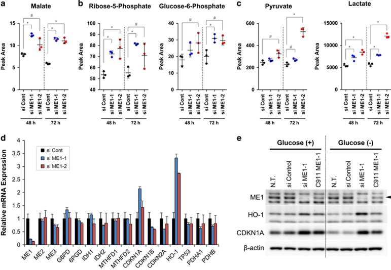
Figure 4.
ME1 knockdown disturbs metabolic homeostasis and induces oxidative stress response in cancer cells. (a–c) Metabolomics analysis was conducted 48 and 72 h after ME1 siRNAs transfection in HCT116 cells. Peak area of malate, ribose-5-phosphate, glucose-6-phosphate, pyruvate and lactate are shown in the dot graph (n=3, t-test; *P<0.01; #P<0.05). (d) Gene expressions of NADPH-producing enzymes, p53-related enzymes, a redox-related gene, and glycolysis-relating enzymes were detected by TaqMan PCR 48 h after ME1 siRNAs transfection in PC3 cells (n=3). (e) ME1, HO-1 and β-actin were detected by western blotting 96 h after control siRNA, ME1 siRNAs, or ME1 C911 transfection in HCT116 cells (ME1 is indicated by a black arrow). After siRNA transfection, cells were cultured in 2 g/l glucose McCoy's 5A media for 72 h, cultured in media with/without glucose for further 24 h, and then the cell lysate was collected for western blotting.

