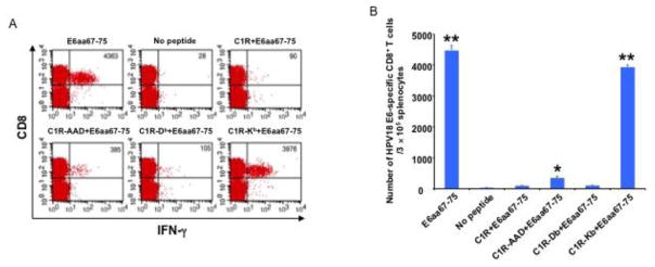
Figure 3. Characterization of MHC class I restriction element for HPV18 E6aa67-75 peptide recognized by CD8+ T cells after DNA vaccination in HLA-A*0201 transgenic C57BL/6 mouse.
5~8 weeks old female HLA-A*0201 transgenic C57BL/6 mice were vaccinated with 50 μg/mouse of pcDNA3-HPV18-E6 DNA through IM injection followed by electroporation. The mice were boosted with the same regimen three times with 4-day interval. 7 days after the last vaccination, splenocytes were harvested from the mice, stimulated with either HPV18 E6aa67-75 peptide (1 μg/ml), or irradiated HPV18 E6aa67-75 peptide loaded C1R, or C1R-AAD, or C1R-Db, or C1R-Kb cells at the presence of GolgiPlug (1 μl/ml) overnight at 37°C. The cells were then collected, washed with PBS+0.5% BSA, surface stained with PE-conjugated anti-mouse CD8a antibody. After wash, the cells were then permeabilized, fixed and intracellularly stained with FITC-conjugated anti-mouse IFN-γ antibody. The cells were acquired with FACSCalibur flow cytometer and analyzed with CellQuest software. A. Representative flow cytometry data. B. Bar graph summary of the flow cytometry data. Data are represented as mean ± SD. p-values were calculated by 2-tailed Student’s t test. * = p<0.05; ** = p<0.01.

