To the Editor
Many studies reporting local recurrence (LR) rates following surgical treatment of lentigo maligna (LM) and lentigo maligna melanoma (LMM) have limited follow-up data and thus the true time to LR is not known.1 The objective of this study was to report long-term follow up of a previously reported cohort of LM and LMM treated with margin-controlled staged excision2 to assess time to LR using a standard definition for LR.
A retrospective chart review was performed for all patients with staged excision and LM/LMM less than 1 mm Breslow depth from May 2000 to November 2006 following institutional review board approval. An institutional cancer database was searched for patient status and last dermatology follow-up date. Follow-up was calculated from surgery date to date of melanoma recurrence, last recorded dermatology visit, or patient death. In addition, each medical record was reviewed for evidence of recurrence. For any patient with subsequent melanoma, all operative reports, pathology slides, and preoperative and postoperative photographs were reviewed by 2 dermatologic surgeons and a dermatopathologist to determine recurrence status. LR was defined as appearance of tumor adjacent to or within the surgical scar, within the area of the initial surgical defect. All suspected recurrences were histologically proven and adequacy of initial excision was confirmed by slide review.
Mean follow-up was 5 years (range 0 to 12 years) with 2 patients lost to follow-up and 22 patients (18.8%) deceased. Of 115 patients with 117 LM/LMM (95% in head and neck location), mean patient age was 66.6 years (range 29 to 88 years) with 61.5% being men. One hundred lesions were primary (85.5%), 9 (7.7%) lesions were recurrent after previous surgical treatment, and 8 (6.8%) lesions were incompletely excised. Seventy-six (65%) were in situ melanoma and 41 (35%) were invasive with mean depth of 0.36 mm. The mean margin excised was 7.1 mm for LM (range 5 to 18 mm) and 10.3 mm for LMM (range 7 to 34 mm), with a mean of 1.67 stages required to excise a lesion.2
Four of 100 primary tumors developed local recurrence (Table I). Two lesions were in situ and 2 were invasive melanoma (Breslow depth 0.1 mm and 0.35 mm) at time of diagnosis (Fig 1). No difference in distribution of recurrence was observed by anatomic location (Fisher’s exact P value = .59). The mean time to LR for primary lesions was 5.9 years (range 1.9 to 8.9 years). Two of 17 patients with previously treated melanomas developed LR at 2.6 and 4.4 years.
Table I.
Local recurrence of primary lentigo maligna
| Site | Depth | Time to first recurrence (months) |
|---|---|---|
| Left ear | 0.35 mm at staged excision | 23 |
| In situ LM at first recurrence (treated by staged excision) | ||
| In situ LM at second recurrence (treated by staged excision) | ||
| Left cheek | In situ LM at staged excision | 55 |
| In situ LM at recurrence | ||
| Right cheek | In situ LM at staged excision | 98 |
| 0.58 mm at recurrence | ||
| Left neck | 0.1 mm at staged excision | 107 |
| 0.8 mm at first recurrence (treated with wide local excision by surgical service) | ||
| 0.6 mm at second recurrence (treated with wide local excision by surgical service) |
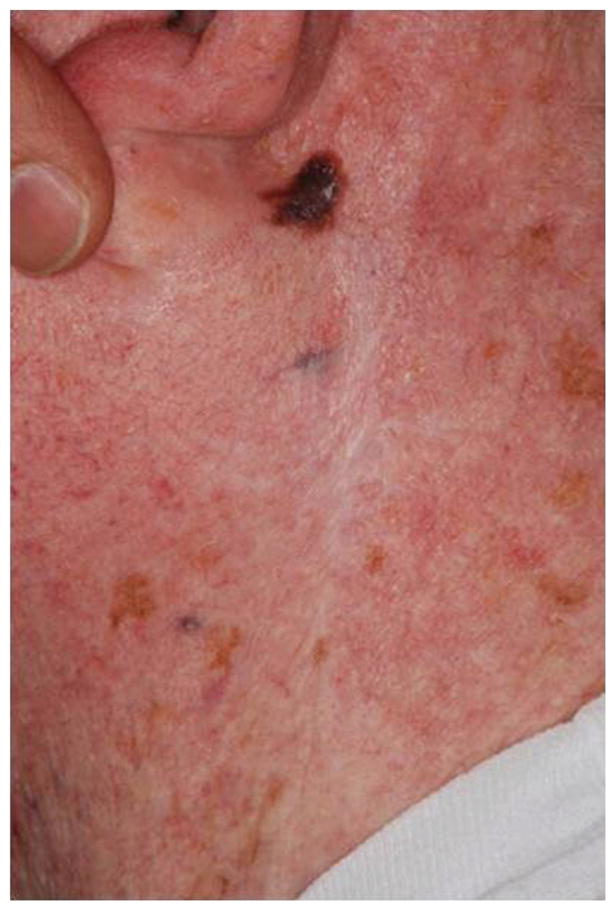
Fig 1.

Recurrent LM along a prior surgical scar, 7 years following initial treatment. The initial lesion was microinvasive with a Breslow depth of 0.1 mm. The recurrence had a depth of 0.8 mm.
Eighteen patients died of unrelated causes. Metastatic melanoma developed in 4 patients. Two patients had prior T2 and T4 invasive melanomas and metastasis was related to these sites. Metastatic spindle cell melanoma of unknown primary developed in 1 patient. One patient was upstaged at time of excision to 2.2 mm melanoma and distant metastasis developed.
In summary, given mean time to LR of 5.9 years for primary LM, follow-up of less than 5 years is inadequate for patients with LM or LMM following surgical treatment. This study may have underestimated the rate of recurrence of LM/LMM, given the mean follow-up of 5 years. Long-term follow-up and time to local recurrence should be considered in future studies evaluating efficacy of therapeutic interventions.
Acknowledgments
Funding sources: None.
Footnotes
Conflicts of interest: None declared.
References
- 1.McLeod M, Choudhary S, Giannakakis G, Nouri K. Surgical treatments for lentigo maligna: a review. Dermatol Surg. 2011;37:1210–1228. doi: 10.1111/j.1524-4725.2011.02042.x. [DOI] [PubMed] [Google Scholar]
- 2.Hazan C, Dusza SW, Delgado R, Busam KJ, Halpern AC, Nehal KS. Staged excision for lentigo maligna and lentigo maligna melanoma: a retrospective analysis of 117 cases. J Am Acad Dermatol. 2008;58:142–148. doi: 10.1016/j.jaad.2007.09.023. http://dx.doi.org/10.1016/j.jaad.2016.02.1150. [DOI] [PubMed] [Google Scholar]


