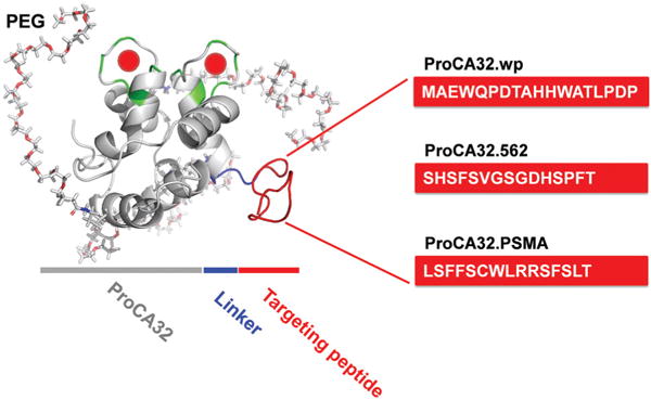
Fig. 1.

Model structure of PSMA-targeted protein MRI contrast agents for the molecular imaging of PSMA in prostate cancer. Each PSMA targeting peptide (red) was fused at the C-terminal of PEGylated ProCA32 (gray) using flexible peptide linkers (blue).
