

Abstract
A study of 186 infertile women was carried out to evaluate the effect of clomiphene citrate on the follicular dynamics. All patients underwent transvaginal sonography over at least 02 consecutive cycles-spontaneous followed by clomiphene stimulated cycle. Clomiphene citrate resulted in higher average follicular diameter on all corresponding days as compared to spontaneous cycles. Multifollicular response was common in clomiphene stimulated cycles.
KEY WORDS: Clomiphene citrate, Follicular dynamics, Infertility, Transvaginal sonography
Introduction
Ovulation is a crucial event in the reproductive cycle of women. Disorders of follicular growth and ovulation are important factors in the etiology of infertility in about 25% of infertile women [1]. Prior to the advent of ultrasound only presumptive evidence of follicular development and ovulation was available. Hackeloer, [2] in 1978, was the first to demonstrate that the developing graffian follicle in the ovary can be monitored in the normal menstrual cycle. Subsequently this technique has been applied to monitor follicular dynamics in stimulated cycles also [3, 4, 5]. Since then ultrasonography has become an essential diagnostic tool in investigating follicular development process and ovulation.
In early eighties a new perspective was offered by the advent of transvaginal sonography (TVS), which allows visualisation of finer ovarian detail. Follicle as small as 2–3 mm can be visualised with TVS. The major advantages of transvaginal ultrasound scanning (TVS) include a more precise localisation and visualisation of the ovary allowing early follicles to be examined and followed throughout the cycle. Full bladder is not necessary with TVS and hence the procedure is more comfortable, easy to schedule technique. High frequency probe used in TVS provides better axial and lateral resolution [6, 7, 8, 9, 10].
The effect of clomiphene citrate (CC) on follicular growth remains debatable. While there are studies reporting larger follicle diameters in CC stimulated cycles [3, 11, 12, 13]; others reported no difference in follicular growth between spontaneous and CC stimulated cycle [14, 15]. The present study is aimed at transvaginal sonographic evaluation of follicular dynamics in spontaneous and clomiphene citrate (CC) stimulated cycles, in the same patients, at a service hospital.
Material and Methods
Between Feb 1998 and Feb 1999, a total of 186 infertile women underwent transvaginal ultrasound examinations as part of their evaluation at our centre. The patients evaluated by TVS were either for intra uterine insemination or timed intercourse. All the ultrasound studies were performed with a 6.5 Mhz real time electronic transducer with 120 scanning angle. Each subject was studied first during a spontaneous cycle and then during a CC stimulated cycle. The stimulation protocol included clomiphene citrate in a dosage of 100 mg/day from day 5 to day 9 (1st day of menstruation being day 1). Folliculometry was done on alternate days from D10 till leading follicular diameter reached a size of 16 mm. Subsequently daily monitoring was done till sonographic evidence of ovulation took place. Each patien was explained briefly the simplicity and accuracy of TVS. Immediately before each examination patients were instructed to empty their bladder. Scanning was done in dorsolithotomy position. Ovaries were located taking iliac vessels as landmarks. Follicles were identified as circular or elliptical echofree structures within the ovaries. Number of follicles were assessed by scanning each ovary from medial to lateral border. Maximum follicular diameter was measured on the inner wall. Follicular diameter was the mean of diameters measures in two planes perpendicular to each other. Ovulation was inferred sonologically by a) disappearance of leading follicle, (b) of mature follicle with development of internal echoes (c) presence of fluid in pouch of Douglas (d) occurrence of ovarian hyperstimulation. One or more of these end points were looked for inferring ovulation. Hyper stimulation was judged to have occurred if cystic ovarian enlargement >5cm in diameter had occurred [1].
After completion of each case examining sonologist recorded the above mentioned data. Retrospective evaluation of the findings were done by sonologists who were blinded to the patients clinical status and the clinical outcome of a particular cycle.
Exclusion criteria: a) Inadequate clinical/sonographic records b) technically inadequate sonograms c) stimulation protocols using gonadotrophins d)ovulation induction with human chorionic gonadotrophins. Statistical significance was compared by means of χ2 test. A ‘p’ value <0.05 was considered significant.
Results
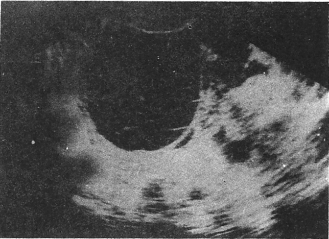
A total of 186 women underwent TVS study in 478 cycles. This included 207 spontaneous cycles and 271 clomiphene citrate (CC) stimulated cycles. Mean age of patients in our study was 27 years ranging from 20–42 years of age. There was a tendency of longer duration of menstrual cycle in CC stimulated cycles. This was noted to be primarily due to an increase in the proliferative phase of the CC stimulated cycles, as compared to spontaneous cycles. Nearly all spontaneous cycles were monofollicular (Fig-1). Multifollicular response was noted in 52% of stimulated cycles (Fig-2). Leading follicular diameter increased steadily in both the groups reaching maximum on the day prior to ovulation. The mean leading follicular diameter was significantly greater in CC stimulated cycles as compared with sponteneous cycles at all points studied. The mean pre-ovulatory diameter in spontaneous cycles was 20mm (Range 15 to 29 mm) and in CC cycles 24 mm (range 16 to 32 mm). The average rate of follicular growth was higher in CC stimulated cycles (2.1 mm/day) as compared to spontaneous cycles (1.9 mm/day). Follicular growth rate was found to be faster in the late proliferative phase in both the groups. There was a tendency towards plateuing of follicular growth immediately before ovulation. Ultrasonic evidence of polycystic ovarian disease, (PCOD) (Fig-3,4) (ovarian enlargement, multiple small subcapsular cysts, increase in cortical stroma) was noted in 33 (17.7%) cases. Sonological evidence of ovarian hyperstimulation syndrome (OHS) was found in 5 (1.84%) cycles (Fig-5). Out of the 5 cases of hyperstimulation, 4 were cases of PCOD. Poor follicular development inspite of stimulation was noted in 19 (7%) cycles. Haemorrhagic follicle (Fig-6) was noted in 22 (8%) of the stimulated cycles, while none was observed in spontaneous cycles. Multiple follicular rupture was observed in 40 (15%) stimulated cycles.
Fig. 1.

Single preovulatory follicle in spontaneous cycle.
Fig. 2.

Multi follicular response in stimulated cycle
Figs. 3 & 4.

Polycystic ovarian disease – note “necklace sign” due to small subcapsular cysts.
Fig. 5.

Ovarian hyperstimulation syndrome
Fig. 6.
Hemorrhagic follice
Discussion
Disorders of ovulation are present in about 25% of infertile women [1]. In many women in this group some form of ovulation induction would be necessary and the timings of ovulation may be critical. Transvaginal ultrasound plays an increasingly important role in monitoring follicular dynamics both in spontaneous and induced cycles. TVS has improved pregnancy rates by (a) distinguishing between presence of one or more mature follicles as opposed to a group of immature follicles, (b) suggesting possibility of multiple ovulations/risks of hyperstimulation, (c) indicating optimum time for hCG administration (d) detection and confirmation of follicular rupture, (e) indicating optimum time for oocyte retrieval in IVF programmes and (f) assessing pelvic pathologic conditions.
The present study confirms that TVS permits accurate determinations of follicular number and size throughout menstrual cycle. Leading follicular diameter has been found to be significantly larger in CC stimulated cycles at all points in this study; confirming earlier reports [3, 11, 12, 13]. However, there are studies which did not find any significant difference in the leading follicular diameter between the spontaneous and CC stimulated cycles [14, 15]. The range of follicular diameter at which ovulation occurred was more or less same in both the groups confirming earlier studies [3, 13, 14]. The mean length of the proliferative phase was longer in CC stimulated cycles as compared to spontaneous cycles. This finding is in agreement with the results of prolonged proliferative phase in the CC cycles[13]. In this study, we have observed a tendency towards plateuing of follicular growth in the immediate pre-ovulatory period, as reported earlier [2]. The number of ovarian hyperstimulation (OHS) cases were low in our study as compared to previous study [1]. This discrepancy could be due to the fact that in the previous study ovulation was stimulated with gonadotrophins and hCG was given to induce ovulation.
Our study is limited by the fact that biochemical correlation with hormonal assessment was not done which would have been ideal. Another limitation of our study is that we did not discriminate various disorders of ovulation. We, indeed, feel that there is a need to study follicular response to gonadotrophins and induction of ovulation with hCG.
In conclusion, we find TVS a very useful tool in evaluating follicular dynamics in spontaneous as well as stimulated cycles. Clomiphene citrate results in higher average follicular diameter, higher maximum diameter of leading follicle and increased multifollicular response as compared to spontaneous cycles.
REFERENCES
- 1.McArdle CR, Seibel M, Weinstein F, Hann LE, Nickerson C, Taymor ML. Induction of ovulation monitored by ultrasound. Radiology. 1983;148:809–812. doi: 10.1148/radiology.148.3.6410452. [DOI] [PubMed] [Google Scholar]
- 2.Hackeloer BJ, Fleming R, Robinson HP, Adam AH, Coutts JRT. Correlation of ultrasonic and endocrinologic assessment of human follicular development. Am J Obstet Gynaecol. 1979;135:122–128. [PubMed] [Google Scholar]
- 3.Ylostalo P, Ronnberg L, Joupilla P. Measurement of ovarian follicle in ovulation induction. Fertil Steril. 1979;135:122–128. doi: 10.1016/s0015-0282(16)44055-0. [DOI] [PubMed] [Google Scholar]
- 4.Smith DH, Picker RH, Sinosich M, Saunders DM. Assessment of ovulation by ultrasound and estradiol levels during spontaneous and induced cycles. Fertil Steril. 1980;33:387–390. [PubMed] [Google Scholar]
- 5.Seibel MM, McArdle CR, Thompson IE, Berger MJ, Taymor ML. The role of ultrasound in ovulation induction: A critical appraisal. Fertil Steril. 1981;36:573–577. doi: 10.1016/s0015-0282(16)45853-x. [DOI] [PubMed] [Google Scholar]
- 6.MeldrumDRChetkowski RJ, Steingold KA, Randle D. Transvaginal ultrasound scanning of ovarian follicles. Fertil Steril. 1984;42:803–805. doi: 10.1016/s0015-0282(16)48212-9. [DOI] [PubMed] [Google Scholar]
- 7.Schwimmer SR, Lebovic J. Transvaginal pelvic ultrasound: Accuracy in follicle and cyst size determination. J Ultrasound Med. 1985;4:61–63. doi: 10.7863/jum.1985.4.2.61. [DOI] [PubMed] [Google Scholar]
- 8.Gonzalez CJ, Curson R, Parsons J. Transabdominal versus transvaginal ultrasound scanning of ovarian follicles: Are they comparable? Fertil Steril. 1988;50:657–659. doi: 10.1016/s0015-0282(16)60202-9. [DOI] [PubMed] [Google Scholar]
- 9.Pache TD, Hop WC, Wladimiroff JW, Fauser BCJ, De Jong FH. Growth patterns non dominant ovarian follicles during normal menstrual cycle. Fertil Steril. 1990;54:638–642. doi: 10.1016/s0015-0282(16)53821-7. [DOI] [PubMed] [Google Scholar]
- 10.Sengoku K, Tamate K, Ishikawa M. Vagino sonography of normal and abnormal follicular development. Transvaginal sonography in infertility. 1998;1:29–37. [Google Scholar]
- 11.Vermesh M, Kletzky OA, Davajan V, Israel R. Monitoring techniques to predict and detect ovulation. Fertil Steril. 1987;47:259–264. [PubMed] [Google Scholar]
- 12.Fossum GT, Vermesh M, Kletzky OA. Biochemical and biophysical indices of follicular development in spontaneous and stimulated ovulatory cycles. Obstet Gynecol. 1990;75:407. [PubMed] [Google Scholar]
- 13.Randall JM, Templeton A. Transvaginal sonographic assessment of follicular and endometrial growth in spontaneous and clomiphene citrate cycles. Fertil Steril. 1991;56:208–212. doi: 10.1016/s0015-0282(16)54473-2. [DOI] [PubMed] [Google Scholar]
- 14.Herlihy C, Pepperell RJ, Robinson HP. Ultrasound timing of human chorionic gonadotrpin administration in clomiphene stimulated cycles. Obstet Gynaecol. 1982;59:40–45. [PubMed] [Google Scholar]
- 15.Marinho AO, Sallam HN, Goessens LKV, Collins WP, Rodeck CH, Campbell S. Real time pelvic ultrasonography during the periovulatory period of patients attending an artificial insemination clinic. Fertil Steril. 1982;37:633–638. doi: 10.1016/s0015-0282(16)46274-6. [DOI] [PubMed] [Google Scholar]