

Abstract
Background
Three types of molecular motors play an important role in the organization, dynamics and transport processes associated with the cytoskeleton. The myosin family of molecular motors move cargo on actin filaments, whereas kinesin and dynein motors move cargo along microtubules. These motors have been highly characterized in non-plant systems and information is becoming available about plant motors. The actin cytoskeleton in plants has been shown to be involved in processes such as transportation, signaling, cell division, cytoplasmic streaming and morphogenesis. The role of myosin in these processes has been established in a few cases but many questions remain to be answered about the number, types and roles of myosins in plants.
Results
Using the motor domain of an Arabidopsis myosin we identified 17 myosin sequences in the Arabidopsis genome. Phylogenetic analysis of the Arabidopsis myosins with non-plant and plant myosins revealed that all the Arabidopsis myosins and other plant myosins fall into two groups - class VIII and class XI. These groups contain exclusively plant or algal myosins with no animal or fungal myosins. Exon/intron data suggest that the myosins are highly conserved and that some may be a result of gene duplication.
Conclusions
Plant myosins are unlike myosins from any other organisms except algae. As a percentage of the total gene number, the number of myosins is small overall in Arabidopsis compared with the other sequenced eukaryotic genomes. There are, however, a large number of class XI myosins. The function of each myosin has yet to be determined.
Background
Movement of eukaryotic cells, intracellular transport, signaling, cell division and cell shape are functions of the cytoskeleton [1, 2, 3, 4]. The cytoskeleton is made up of three types of filaments: actin filaments, intermediate filaments and microtubules. Three groups of proteins called molecular motors utilize energy from the hydrolysis of ATP to move in association with the cytoskeleton: kinesins, dyneins and myosins [1, 5, 6]. Kinesins and dyneins move along microtubules [5, 7] and actin is utilized by myosin for motility [8, 9].
Molecular motors in non-plant systems have been extensively characterized but less is known about the presence and functions of these motors in plant cells. Using antibodies to mouse dynein, two 400 kDa proteins were identified in tobacco pollen during pollen germination [10] suggesting the presence of dynein in pollen tubes. To date, no report has been published on the presence of dynein at the molecular level. Using animal dynein sequences to search the Arabidopsis database TAIR (The Arabidopsis Information Resource) [11], no sequences similar to heavy or intermediate chains were found. However, some sequences showing similarity to light chains are present in the database. Kinesins have been identified in Arabidopsis and other plant systems [12, 13, 14, 15, 16] and their movement along microtubules has been analyzed [16, 17, 18, 19]. Kinesins are a superfamily of molecular motors containing at least nine subfamilies [7, 20]. Plant kinesins are represented in all but two of the families. Using the amino-acid sequence of the motor domain of a plant kinesin, a search of the Arabidopsis genome yielded 61 kinesin-like proteins [21]. This is the largest number of kinesins in an organism per thousand genes compared to yeast, Drosophila melanogaster and Caenorhabditis elegans.
Phylogenetic analysis of known myosins in various organisms has resulted in the classification of myosins into several groups. The Myosin Home Page (MHP) [22] has a phylogenetic tree with 143 myosins classified into 17 classes. However, an analysis of the myosin superfamily in Drosophila, concluded that two new mammalian myosins and a Drosophila myosin make up a new class of myosins, class XVIII [23]. These myosins have a unique amino-terminal PDZ domain. The classes have been named according to the order in which each class was first discovered except for myosins I and II. Myosin II is the conventional myosin, which was discovered 60 years ago [8]. The next myosin identified was myosin I and then in order of class name. Myosins have three domains in common; a motor domain that interacts with actin and binds ATP, a neck domain that binds light chains or calmodulin and a tail domain. The tail domain varies by class. Phylogenetic analysis is often based on the motor domain of the myosins. However, using the full-length sequence results in nearly the same grouping, indicating that the heads and tails have evolved together [23, 24, 25, 26]. A study using the head (motor domain), neck and tail domains separately for phylogenetic analysis or the head and neck/tail showed that this is generally true [27]. The neck domain consists of one or more helical sequences termed the IQ motif, which has the consensus sequence IQXXXRGXXXR [28]. The IQ motif binds the conventional myosin II light chains and calmodulin or calmodulin-like proteins in other myosins [29]. Unlike most calmodulin-binding proteins, myosins bind calmodulin in the absence of Ca2+.
As actin is utilized by myosin for motility, the possible functions of myosin in plants are closely linked to the functions of actin. The actin cytoskeleton has been shown to be involved in many processes in plants including transportation, signaling, cell division, cytoplasmic streaming and morphogenesis [2, 3]. Much of the cytoplasmic streaming work has been done in algal cells and the direct involvement of actin and myosin has been shown [30, 31]. Genetic, biochemical and cell biological studies with trichomes during the past four years have provided interesting insights into the role of the cytoskeleton in trichome morphogenesis. These studies indicate that actin and the microtubule cytoskeleton play a pivotal role in cell expansion and branching during trichome development [32].
Localization studies and visualization of the actin cytoskeleton in live cells with an actin-binding protein tagged with green fluorescent protein (GFP) indicate that the organization of F-actin changes during trichome morphogenesis [33, 34]. Chemicals that promote depolymerization or stabilization of the actin cytoskeleton did not effect branching but produced distorted trichomes. The morphology of these trichomes is similar to that observed in a 'distorted' class of mutants, suggesting that at least some of the affected genes are likely to code for proteins involved in actin organization/dynamics (for example myosins, actin-depolymerizing factors, actin-binding proteins). There is also evidence that the actin cytoskeleton is involved in mitosis and during separation of daughter cells after the successful segregation of chromosomes into daughter nuclei [3]. The actin cytoskeleton is also involved in pollen tube growth, and calcium regulation has also been shown to be involved [35, 36].
Myosins have been identified in plants both biochemically [37, 38, 39, 40] and at the molecular level [41, 42, 43]. Immunological detection of myosins using antibodies against animal myosin identified proteins of various sizes from different plants [44, 45, 46]. Immunofluorescence studies localized myosin to the surface of organelles, the vegetative nuclei and generative cells in pollen grains and tubes [39], to the active streaming lanes and cortical surface in pollen tubes [40] and, more recently, to plasmodesmata in root tissues [38, 47]. Motility assays [48] and ATPase assays [48, 49, 50] using myosin-like proteins isolated from plants have also demonstrated the presence of myosins in plants.
Since 1993, five partial or full-length myosins from Arabidopsis have been characterized at the molecular level. Using PCR-based approaches, Knight and Kendrick-Jones [43] cloned a myosin they called ATM (Arabidopsis thaliana myosin), Kinkema and Schiefelbein [41] cloned the myosin MYA1 and Kinkema et al. [42] cloned another full-length myosin, ATM2, and two partial length myosins MYA2 and MYA3. Kinekema et al. [42] also identified three PCR products that coded for unique myosin motor domain sequence. Phylogenetic analysis using these myosins indicated that the ATM myosins were a unique class and they were named class VIII. The MYA myosins are somewhat related to class V myosins but as other analyses have been done, these myosins were also assigned to a new class, class XI [8, 42].
Myosins have been identified in Zea mays, two of which belonged to class XI and one to class VIII [51]. PCR fragments for fern myosins have been reported [52, 53] and sequences are available for myosins from Helianthus annuus (0. Vugrek and D. Menzel, unpublished data). Two fern (Anemia phyllitidis) PCR products and the H. annuus myosins also fall either into class VIII or class XI myosins [22, 42]. Two algal myosins are also members of the class XI myosins, one from Chara corallina and one from Chlamydomonas reinhardtii [22, 54]. A third class of myosins (XIII) is composed of two algal myosins from Acetabularia cliftonii. No animal myosins are in any of these classes and no plant myosins are in any other myosin class. However, the cellular slime mold Dictyostelium discoideum has one myosin (Dd MyoJ), which is alternatively grouped with class V or class XI [27].
Other organisms have myosins from more than two classes. The yeast Saccharomyces cerevisiae has five myosins in three different classes. Caenorhabditis elegans has myosins in seven classes and Drosophila melanogaster in nine. Do plants have only two classes of myosins? How many myosins are in a plant genome? What are the similarities and differences between plant and non-plant myosins that might help establish a function for the myosins? Until the recent completion of the sequencing of the Arabidopsis genome [55], answers to these questions were not known. It is now possible to determine how many myosins are in the Arabidopsis genome and to see if any plant myosins fall into other myosin classes. As the myosin motor domain is highly conserved, the sequence from one myosin motor can be used to search a database for all other myosins. We used the motor domain from MYA1 to search the Arabidopsis database [11] for sequences with similarity to this domain. We identified 17 Arabidopsis myosins, including the 5 reported myosins, in the Arabidopsis genome. Phylogenetic analysis using non-plant and plant myosins showed that all 17 fall into either myosin class VIII or XI. Only 4 are in class VIII and 13 in class XI. An analysis of their exon/intron junctions and sequence similarities indicates that all myosins are highly conserved and some may represent gene duplication events.
Results
Identification of Arabidopsis myosins
Using the amino-acid sequence of the conserved motor domain of the plant myosin MYA1 [41], databases were searched using BLASTP and TBLASTN at TAIR [11]. Other searches using the amino-acid sequence of motor domains from representatives of other classes of myosins were also done but they did not reveal any other myosin sequences. Sixteen unique sequences were obtained that contain a myosin motor domain as identified by the SMART (Smart Modular Architecture Research Tool) program [56]. The sequences obtained in this search were compared to the Munich Information Center for Protein Sequences (MIPS) [57] list of myosin domains in Arabidopsis. MIPS lists 16 Arabidopsis sequences showing myosin domains. A check of these showed that 13 of the sequences were myosins identified in our search and one was a myosin not available in the NCBI (National Center for Biotechnology Information) protein database [58]. Two are not full-length myosins. One is a putative helicase (At1g26370) with no myosin motor domain and one is a possible pseudogene (At1g42680) with only 162 amino acids that have some similarity to the myosin motor domain. MIPS does not list three myosins identified in our search (At XIG, At XIF and At XI-I). Table 1 lists the myosins by names as given in the phylogenetic tree constructed by Hodge and Cope [59] and as assigned by us. There are a total of 17 myosin genes in Arabidopsis. In comparison, S. cerevisiae, Schizosaccharomyces pombe, C. elegans and D. melanogaster have 5, 5, 20 and 13 myosins, respectively (Figure 1) [60, 61]. Arabidopsis has the lowest percentage (0.068%) of myosin genes out of the total number of genes, as compared to S. cerevisiae and S. pombe with 0.080% and 0.093%, respectively, C. elegans with 0.11% and D. melanogaster with 0.096% (see Figure 1).
Table 1.
Myosin-like proteins in Arabidopsis
| Name | Number of | Protein ID | Gene code | Old name | Class | Domains | Reference |
| amino acids | |||||||
| 1. At ATM | 1166 | 479413 | AT3g19960 | (ATM1)* | VIII | MD,CC,IQ | [43] |
| 11994771 | MZE19.1 | AtDB, MIPS | |||||
| 2. At ATM2 | 1111 | 9759501 | AT5g54280 | MDK4.10 | VIII | MD,CC,IQ | AtDB, MIPS |
| 1101† | 499045 | ATM2/AtMYOS1 | [42] | ||||
| 3. At VIIIA | 1085 | 5734787 | AT1g50360 | F14I3.6 | VIII | MD,CC,IQ | AtDB, MIPS |
| 4. At VIIIB | 1126 | 3269298 | AT4g27370 | M4I22.180 | VIII | MD,CC,IQ | AtDB, MIPS |
| 5. At MYA1 | 1520 | 1076348 | AT1g17580 | (AtMYA1)* | XI | MD,CC,IQ | [41] |
| 1599‡ | 8778462 | F1L3.28 | AtDB, MIPS | ||||
| 6. At MYA2 | 1505‡ | 2129653 | AT5g43900 | F6B6.4 | XI | MD,IQ | AtDB, MIPS |
| 1515 | 8953751 | (AtMYA2)* | [42] | ||||
| 7. At XIA | 1730 | 2494118 | AT1g04600 | T1G11.15 | XI | MD,CC,IQ | AtDB, MIPS |
| 8. At XIB | 1519 | 3142302 | AT1g04160 | F20D22.7 | XI | MD,IQ | AtDB, MIPS |
| 9. At XIC | 1572 | 3063460 | AT1g08730 | F22O13.22 | XI | MD,CC,IQ | AtDB, MIPS |
| 10. At XID | 1611 | 2924770 | AT2g33240 | F25I18.2 | XI | MD,CC,IQ | AtDB, MIPS |
| 11. At XIE | 1529 | 3776579 | AT1g54560 | T22H22.1 | XI | MD,CC,IQ | AtDB, MIPS |
| 12. At XIF | 1556§ | 4887746 | AT2g31900 | F20M17.6 | XI | MD,IQ | AtDB, MIPS |
| 13. At XIG | 1502 | 4512706 | AT2g20290 | F11A3.16 | XI | MD,CC,IQ | AtDB, MIPS |
| 14. At XIH | 1452§ | 4218127 | AT4g28710 | F16A16.180 | XI | MD,CC,IQ | AtDB, MIPS |
| 15. At XI-I | 1374 | 4455334 | AT4g33200 | F4I10.130 | XI | MD,CC,IQ | AtDB, MIPS |
| 16. At XIJ | 1242 | 11276963 | AT3g58160 | F9D24.70 | XI | MD,CC,IQ | AtDB, MIPS |
| 963† | 602328 | (AtMYOS3)*, | [42] | ||||
| 998† | 629533 | (AtMYA3)* | [42] | ||||
| 17. At XIK | 1544 | AT5g20490 | F7C8.80 | XI | MD,CC,IQ | MIPS |
*Name as reported in the literature. †Number of amino acids previously reported for partial sequence. ‡Number of amino acids predicted by NCBI. §Edited by authors for full-length sequence: AtDB, Arabidopsis database; MIPS, Munich Information Center for Protein Sequences; MD, motor domain; CC, coiled-coil region; IQ, putative calmodulin-binding motif.
Figure 1.
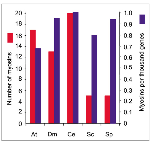
The numbers of myosins in eukaryotic sequenced genomes. The number of myosins in each organism is on the left (red column) and the number per thousand for each organism is on the right (blue column). At, Arabidopsis thaliana; Dm, Drosophila melanogaster; Ce, Caenorhabditis elegans; Sc, Saccharomyces cerevisiae; Sp, Schizosaccharomyces pombe.
Only 5 of the 17 Arabidopsis myosins have been reported in the literature [41, 42, 43]. The other 12 are sequences obtained from the Arabidopsis database sequenced as part of the Arabidopsis Genome Sequencing Project. These sequences are, therefore, predicted sequences that have not been verified by complete cDNAs. The average sequence length of the Arabidopsis myosins is 1,400 residues, with the shortest sequence prediction being 1,085 (At VIIIA) amino acids and the longest 1,730 (At XIA). Some of the intron/exon predictions may not be correct, which could reduce or increase the size of the predicted proteins and so the sizes may change as more characterization is done for each myosin. A case in point is the cDNA that was isolated by Kinkema and Schiefelbein [41] for At MYA1 (At MYA1) which codes for 1,520 amino acids, whereas the predicted protein has 1,599 because of differences in intron prediction.
Using the Arabidopsis Sequence Map Overview of TAIR [62], the location of each myosin was determined (Figure 2). The myosin genes are distributed throughout the Arabidopsis genome. The chromosome lengths are based on the centimorgan (cM) scale as shown on the TAIR Map Overview [62]. The maps reported with the announcement of the Arabidopsis genome sequence show somewhat different lengths than the TAIR maps [55].
Figure 2.
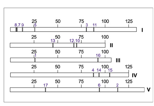
Location of myosins on the Arabidopsis chromosomes. Roman numerals represent chromosome numbers. Large numbers indicate chromosome length in cM. Small blue numbers are the myosin numbers from Table 1.
Phylogenetic analysis
All Arabidopsis myosins and a selection of myosins from other organisms representing each of the myosin classes were aligned using the motor domain sequence as determined by the SMART program [56]. The alignment was done in Megalign by the CLUSTAL method and a phylogenetic tree was generated using the Bootstrap (100 replicates) method with a heuristic search of the PAUP 4.0b6 program (Figure 3). The Arabidopsis myosins all group into two classes along with other plant myosins - class VIII and class XI. No animal myosins group with the plant myosins and no plant myosins group into any of the animal myosins. An algal (Chara corallina) myosin, Cc ccm, does group with the plant class XI myosins but is on a separate branch from any other class XI myosin (Figure 3). The D. discoideum myosin Dd myoJ did not fall into a class with any of the plant myosins. In fact, three D. discoideum myosins (Dd myoI, Dd myoJ, and Dd myoM) did not fall into any of the classes (Figure 3). The phylogenetic trees of Hodge and Cope and the tree on the myosin home page [22, 59] show the Dd myoI branching from class VII myosins. A heuristic search without bootstrapping also showed the Dd myoI myosin as a branch from class VII and domain analysis shows that Dd myoI has the MyTH4 domain found in other class VII myosins. Other phylogenetic anaylses have placed Dd myoJ as a branch off class XI myosins from plants [22, 59]. However, the phylogenetic tree generated from full-length sequences of plant myosins and Dd myoJ (see below) also shows that Dd myoJ is separate from the plant myosins.
Figure 3.
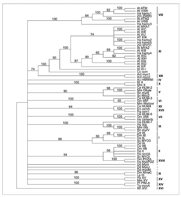
Phylogenetic tree. Alignment of the motor domain of representative myosins and all Arabidopsis myosins was done in Megalign by the CLUSTAL method and a phylogenetic tree was generated using the bootstrap method with a heuristic search of the PAUP 4.0b6 program. The myosin groups, as defined by Hodge and Cope [59] and Yamashita et al. [23], are identified on the right in roman numerals. Myosins from the following organisms were used: Ac, Acanthamoeba castellani; Acl, Acetabularia cliftoni; At, Arabidopsis thaliana; Cc, Chara corallina, Ha, Helianthus annuus; Zm, Zea mays; Bt, Bos taurus; Mm, Mus musculus; Ce, Caenorhabditis elegans; Dm, Drosophila melanogaster; Rn, Rattus norvegicus; Sc, Saccharomyces cerevisiae; Hs, Homo sapiens; Dd, Dictyostelium discoideum; Lp, Limulus polyphemus; En, Emericella nidulans; Pg, Pyricularia grisea; Pf, Plasmodium falciparum; and Tg, Toxoplasma gondii. The number at the branches indicates the number of times the dichotomy was supported out of 100 bootstrap tries.
Myosins from another alga, Acetabularia cliftonii, are classified into a separate group (XIII) and one myosin each from the fungi Emericella nidulans and Pyricularia grisea are also assigned to a separate class (XVII). A second alignment was done using the full-length sequences for all Arabidopsis and other known full-length plant myosins with a human heavy-chain myosin (Hs Ib) as an outgroup. The two classes of plant myosins are clearly seen (Figure 4). Among the class XI myosins the similarity ranges from 40-85% (full length) and 61-91% (motor domain). The similarity between the class VIII myosins ranges from 50-83% (full length) and 64-92% (motor domain). When class VIII myosins are compared to class XI myosins the similarity only ranges from 22-29% (full-length) and 35-42% (motor domain). Thirteen Arabidopsis myosins group into class XI. Two subgroups branch off in this class with three outliers (Figure 4). One subgroup consists of two pairs of Arabidopsis myosins, At XIB/At MYA2 and At XIG/At XIH, which are most similar to the sunflower myosin Hahamy4 and then another pair of Arabidopsis myosins, At XID/At XIA. The other subgroup consists of the Arabidopsis myosin pair At XIC/At XIE and two unpaired Arabidopsis myosins, At XIK and At MYA1, that are most closely related to sunflower myosins Hahamy2 and Hahamy5 and to the maize myosin ZmMYO1. At XIJ, AT XIF and At XI-I are on separate branches that group with the other class XI myosins but not within the two subgroups. There are four class VIII Arabidopsis myosins that form two pairs, At ATM/At VIIIA and At VIIIB/At ATM2. The first pair group with class VIII myosins from Z. mays and H. annuus whereas the second pair are on a separate branch.
Figure 4.
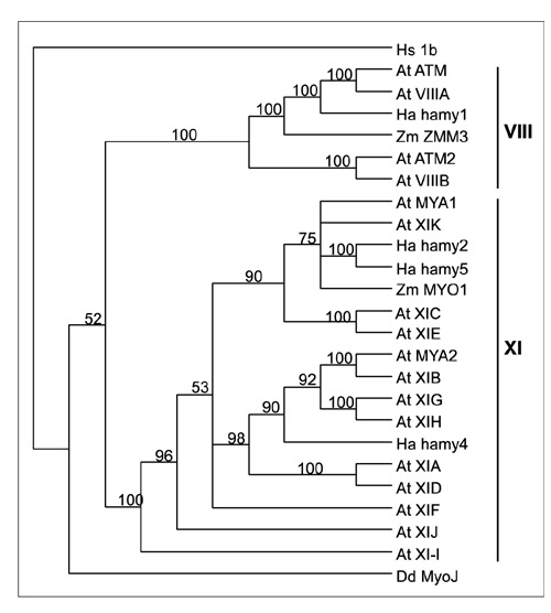
Phylogenetic tree for plant myosins. Alignment of the full-length Arabidopsis myosins, other full-length plant myosins available in the NCBI database and Dd myoJ was done in Megalign by the CLUSTAL method and a phylogenetic tree was generated using the bootstrap method with a heuristic search of the PAUP 4.0b4a (PPC) program. A human myosin (Hs 1b) was used as an outgroup. At, Arabidopsis thaliana; Dd, Dictyostelium discoideum; Ha, Helianthus annuus; Zm, Zea mays. The number at the branches indicates the number of times the dichotomy was supported out of 100 bootstrap tries.
Characterization of the Arabidopsis myosins
Figure 5 shows schematic diagrams of each myosin. The motor domain in all cases is in the amino-terminal region. The motor domain starts at about 50-55 residues for the class XI myosins whereas the class VIII myosins have a longer amino-terminal region before the motor domain (99-159 residues). The IQ domains usually follow right after the motor domain but are separated slightly from the motor domain in At XID, At XI-I, and At XIK. There are three or four IQ domains in class VIII myosins and five or six in class XI, except for At XIK, which has only four. There are coiled-coil domains, that differ in length and number, in all the myosins. They often follow directly after the IQ domains, but in some cases there is intervening sequence. Based on the presence of the coiled-coil domains, the Arabidopsis myosins are probably dimeric [26]. The class XI myosins are much longer than the class VIII myosins with the difference being in the length of the carboxy-terminal region following the conserved domains found in myosins.
Figure 5.
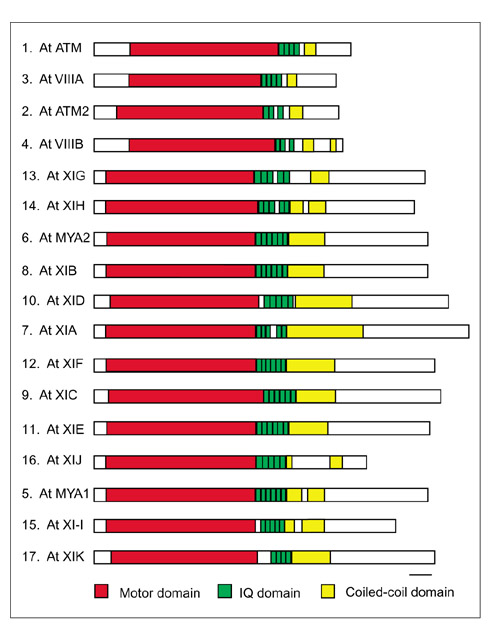
Schematic diagram of Arabidopsis myosins. The numbers refer to the number in Table 1. The motor domain, IQ domains, and coiled-coil domains are as indicated in the key. The first four myosins are in class VIII and the following 13 are in class XI. The bar represents 100 amino acids.
Besides the motor, IQ and coiled-coil domains, other domains have been identified in myosins from classes other than the plant classes VIII and XI. These include SH3 domains (Src homology 3 domains, that bind to target proteins), MYTH4 (a domain of unknown function found in a few classes of myosins), a zinc-binding domain, a pleckstrin homology domain, FERM/talin (band 4.1/ezrin/radixin/moesin), GPA-rich domains and a protein kinase domain [8, 22, 26]. These domains are involved in protein interactions and presumably give specificity to the action of the myosin. Except for the IQ and coiled-coil domains, the SMART program used to identify the motor domain of the myosin sequences did not identify any domains other than a few with scores less significant than the required threshold.
Myosins have 131 highly conserved residues spread throughout the motor domain that define a core consensus sequence [26]. Comparison of an alignment of Arabidopsis myosin motor domains to these conserved sequences shows a great deal of conservation among them (data not shown). One example is the ATP-binding site which consists of GESGAGKT (179-187 in Dictyostelium myosin II, DmyoII) and NxNSSR-FGK (233-241, DmyoII). With the exception of only one residue these are conserved in all 17 Arabidopsis myosins. The conformational state of myosin changes with ATP hydrolysis and a very conserved region implicated in this process has the conserved sequence LDIxGFExFxxN(S/G)(F/L)EQxxINxxNExLQQxF (453-482, DmyoII) [26]. The plant sequences are very conserved through this region. The sequence in this region is LDIYGFExFxxNSFEQxCINE(K/R)LQQHF (the first x is S in all but one myosin, the fourth x is F in all but one myosin). Cope et al. [26] suggest that release of the γ-phosphate of ATP may be through a hole in the structure centered around an absolutely conserved arginine residue (residue 654, DmyoII) which is also absolutely conserved in all Arabidopsis myosins. The presence of these highly conserved residues in plant myosins suggests that they are capable of motor function. In fact, in vitro motility studies with a purified myosin from Chara (myosin XI, Cc ccm in Figure 3) have confirmed that it is indeed an actin-based motor [54]. A loop present in the motor domain called the HCM (mutations in this loop cause hypertrophic cardiomyopathy) is the location of a phosphorylatable serine (S) or threonine (T) in certain amoeboid myosin I molecules and myosin VI molecules. This S or T residue is 16 residues upstream from the conserved DALAK sequence. The enzyme activity of the amoeboid myosins depends on phosphorylation of this site, but although phosphorylation of the myosin VI T residue has been demonstrated, the regulation of enzyme activity has not [8, 63]. Most other myosins have a constitutively negatively charged amino acid, either aspartic acid (D) or glutamic acid (E) at this site. This site has been named the TEDS rule site on the basis of these amino acids [8]. The Arabidopsis and other plant myosins all have aspartic acid, glutamic acid or glycine residue at this site, suggesting that they are not regulated by phosphorylation at this site. However, three residues upstream (19 from DALAK), all the class XI myosins have a threonine residue.
The site for each predicted or actual intron was located and is shown schematically in Figure 6. The intron locations were determined from the information at MIPS [57]. The length of each exon and the domain(s) they code for are shown in Tables 2 and 3 for class VIII and class XI myosins, respectively. The exons vary in length from 12 to greater than 672 nucleotides (the length of the beginning and last exons for each gene are not known as the predicted sizes include only the protein-coding nucleotides) with an average of 122 nucleotides. The four class VIII myosins have seven exons of the same length in the same order within the myosin motor domain (Table 2). The motor domain starts in the third exon of each class VIII myosin. The start of the IQ domains and the coiled-coil domains is more variable except for the At ATM2/At VIIIB pair. The class XI myosins also have many exons that are of the same length and in the same order but that differ from the class VIII pattern (Table 3). The exons coding for the motor domain sequence are most conserved in length. Most class XI myosins motor domains start in the third exon and end in the twentieth. Six of the class XI myosins have an intron after the start codon. Most differences in exon length are in the carboxy-terminal regions (Figure 6 and Table 3). However, even in the carboxy-terminal region there are some exon lengths conserved between some or all of the myosins. The two XI myosins with the closest similarity are At XIB and At MYA2. A Clustal alignment at Pole Bio-Informatique Lyonnais [64] showed 83.88% identity, 8.19% strong similarity and 2.36% weak similarity between these two myosins. Their motor domains are 91.6% identical. Twenty-three of their introns are at the same location in the motor domain area and then following a few different size exons, there are similar sized exons again. They are located on chromosomes I and V, respectively. It is possible that this pair is a result of gene duplication. Class VIII myosins At ATM and At VIIIA have 13 exons of the same length. Their full-length sequences are 79% identical with another 6.72% strongly similar and 3.52% weakly similar. Their motor domains have 93% similarity. At ATM is on chromosome III whereas At VIIIA is on chromosome I. This again may have resulted from a gene duplication. Analysis of the total Arabidopsis genome revealed that a whole genome duplication occurred, followed by subsequent gene loss and extensive local gene duplications [55]. The duplicated segments represent 58% of the Arabidopsis genome. The S. cerevisiae genome has also had a complete ancient genome duplication and 30% of the genes form duplicate pairs. Duplicated genes account for 48% of the total genes of C. elegans and Drosophila [60].
Figure 6.
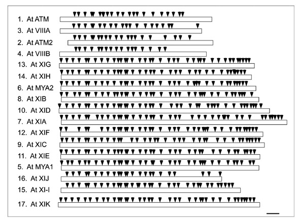
Location of the introns. The numbers refer to the number in Table 1. Arrowheads indicate the location of each intron along the length of the myosin. The bar represents 100 amino acids.
Table 2.
Analysis of exon sizes in class VIII myosins and the domain coded by each exon
| At ATM | At VIIIA | At ATM2 | At VIIIB | |||||
| Number | Size | Domain | Size | Domain | Size | Domain | Size | Domain |
| 1 | 339 | N | 315 | N | 159 | N | 333 | N |
| 2 | 102 | N | 132 | N | 102 | N | 118 | N |
| 3 | 144 | N,M | 144 | N,M | 144 | N,M | 131 | N,M |
| 4 | 151 | M | 151 | M | 151 | M | 155 | M |
| 5 | 28 | M | 28 | M | 25 | M | 169 | M |
| 6 | 166 | M | 158 | M | 129 | M | 64 | M |
| 7 | 64 | M | 104 | M | 64 | M | 99 | M |
| 8 | 14 | M | 139 | M | 99 | M | 104 | M |
| 9 | 84 | M | 119 | M | 104 | M | 139 | M |
| 10 | 104 | M | 153 | M | 139 | M | 119 | M |
| 11 | 139 | M | 90 | M | 119 | M | 153 | M |
| 12 | 119 | M | 78 | M | 153 | M | 90 | M |
| 13 | 153 | M | 159 | M | 90 | M | 78 | M |
| 14 | 90 | M | 207 | M | 78 | M | 159 | M |
| 15 | 78 | M | 144 | M | 159 | M | 186 | M |
| 16 | 159 | M | 114 | M | 186 | M | 206 | M |
| 17 | 207 | M | 130 | M,I | 342 | M | 136 | M |
| 18 | 206 | M | 147 | I | 244 | M,I | 130 | M,I |
| 19 | 136 | M | 68 | C | 116 | I | 108 | I |
| 20 | 130 | M,I | 595 | C,T | 213 | I,C | 140 | I,C |
| 21 | 147 | I | 83 | T | 480 | C,T | 189 | C |
| 22 | 68 | I,C | 375 | C,T | ||||
| 23 | 672 | C,T | ||||||
N, amino-terminal sequence; M, motor domain; I, IQ domain; C, coiled-coil domain; T, tail domain. The size of the first and last exons in each gene reflects only the size of the coding region.
Table 3.
Analysis of exon sizes in class XI myosins and the domain coded by each exon
| At XIG | At XIH | At MYA2 | At XIB | At XID | At XIA | At XIF | ||||||||
| No. | Size | Domain | Size | Domain | Size | Domain | Size | Domain | Size | Domain | Size | Domain | Size | Domain |
| 1 | 36 | N | 3 | N | 3 | N | 3 | N | 3 | N | 3 | N | 3 | N |
| 2 | 126 | N | 139 | N | 129 | N | 129 | N | 171 | N | 126 | N | 129 | N |
| 3 | 144 | N,M | 131 | N,M | 144 | N,M | 144 | N,M | 144 | N,M | 144 | N,M | 144 | N,M |
| 4 | 146 | M | 146 | M | 146 | M | 146 | M | 146 | M | 146 | M | 146 | M |
| 5 | 157 | M | 157 | M | 157 | M | 160 | M | 157 | M | 157 | M | 157 | M |
| 6 | 59 | M | 59 | M | 59 | M | 59 | M | 59 | M | 59 | M | 59 | M |
| 7 | 160 | M | 160 | M | 160 | M | 160 | M | 160 | M | 160 | M | 160 | M |
| 8 | 150 | M | 150 | M | 150 | M | 150 | M | 150 | M | 150 | M | 150 | M |
| 9 | 134 | M | 137 | M | 137 | M | 136 | M | 137 | M | 137 | M | 137 | M |
| 10 | 147 | M | 147 | M | 147 | M | 147 | M | 147 | M | 147 | M | 147 | M |
| 11 | 102 | M | 102 | M | 102 | M | 102 | M | 102 | M | 102 | M | 102 | M |
| 12 | 58 | M | 58 | M | 58 | M | 58 | M | 58 | M | 58 | M | 58 | M |
| 13 | 102 | M | 102 | M | 102 | M | 102 | M | 102 | M | 102 | M | 102 | M |
| 14 | 38 | M | 38 | M | 38 | M | 38 | M | 38 | M | 38 | M | 38 | M |
| 15 | 127 | M | 127 | M | 127 | M | 127 | M | 127 | M | 127 | M | 127 | M |
| 16 | 171 | M | 171 | M | 168 | M | 168 | M | 171 | M | 171 | M | 171 | M |
| 17 | 132 | M | 132 | M | 132 | M | 132 | M | 132 | M | 132 | M | 132 | M |
| 18 | 110 | M | 110 | M | 110 | M | 110 | M | 110 | M | 110 | M | 107 | M |
| 19 | 61 | M | 82 | M | 61 | M | 61 | M | 61 | M | 61 | M | 61 | M |
| 20 | 178 | M,I | 178 | M,I | 178 | M,I | 178 | M,I | 178 | M,I | 178 | M,I | 178 | M,I |
| 21 | 194 | I | 206 | I | 206 | I | 206 | I | 251 | I | 206 | I | 206 | I |
| 22 | 120 | I | 120 | I | 120 | I,C | 120 | I,C | 120 | I,C | 120 | I,C | 120 | I,C |
| 23 | 99 | U | 99 | U | 99 | C | 99 | C | 99 | C | 99 | C | 99 | C |
| 24 | 213 | C | 213 | C | 213 | C | 213 | C | 213 | C | 288 | C | 216 | C |
| 25 | 140 | C,T | 140 | C | 140 | C | 140 | C | 153 | C | 153 | C | 140 | C |
| 26 | 112 | T | 94 | C,T | 12 | C | 115 | C | 54 | C | 150 | C | 102 | C |
| 27 | 45 | T | 168 | T | 45 | C,T | 45 | C,T | 203 | C | 165 | C | 109 | C,T |
| 28 | 84 | T | 144 | T | 63 | T | 51 | T | 94 | C,T | 140 | C | 45 | T |
| 29 | 198 | T | 201 | T | 171 | T | 171 | T | 60 | T | 115 | C,T | 60 | T |
| 30 | 144 | T | 138 | T | 153 | T | 150 | T | 78 | T | 21 | T | 171 | T |
| 31 | 162 | T | 71 | T | 201 | T | 192 | T | 182 | T | 78 | T | 156 | T |
| 32 | 111 | T | 46 | T | 129 | T | 129 | T | 187 | T | 171 | T | 207 | T |
| 33 | 71 | T | 57 | T | 71 | T | 71 | T | 177 | T | 153 | T | 150 | T |
| 34 | 100 | T | 57 | T | 97 | T | 97 | T | 78 | T | 177 | T | 71 | T |
| 35 | 57 | T | 81 | T | 57 | T | 57 | T | 50 | T | 291 | T | 100 | T |
| 36 | 57 | T | 83 | T | 57 | T | 57 | T | 97 | T | 71 | T | 57 | T |
| 37 | 81 | T | 112 | T | 81 | T | 164 | T | 57 | T | 100 | T | 57 | T |
| 38 | 65 | T | 83 | T | 169 | T | 57 | T | 57 | T | 81 | T | ||
| 39 | 118 | T | 112 | T | 81 | T | 57 | T | 83 | T | ||||
| 40 | 77 | T | 81 | T | 133 | T | ||||||||
| 41 | 115 | T | 77 | T | ||||||||||
| 42 | 115 | T | ||||||||||||
| At XIC | At XIE | At XIJ | At MYA1 | At XI-I | At XIK | |||||||||
| No. | Size | Domain | Size | Domain | Size | Domain | Size | Domain | Size | Domain | Size | Domain | ||
| 1 | 52 | N | 12 | N | 126 | N | 180 | N,M | 144 | N | 55 | N | ||
| 2 | 104 | N | 129 | N | 144 | N,M | 138 | M | 126 | N,M | 119 | N | ||
| 3 | 144 | N,M | 144 | N,M | 146 | M | 146 | M | 146 | M | 144 | N,M | ||
| 4 | 146 | M | 146 | M | 157 | M | 157 | M | 157 | M | 146 | M | ||
| 5 | 157 | M | 157 | M | 59 | M | 92 | M | 59 | M | 157 | M | ||
| 6 | 59 | M | 59 | M | 160 | M | 160 | M | 156 | M | 110 | M | ||
| 7 | 160 | M | 160 | M | 150 | M | 150 | M | 150 | M | 160 | M | ||
| 8 | 150 | M | 150 | M | 137 | M | 137 | M | 137 | M | 150 | M | ||
| 9 | 137 | M | 137 | M | 147 | M | 147 | M | 147 | M | 137 | M | ||
| 10 | 147 | M | 147 | M | 102 | M | 102 | M | 102 | M | 111 | M | ||
| 11 | 102 | M | 102 | M | 58 | M | 58 | M | 58 | M | 102 | M | ||
| 12 | 58 | M | 58 | M | 102 | M | 102 | M | 102 | M | 58 | M | ||
| 13 | 242 | M | 102 | M | 38 | M | 38 | M | 38 | M | 102 | M | ||
| 14 | 127 | M | 38 | M | 127 | M | 127 | M | 131 | M | 38 | M | ||
| 15 | 171 | M | 127 | M | 168 | M | 171 | M | 122 | M | 127 | M | ||
| 16 | 132 | M | 171 | M | 132 | M | 132 | M | 36 | M | 159 | M | ||
| 17 | 110 | M | 132 | M | 110 | M | 110 | M | 132 | M | 108 | M | ||
| 18 | 61 | M | 110 | M | 61 | M | 61 | M | 110 | M | 110 | M | ||
| 19 | 178 | M,I | 61 | M | 178 | M,I | 313 | M | 61 | M | 61 | M | ||
| 20 | 206 | I | 178 | MI | 206 | I | 206 | M,I | 178 | M,I | 178 | M | ||
| 21 | 120 | I | 206 | I | 120 | I,C | 120 | I | 206 | I | 239 | I | ||
| 22 | 99 | C | 120 | I,C | 651 | C | 99 | I,C | 120 | I,C | 120 | I | ||
| 23 | 222 | C | 99 | C | 140 | C,T | 219 | C | 99 | C | 99 | C | ||
| 24 | 140 | C | 222 | C | 257 | T | 140 | C | 222 | C | 222 | C | ||
| 25 | 112 | C,T | 140 | C | 53 | T | 139 | C,T | 140 | C | 140 | C | ||
| 26 | 48 | T | 112 | C,T | 51 | T | 100 | C,T | 118 | C | ||||
| 27 | 255 | T | 48 | T | 51 | T | 51 | T | 51 | C,T | ||||
| 28 | 156 | T | 255 | T | 171 | T | 171 | T | 72 | T | ||||
| 29 | 207 | T | 156 | T | 156 | T | 63 | T | 171 | T | ||||
| 30 | 144 | T | 195 | T | 210 | T | 177 | T | 156 | T | ||||
| 31 | 71 | T | 144 | T | 147 | T | 71 | T | 207 | T | ||||
| 32 | 100 | T | 71 | T | 71 | T | 100 | T | 138 | T | ||||
| 33 | 57 | T | 157 | T | 100 | T | 81 | T | 75 | T | ||||
| 34 | 57 | T | 57 | T | 114 | T | 83 | T | 81 | T | ||||
| 35 | 81 | T | 81 | T | 85 | T | 151 | T | 57 | T | ||||
| 36 | 83 | T | 83 | T | 76 | T | 57 | T | ||||||
| 37 | 124 | T | 124 | T | 124 | T | 81 | T | ||||||
| 38 | 83 | T | ||||||||||||
| 39 | 136 | T | ||||||||||||
| 40 | ||||||||||||||
| 41 | ||||||||||||||
| 42 | ||||||||||||||
N, Amino-terminal sequence; M, motor domain; I, IQ domain; C, coiled-coil domain; U, undefined; T, tail domain. The size of the first and last exons in each gene reflects only the size of the coding region.
If the gene pairs are the result of duplication, it is interesting to note that while exon lengths have been conserved, intron lengths have not. The intron lengths are shown in Table 4. No pattern can be seen in intron lengths between any of the myosins. The average intron length is 131 nucleotides with the shortest intron at 47 nucleotides and the longest at 860. At XI-I has the highest average, 272 nucleotides. It contains the 860-nucleotide intron and three others that are over 500 nucleotides. In a study of 998 introns only 3.3% of the introns were longer than 500 nucleotides with sizes ranging from 59 to 1242 nucleotides [65]. This makes At XI-I unusual in having four out of 33 introns (12%) longer than 500 nucleotides. Only two other myosins had an intron over 500 nucleotides. Of the total 557 splice sites that were identified in the Arabidopsis myosins only six (a little more than 1%) were over 500 nucleotides with four out of the six being in one myosin. Hunt et al. found that a SV40 small-t intron only 66 nucleotides in length was spliced efficiently in tobacco cells [66]. Several of the introns in the myosins are between 66 and 70 nucleotides and so may be long enough to be spliced. Only one is in a cloned myosin known to be spliced at that site (At XIJ). There is also a predicted intron of only 47 nucleotides in length (At XID) which is thought to be too short for efficient splicing. Brown et al. [65] found three introns less than 66 nucleotides in length in known expressed proteins, but none of them was less than 59 nucleotides. Until the expression of At XID is studied, no conclusion can be made as to the validity of this intron prediction. The significance of the range and variability of intron length is not known. In Arabidopsis, in general, the range is even greater (47-6,442) [11].
Table 4.
Intron size and sequence of 5' and 3' splice sites
| At ATM | At VIIIA | At ATM2 | At VIIIB | |||||||||
| No. | Size | 5' site | 3' site | Size | 5' site | 3' site | Size | 5' site | 3' site | Size | 5' site | 3' site |
| 1 | 137 | AG GTATTC | TTTAG AT | 107 | AG GTATTG | TAGAG GC | 310 | AG GTAATT | TTCAG AA | 179 | AG GTAAAT | GCCAG AA |
| 2 | 84 | AA GTAAGT | AACAG GT | 88 | AA GTAAGT | AACAG GT | 95 | AT GTGAGT | CAAAG GT | 81 | AA GTTCTT | AGTAG CA |
| 3 | 124 | AT GTAAGT | GCTAG AC | 126 | AT GTAAAT | GCTAG AC | 91 | AT GTGAGT | TACAG AG | 84 | TA GTAAGT | TTTAG AG |
| 4 | 109 | CG GTGGGT | TCCAG AT | 92 | AG GTTGGA | TTCAG TC | 113 | AG GTGAGG | AGAAG AG | 226 | GA GTGAAA | CTTAG TC |
| 5 | 247 | AG GTTAGT | TCCAG CG | 302 | AG GTTAGT | TCCAG TG | 121 | AG GTACGG | TATAG AG | 159 | TC GTGAGT | TGCAG GG |
| 6 | 114 | TT GTAAGC | TACAG GG | 643 | TT GTAAGC | GACAG GG | 152 | TT GTGAGA | CACAG GT | 194 | TT GTAAGA | AGTAG TC |
| 7 | 103 | CT GTAAGT | TGCAG TT | 89 | AG GTAACT | TTCAG GA | 205 | TT GTAAGT | GGTAG TC | 196 | AG GTAACA | TGCAG AG |
| 8 | 101 | AG GTAGCT | AACAG TC | 201 | AA GTATGG | TCCAG GT | 151 | AG GTAACA | TGTAG AG | 100 | TG GTACTT | TATAG GA |
| 9 | 376 | AG GTATGG | TGCAG AG | 170 | AG GTAGGC | ACCAG GC | 102 | TG GTAATT | TGCAG GA | 98 | AG GTAGAG | TACAG CT |
| 10 | 101 | AG GTAATT | TGCAG GA | 135 | AT GTATGC | TGCAG AA | 78 | AG GTAGAA | TACAG CT | 97 | TG GTTTGT | TTCAG GC |
| 11 | 295 | AA GTAAGC | TTCAG GT | 114 | AG CTAACG | TCCAG GA | 94 | AG GTAATG | TTAAG GT | 75 | AG GTTCGT | TTTAG GA |
| 12 | 326 | AG GTATAT | TTCAG GC | 207 | AG GTAATG | TGCAG AA | 89 | AG GTTAGT | TTCAG AA | 123 | TG GTGATC | TTCAG GA |
| 13 | 197 | AT GTATGT | TGCAG AA | 146 | TG GTAATA | CTCAG GT | 82 | AG GTGGTT | CTCAG GA | 139 | TG GTAAGT | TGCAG AA |
| 14 | 136 | AG GTAAAG | TTCAG GA | 192 | AG GTTGGG | TTCAG GG | 95 | AG GTAATT | AGCAG AA | 126 | AG GTCAGT | AATAG GT |
| 15 | 160 | AG GTATAT | TGCAG AA | 211 | AG GTCGTT | TGGAG AA | 125 | AG GTCAGT | TACAG GT | 111 | TG GTGACA | TACAG GC |
| 16 | 122 | AG GTAACA | ATCAG GT | 86 | TG GTACTT | TGCAG AT | 87 | AG GTAAAG | TACAG GG | 104 | TG GTTTGG | AGTAG AT |
| 17 | 228 | AG GTGAGT | TCCAG AG | 85 | TA GTATTG | TTCAG TT | 87 | AA GTAAGC | CATAG AT | 82 | AT GTAAGT | GATAG AT |
| 18 | 87 | AG GTGACA | TGCAG AT | 103 | TG GTAAAA | TGTAG CA | 82 | TG GTAAGC | TGCAG CG | 109 | TA GTAATC | TACAG AT |
| 19 | 77 | AG GTATAA | TGCAG AT | 88 | TG GTCCTC | TGTAG TG | 82 | AG GTACTT | TTCAG GA | 85 | TA GTAAAT | TGTAG TG |
| 20 | 112 | AT GTATAA | TTCAG TT | 83 | AG GTGGTT | TTGAG AC | 88 | AG GTCAAA | TGCAG AT | 70 | GC GTCTCT | TTGAG GT |
| 21 | 250 | AG GTAAAA | TGCAG CA | 80 | AG GTAAGT | TGCAG AT | ||||||
| 22 | 111 | AG GTAAAA | CGCAG AC | |||||||||
| At XIG | At XIH | At MYA2 | At XIB | |||||||||
| No. | Size | 5' site | 3' site | Size | 5' site | 3' site | Size | 5' site | 3' site | Size | 5' site | 3' site |
| 1 | 168 | TG GTTATT | TTCAG CG | 365 | AT GTGAGA | TGCAG GC | 330 | TG GTAAGA | TACAG GT | 618 | TG GTAAAA | TGCAG GT |
| 2 | 103 | CG GTATGT | TTCAG GT | 135 | CA GTTTGA | TAAAG TT | 100 | AT GTATGT | TTCAG GT | 127 | AA GTATGT | CACAG GT |
| 3 | 92 | AT GTGAGT | ACTAG AC | 137 | AG GTGAGT | TCCAG AC | 74 | AT GTGAGT | TTCAG AC | 143 | AT GTGAGT | TTCAG AC |
| 4 | 90 | AG GTGCTT | TATAG AC | 96 | AG GTGCCT | GGTAG AC | 102 | AG GTAATT | TGCAG AC | 87 | AG GTAATT | TGCAG AC |
| 5 | 105 | AG GTAACT | TGCAG TC | 98 | AG GTTATC | TGCAG TC | 300 | AG GTGAAA | TTCAG TC | 201 | AG GTGAAA | TACAG TC |
| 6 | 120 | AG GTGAAT | TGCAG TC | 123 | AG GTGTAT | TGCAG TC | 76 | AG GTAACC | TATAG TC | 101 | AG GTAAGG | TATAG TC |
| 7 | 274 | AG GTACAT | GACAG GA | 289 | AG GTACAT | ATCAG GA | 125 | AA GTAAGT | TACAG GA | 93 | AA GTAAGT | TTCAG GA |
| 8 | 76 | AG GTAGTT | GTCAG GA | 83 | AG GTAACT | GTCAG GA | 95 | AG GTAGTT | TTCAG GA | 81 | AG GTACCT | TTTAG GA |
| 9 | 115 | AT GTGTGT | TGCAG GT | 101 | TA GTGAGT | GTCAG GT | 103 | AG GTAAAT | TCCAG CT | 89 | AT GTAAAT | TGCAG GT |
| 10 | 111 | TG GTATGT | TGTAG GA | 107 | TG GTATGT | TTCAG GA | 111 | TG GTGGGT | TGCAG GC | 125 | TG GTGAGT | TGCAG GC |
| 11 | 300 | AG GTGCAT | TTCAG TT | 284 | AG GTGCTT | TGCAG TT | 355 | AG GTGCTT | TGCAG TT | 417 | AG GTGCTT | TGCAG TT |
| 12 | 84 | AG GTTTGT | GGCAG CA | 88 | AG GTTTGT | GGCAG CA | 91 | AG GTTTGA | TGCAG CA | 91 | AG GTTTTG | TGCAG CA |
| 13 | 97 | AG GTAACT | TTCAG AA | 80 | AG GTTAGT | CTCAG AA | 234 | GA GTCTGT | TTCAG AA | 243 | AG GTTATC | TTCAG AA |
| 14 | 82 | TG GTAAGC | TGCAG CA | 87 | TG GTATGA | TGCAG CA | 153 | TG GTGAGT | TGCAG CA | 123 | AG GTGAGT | TGCAG CA |
| 15 | 99 | AT GTGAGT | TTCAG GT | 104 | TA GTGAGT | TTCAG GT | 117 | AT GTGAGT | TCCAG GT | 121 | AT GTGAGC | TCCAG GT |
| 16 | 85 | AG GTGCAG | TGCAG CA | 82 | AG GTGCAG | TGCAG CA | 87 | AG GTAAGT | TTCAG CA | 91 | AG GTGAGT | TGCAG CA |
| 17 | 92 | GG GTGAGA | TTTAG GG | 87 | GG GTGGGA | TTCAG GG | 91 | GG GTGCGA | TTTAG GG | 98 | GG GTGCGA | CACAG GG |
| 18 | 86 | AG GTATGC | GCTAG TT | 79 | AG GTTCCC | TCTAG TA | 77 | AA GTAAGA | AATAG CT | 88 | AA GTAAGA | ACTAG TT |
| 19 | 75 | AG GTACTT | CACAG AT | 113 | AA GTACGT | TCCAG AT | 87 | AG GTAATT | TGTAG AT | 93 | AG GTAATT | TGTAG AT |
| 20 | 99 | AG GTATCT | AACAG GT | 86 | AG GTACTT | TGTAG GT | 117 | AG GTATTT | GTCAG GT | 88 | AG GTATTT | TTCAG GT |
| 21 | 147 | AG GTGGAG | CAGAG CC | 147 | AG GTGCTG | TACAG AG | 159 | AG GTACAC | TATAG AC | 170 | AG GTATGA | TACAG AC |
| 22 | 130 | CG GTGTGC | TGCAG GA | 296 | TG GTGAGC | TGCAG GC | 122 | TG GTGAGA | CCTAG GC | 150 | AG GTGAGA | CACAG GC |
| 23 | 117 | GG GTCAGA | TGTAG GT | 120 | GG GTAAGT | TTTAG AC | 125 | GG GTGTGA | TGCAG AC | 105 | GG GTGAGT | TGCAG AC |
| 24 | 107 | AG GTAGGG | TGCAG TC | 119 | AG GTAGGA | TTCAG TC | 150 | AG GTTTGT | TACAG AG | 120 | AG GTGGGT | TGCAG GG |
| 25 | 99 | AA GTATTC | TGCAG TC | 94 | GA GTACCC | TGCAG AC | 89 | TG GTATCC | TCCAG GC | 87 | AG GTACTG | TGCAG GC |
| 26 | 84 | AG GTAGAC | TTTAG AA | 392 | CA GTTAAG | AGGAG AA | 89 | AG GTAGAA | TGTAG AA | 90 | AG GTAGAA | TGCAG AA |
| 27 | 85 | CA GTGTAA | TGCAG GG | 133 | AG GTACTG | ATCAG GA | 104 | AT GTATAT | TCCAG GA | 106 | TA GTAGGG | TTCAG GA |
| 28 | 152 | AT GTATGT | TGAAG AG | 89 | TG GTATAT | ACCAG GG | 82 | TT GTATGT | TGCAG AT | 82 | TT GTACTG | TGCAG GA |
| 29 | 85 | AG GTACTA | TTTAG GA | 105 | AG GTCAGC | TCTAG GC | 181 | AG GTAATT | TTCAG AA | 316 | TG GTAAAT | TTCAG AA |
| 30 | 97 | AG GTATAT | AACAG GG | 73 | TT GTATGG | TTCAG GT | 103 | TG GTTTGT | ACCAG AG | 86 | TG GTATTT | ACCAG AG |
| 31 | 83 | AG GTGACA | TCTAG GC | 81 | AG GTGAGA | TGTAG CC | 95 | AG GTTCCT | TTCAG GC | 158 | TG GTTTCA | TTCAG GC |
| 32 | 78 | TT GTATGT | TACAG GT | 150 | TT GTAAAA | TGCAG TA | 85 | AT GTAAGG | TCCAG GT | 77 | AT GTAAGG | TACAG GT |
| 33 | 91 | AG GTGAGA | TGCAG CC | 128 | TG GTATGT | AACAG GT | 78 | AG GTAAGT | TACAG TC | 169 | AG GTAAAT | AATAG CC |
| 34 | 81 | AG GTAATC | GATAG TA | 100 | CT GTGAGT | TGCAG AT | 95 | AA GTAAAA | GGCAG TA | 74 | AA GTAAGT | TGCAG TA |
| 35 | 104 | TG GTATGT | AACAG CT | 92 | AT GTATGC | AACAG GT | 165 | AG GTATGT | TGCAG GT | 90 | TG GTATGT | ATCAG GT |
| 36 | 88 | CA GTAAGT | CTCAG AA | 101 | AG GTAACA | CTTAG CA | 88 | CG GTAAGG | TACAG GT | 83 | CG GTAAAG | TACAG AT |
| 37 | 89 | AT GTAAGC | AATAG GT | 103 | AA GTACCT | TGCAG GT | 86 | AG GTAACT | AATAG AC | |||
| 38 | 108 | AG GTAAGT | CACAG CA | 156 | AG GTGAAA | GACAG CA | ||||||
| At XID | At XIA | At XIF | At XIC | |||||||||
| No. | Size | 5' site | 3' site | Size | 5' site | 3' site | Size | 5' site | 3' site | Size | 5' site | 3' site |
| 1 | 228 | TG GTACGA | ATCAG GC | 430 | TG GTACGA | TGCAG GC | 89 | TG GTAAGC | GTTAG GG | 143 | AG GTTAGT | TGTAG GT |
| 2 | 47 | AG GTACCT | TGTAG GT | 215 | CG GTAAGA | CTTAG GT | 169 | CA GTAAGA | TACAG GT | 93 | AG GTCCAG | TATAG GT |
| 3 | 173 | AT GTACGC | TACAG AC | 134 | TA GTAAGC | TCCAG AC | 100 | TA GTCAGT | CGCAG AC | 82 | AT GTTTTG | GACAG AC |
| 4 | 89 | AG GTAATC | TTTAG AA | 91 | AG GTAACT | TTCAG GA | 81 | TG GTAAAA | ACTAG GG | 95 | AG GTGAGT | CTCAG GG |
| 5 | 109 | AG GTAGAT | TGCAG TC | 112 | AG GTAATG | TGCAG TC | 71 | AG GTGAGT | TATAG TC | 93 | AA GTAATG | TCCAG TC |
| 6 | 90 | AG GTGGAA | TGCAG TC | 93 | AG GTGGAG | TGCAG TC | 96 | AG GTGGTG | GACAG TC | 83 | AG GTGAAG | CTCAG TC |
| 7 | 117 | AG GTAAAC | TTCAG GA | 101 | AG GTAAGC | TTCAG GA | 84 | AG GTAAGT | TTCAG GA | 72 | AG GTACGT | AGCAG GA |
| 8 | 68 | AG GTACCT | TGTAG GA | 66 | AG GTACTT | TGTAG GA | 76 | TG GTTTGT | TTTAG GA | 101 | AG GTCAGT | AACAG GA |
| 9 | 84 | AT GTATAT | GGTAG GT | 86 | TA GTAAAT | TGCAG GT | 79 | TG GTATCT | CGTAG GT | 174 | AT GTAAAA | TTCAG GT |
| 10 | 90 | GG GTAGGT | CCCAG GC | 80 | TG GTAGAT | TTAAG GA | 264 | TG GTATGT | GACAG GA | 74 | TG GTAAGT | TCTAG TA |
| 11 | 309 | AG FTFCTT | TGCAG TT | 297 | AG GTGCTT | TGCAG TT | 79 | AG GTAGAC | CAAAG TT | 76 | AG GTAAAT | TGCAG TT |
| 12 | 93 | AG GTTGGA | TACAG CA | 74 | AG GTTGGA | TACAG CA | 72 | AG GTAGAA | TGCAG CA | 71 | AG GTATTG | TTCAG CA |
| 13 | 113 | AG GTAAGT | GTCAG AA | 99 | AG GTTAGT | GTCAG AA | 97 | AG GTATAA | TTCAG AA | 84 | TG GTAAAG | TTCAG CA |
| 14 | 86 | TG GTAATG | TACAG TA | 84 | TG GTAATG | TGCAG CA | 106 | TG GTAAGT | TGCAG CA | 74 | AA GTAGGT | TCCAG GT |
| 15 | 105 | AT GTTAGT | TTCAG GT | 82 | AT GTTAGT | TCCAG GT | 78 | AT GTGAGA | TCCAG GT | 154 | AG GTAGGG | TGCAG CT |
| 16 | 78 | AG GTCTAC | TACAG CA | 214 | AG GTCTGA | TACAG CA | 70 | AG GTAAGC | CCCAG CA | 135 | GT GTAAGT | TCTAG GG |
| 17 | 102 | GG GTAAGC | CTCAG GG | 105 | GG GTAAGC | TTCAG GG | 90 | GA GTAAGC | AACAG GG | 92 | AG GTAAGT | AACAG CT |
| 18 | 111 | AG GTAGAT | TATAG CT | 128 | AG GTAGCT | AATAG CT | 102 | GG GTAAAA | GACAG AT | 120 | AG GTAACG | TGCAG AT |
| 19 | 152 | AG GTGCGT | CACAG AT | 202 | AG GTGCAG | CATAG AT | 101 | AG GTATGT | TTCAG AT | 114 | AG GTGAGC | TGTAG GA |
| 20 | 92 | AG GTAATA | TTCAG GA | 83 | AT GTTATA | TTTAG GT | 175 | AG GTTTTT | TGTAG CA | 88 | AG GTTTAG | GGCAG GC |
| 21 | 69 | TC GTATCT | CACAG AG | 113 | AA GTAAGT | CGCAG AG | 292 | AG GTACTA | AACAG AG | 296 | TG GTACAA | TTCAG GC |
| 22 | 280 | TG GTGACT | TCCAG GC | 256 | TG GTAATC | TTCAG GC | 148 | TG GTAAGT | CAAAG GC | 79 | GG GTATTT | TATAG GG |
| 23 | 86 | GG GTACAC | TGCAG AT | 126 | GG GTACAC | TGCAG AT | 73 | AG GTATTG | ATTAG GC | 114 | AG GTACTT | AACAG GT |
| 24 | 72 | AG GTAAGG | CTAAG GA | 122 | AG GTTAGT | AAAAG GT | 68 | AG GTAAGT | TGTAG GT | 105 | AG GTAAGA | ATCAG GA |
| 25 | 120 | CC GTCATT | CGTAG GC | 114 | AG GTAAGA | CTTAG GC | 86 | AG GTATAC | TCCAG AT | 96 | AG GTAAAC | TACAG AG |
| 26 | 432 | AC GTAACA | TACAG GA | 117 | AG GTAATC | CTTAG GC | 176 | AG GTACGG | ATCAG CC | 92 | TG GTAAAT | ATCAG GA |
| 27 | 118 | AG GTTATC | TTTAG GC | 87 | TA GTTAGT | AACAG GA | 84 | AG GTGCAA | TGCAG AA | 88 | AG GTTGGC | CTCAG AC |
| 28 | 77 | AG GTGTCA | TCAAG AA | 120 | AG GTTTTG | TTTAG GC | 70 | AG GTACGA | TTCAG GA | 113 | AG GTGATG | ATTAG AG |
| 29 | 96 | AT GTAAGT | TACAG GA | 79 | CG GTAAAT | TGCAG CC | 121 | AG GTATTA | GACAG GA | 87 | AG GTATGC | AATAG GC |
| 30 | 86 | AT GTATGT | TGCAG GA | 105 | AG GTAAGT | TACAG GA | 93 | AG GTAATA | AGAAG GG | 85 | AT GTGAGT | TTTAG GT |
| 31 | 78 | AA GTTTAA | CTCAG AA | 88 | TA GTATGT | AGCAG GA | 75 | AA GTAAGC | TGTAG GG | 103 | AG GTTTTT | AACAG CC |
| 32 | 121 | AG GTAACA | TTTAG GG | 164 | AG GTAACC | TTCAG AA | 93 | AT GTTAGT | AACAG GC | 70 | AG GTATCT | TTCAG TA |
| 33 | 360 | AG GTAGAA | CTGAG GA | 147 | TG GTAACG | TTTAG GG | 85 | AT GTAAAA | TCCAG GT | 79 | TG GTAACC | TACAG GT |
| 34 | 109 | AC GTAAGA | CTCAG AA | 92 | TG GTATAC | TTCAG AG | 82 | AG GTACAA | GGCAG TT | 148 | CG GTAAGT | GACAG GT |
| 35 | 97 | AG GTAAAA | TGCAG CC | 67 | AC GTAAGA | TTCAG GT | 97 | AG GTAGGC | TACAG GC | 97 | AC GTAAGT | AATAG GT |
| 36 | 87 | AT GTAAGT | TGCAG TT | 97 | TG GTTATT | TGCAG TC | 84 | TG GTATAG | TACAG GT | 74 | AG GTTGTT | TGCAG CA |
| 37 | 98 | TG GTCAGT | TCCAG GT | 76 | AG GTAAAA | TGCAG TT | 230 | CG GTAAAG | CTCAG GT | |||
| 38 | 125 | CG GTAACT | CTCAG GC | 78 | TG GTTTGT | TTCAG GT | 123 | AG GTAAGT | AATAG GT | |||
| 39 | 84 | AC GTATGT | TGCAG GT | 206 | CG GTAAGT | GTCAG GT | 70 | AG GTACGC | TTCAG CA | |||
| 40 | 91 | AG GTATTG | CTCAG CA | 79 | AG GTACAT | TGCAG GT | ||||||
| 41 | 84 | AG GTACTG | AACAG CA | |||||||||
| At XIE | At XIJ | At MYA1 | At XI-I | |||||||||
| No. | Size | 5' site | 3' site | Size | 5' site | 3' site | Size | 5' site | 3' site | Size | 5' site | 3' site |
| 1 | 111 | CA GTGACT | TGCAG GG | 120 | AT GTAAA | GTCAG GT | 330 | TG GTAAGA | TACAG GT | 134 | AG GTCTGA | AAAAG CT |
| 2 | 86 | AG GTGAGT | TGTAG AT | 117 | AT GTAAGA | GACAG AC | 100 | AT GTATGT | TTCAG GT | 860 | AT GTGAAC | TTCAG AC |
| 3 | 80 | AT GTTAGT | GACAG AC | 85 | AG GTGATT | AACAG GG | 74 | AT GTGAGT | TTCAG AC | 95 | AG GTGATC | CCCAG AG |
| 4 | 80 | AG GTGCTC | TTCAG GG | 292 | AA GTAAGT | TACAG TC | 102 | AG GTAATT | TGCAG AC | 181 | AA GTAAGA | TGCAG TC |
| 5 | 116 | AA GTATGA | GGCAG TC | 135 | AG GTAAAC | TACAG CC | 300 | AG GTGAAA | TTCAG TC | 241 | AG GTGGGT | TTCAG CC |
| 6 | 85 | AG GTGAAA | GTCAG AT | 72 | AG GTAGGT | TGCAG GA | 76 | AG GTAACC | TATAG TC | 149 | AT GTAATT | CTTAG GA |
| 7 | 75 | AG GTATAC | ACTAG CA | 88 | AG GTTTGC | TTCAG GA | 25 | AA GTAAGT | TACAG GA | 90 | AG GTATAA | ATCAG GA |
| 8 | 79 | AG GTAAGC | AACAG GA | 67 | AT GTAATA | TTTAG GT | 95 | AG GTAGTT | TTCAG GA | 91 | AA GTACAT | ATCAG GT |
| 9 | 76 | AT GTAAGT | TTTAG GT | 91 | TG GTAAAT | TCCAG GT | 103 | AG GTAAAT | TCCAG CT | 94 | TG GTTTGC | GTCAG GC |
| 10 | 101 | TG GTAAGT | TGCAG GT | 315 | AG GTGATG | TGCAG TT | 111 | TG GTGGGT | TGCAG GC | 135 | AG GTTAGC | TGCAG TT |
| 11 | 86 | AG GTAAGG | TGCAG TT | 81 | AG GTATGA | TACAG CA | 355 | AG GTGCTT | TGCAG TT | 83 | AG GTAATA | TTCAG CA |
| 12 | 88 | AG GTAATT | TTCAG CA | 440 | AG GTTTGT | TGCAG AA | 91 | AG GTTTGA | TGCAG CA | 717 | AG GTCGTT | TGCAG AA |
| 13 | 115 | AG GTTATT | AGCAG AA | 110 | TG GTATAA | TGCAG CA | 234 | GA GTCTGT | TTCAG AA | 85 | TG GTACAA | TGCAG CA |
| 14 | 91 | TG GTAATA | TTCAG CA | 88 | AT GTAAGT | TTCAG GT | 153 | TG GTGAGT | TGCAG CA | 98 | AA GTCTTG | TGAAG CC |
| 15 | 103 | AA GTAAGT | TTCAG GT | 138 | AG GTGACT | TGCAG CT | 117 | AT GTGAGT | TCCAG GT | 127 | AG GTAGAG | TTTAG CA |
| 16 | 70 | AG GTAGAT | GATAG TT | 75 | GG GTCTGT | TGCAG GG | 87 | AG GTAAGT | TTCAG CA | 547 | GG GTTAGT | GATAG CC |
| 17 | 107 | GT GTAAGT | TGTAG GG | 106 | GA GTATGT | ATCAG GT | 91 | GG GTGCGA | TTTAG GG | 302 | AG GTACGA | TGCAG CA |
| 18 | 85 | AA GTAAGT | AACAG CT | 154 | AG GTAAAG | TGCAG AT | 77 | AA GTAAGA | AATAG CT | 95 | AG GTATGG | CACAG CT |
| 19 | 92 | AG GTTTTT | TGCAG GT | 99 | AG GTGAGG | TTTAG GA | 87 | AG GTAATT | TGTAG AT | 269 | AG GTTCCT | GCAAG GA |
| 20 | 157 | AG GTGAAC | TATAG GA | 99 | AG GTTCTA | TGCAG GC | 117 | AG GTATTT | GTCAG GT | 180 | AG GTACTT | TTTAG GC |
| 21 | 88 | AG GTTTTA | TGCAG GC | 119 | AG GTATTG | TATAG GC | 159 | AG GTACAC | TATAG AC | 96 | AG GTATGA | TGCAG GT |
| 22 | 184 | TG GTACGT | TTCAG GC | 134 | AG GTAATG | TTCAG GC | 122 | TG GTGAGA | CCTAG GC | 80 | GA GTATGT | TACAG AC |
| 23 | 90 | GG GTATTT | GTCAG GT | 130 | AG GTATTA | TCCAG GT | 125 | GG GTGTGA | TGCAG AC | 701 | AG GTAATT | CACAG AA |
| 24 | 164 | AG GTACTC | AACAG GC | 197 | AG GTCAGT | TGCAG GA | 150 | AG GTTTGT | TACAG AG | 88 | AG GTTTGT | TTCAG TC |
| 25 | 125 | AG GTAAGT | GTCAG GC | 89 | TG GTATCC | TCCAG GC | 277 | AA GTATGT | AGCAG AA | |||
| 26 | 95 | AG GTACGG | AACAG GT | 89 | AG GTAGAA | TGTAG AA | 620 | TT GTAAGT | ATCAG GA | |||
| 27 | 101 | TG GTAAGT | ATCAG GA | 104 | AT GTATAT | TCCAG GA | 220 | AG GTGATC | TGCAG AG | |||
| 28 | 91 | AG GTTTGT | TTCAG AC | 82 | TT GTATGT | TGCAG AT | 129 | AT GTGAGT | ACCAG GG | |||
| 29 | 85 | AG GTGTGT | TCTAG AG | 181 | AG GTAATT | TTCAG AA | 466 | AG GTGAGA | GATAG GT | |||
| 30 | 90 | AG GTATAT | AATAG GC | 103 | TG GTTTGT | ACCAG AG | 89 | AG GTAAAT | TTCAG TC | |||
| 31 | 86 | AC GTGAGT | CTTAG GT | 95 | AG GTTCCT | TTCAG GC | 399 | AG GTACAC | TATAG GT | |||
| 32 | 79 | AG GTCTGT | TACAG TC | 85 | AT GTAAGG | TCCAG GT | 88 | AG GTGAGT | TGTAG GT | |||
| 33 | 92 | AG GTACAT | TGCAG GT | 78 | AG GTAAGT | TACAG TC | 326 | AG GTATTA | TGCAG CA | |||
| 34 | 78 | CG GTAAGT | TGCAG GT | 95 | AA GTAAAA | GGCAG TA | ||||||
| 35 | 80 | AC GTAAGT | GATAG GT | 165 | AG GTATGT | TGCAG GT | ||||||
| 36 | 99 | AG GTTAGT | GGCAG TA | 88 | CG GTAAGG | TACAG GT | ||||||
| 37 | 103 | AA GTACCT | TGCAG GT | |||||||||
| 38 | 156 | AG GTGAAA | GACAG CA | |||||||||
| At XIK | ||||||||||||
| No. | Size | 5' site | 3' site | No. | Size | 5' site | 3' site | No. | Size | 5' site | 3' site | |
| 1 | 237 | AA GTGAGT | CCCAG TC | 14 | 157 | TG GTAGGC | TGCAG TA | 27 | 98 | CG GTAAGG | CACAG GA | |
| 2 | 269 | CC GTAAGT | TTCAG GT | 15 | 87 | AG GTATAA | ATCAG GC | 28 | 110 | AG GTATCA | TGCAG GA | |
| 3 | 105 | AT GTAAGT | CGCAG AC | 16 | 319 | AG GTATGC | TTCAG GT | 29 | 118 | AA GTAAGT | ACCAG GT | |
| 4 | 102 | AG GTTATT | GGTAG GG | 17 | 148 | AC GTAATT | TTAAG GG | 30 | 99 | AA GTAAGA | AATAG GG | |
| 5 | 115 | TG GTGAGG | GAGAG GC | 18 | 150 | AA GTAAGT | TGCAG TT | 31 | 276 | AG GTAATT | TATAG GC | |
| 6 | 356 | AG GTACGT | TGCAG AC | 19 | 87 | AA GTAAGC | TCCAG TT | 32 | 90 | TG GTAAAA | TACAG GC | |
| 7 | 105 | AG GTATTG | TGTAG GA | 20 | 193 | AG GTATCT | TGGAG TT | 33 | 110 | TA GTTTCA | GTGAG TG | |
| 8 | 85 | AG GTCAGT | ATCAG GA | 21 | 125 | AG GTAATT | TTTAG GC | 34 | 91 | AA GTAAGC | TACAG TA | |
| 9 | 84 | AG GTATGT | AAAG GT | 22 | 84 | AG GTTCGG | ATCAG GC | 35 | 93 | TG GTAAAA | TTCAG GT | |
| 10 | 229 | GC GTTAGC | TTCAG GC | 23 | 74 | GA GTAAGT | TATAG TC | 36 | 94 | CG GTATTT | TTCAG GT | |
| 11 | 81 | AG GTAAAG | CTCAG CT | 24 | 121 | AG GTATGT | TACAG GC | 37 | 79 | AT GTATGT | CATAG GT | |
| 12 | 87 | AG GTCCGT | AACAG CA | 25 | 202 | AG GTTCGT | TTCAG AC | 38 | 81 | AG GTAACC | CGCAG CA | |
| 13 | 91 | AG GTGTCC | TTCAG AA | 26 | 97 | CG GTGCCT | TTCAG AG | |||||
The consensus nucleotide sequences for the 5' and 3' splice sites are A-2G-1 G+1T+2A+3A+4G+5T+6 and T-5G-4C-3A-2G-1G+1T+2, respectively [65]. The most conserved sequences are the 5' consensus G (100%) T (99%) at the +1, +2 positions, respectively, and the 3' A(100%) G(100%) at the -2, -1 positions, respectively. The splice sites in the reported myosins and the predicted myosins (Table 4) all contain the 5' GT and 3' AG sequences. The sequences in the Arabidopsis myosins upstream and downstream of these two very conserved sites varied as a reflection of the less conserved nature of these nucleotides (Table 4). However, these predicted sites at the 5' and 3' splice sites need to be confirmed experimentally.
Discussion
Only two classes of myosins are present in Arabidopsis. A study of myosins in lily and tobacco pollen tubes using antibodies to three animal-type myosins IA and IB, II and V suggested the presence of three types of myosins in these plants [40]. However, no type I, II or V myosins have been found in any plant and only two types (VIII and XI) have been identified. Class XI are somewhat similar to class V myosins [42] and this may explain the reaction with the type V antibody. Possibly the other reactions were due to similarities in the myosin motor domain. Phylogenetic analysis of Arabidopsis myosins along with other plant myosins suggests that most class XI myosins (except three) fall into two subgroups (Figure 4).
The Arabidopsis myosins have anywhere from three to six IQ domains. The IQ domain in non-plant myosins has been shown to bind to calmodulin in a calcium-independent manner. The regulation of myosin action is thought to be due to calmodulin interaction. In plants, two myosin heavy chains have been shown to associate with calmodulin [37, 67]. A myosin-containing protein fraction from tobacco BY2 cells was used in motility assays with F-actin. Concentrations of Ca2+ higher than 10-6 M caused a significant reduction in F-actin sliding [37]. Another study with myosin isolated from lily pollen, also demonstrated a co-precipitation of myosin and calmodulin and a similar effect of Ca2+concentration [67]. Not only did concentrations above 10-6M cause inhibition of myosin activity, but the effects of concentrations higher than 10-5 M were not reversible upon Ca2+ removal. These studies provide evidence that plant myosins bind calmodulin in the absence of Ca2+ and are active when calmodulin is bound and inactivated when the Ca2+ concentration is increased. They also found that when the myosin fraction was pretreated with CaCl2 calmodulin did not bind the myosin, suggesting that calmodulin dissociates from myosin at high concentrations of Ca2+. The myosins in the above studies have not been cloned, and binding to specific IQ domains has not been established. However, the presence of IQ domains in Arabidopsis and other plant myosins suggests that these are the sites of Ca2+ regulation. It would be interesting to investigate the possible phosphorylation of the threonine residue which is three residues upstream from the TEDS rule site in class XI myosins and to see if enzyme activity is regulated by phosphorylation of this residue.
Myosins are involved in a wide range of cellular functions. They have been shown to be involved in movement, translocation, cell division, organelle transport, G-protein-linked signal cascade and maintenance of structure within cells [26]. Insight into the function of plant myosins has been gained by studies in algae. Cytoplasmic streaming is responsible for movement of organelles and vesicles and of generative cells and vegetative nuclei in pollen tubes. Physiological studies in Chara have shown that an increase in Ca2+ concentration causes cytoplasmic streaming to stop [68]. A myosin isolated from the alga Chara corallina was shown to be responsible for cytoplasmic streaming [30, 69, 70]. The myosin was cloned and characterized and found to be a class XI myosin related to the Arabidopsis MYA myosins [54].
Myosins in plants have also been shown to be involved in cytoplasmic streaming. Using immunofluorescence, myosin was localized to vesicles, organelles and generative cells and vegetative nuclei in grass pollen tubes [39]. A myosin isolated from lily pollen has been shown to be responsible for cytoplasmic streaming in pollen tubes and two myosins were identified in tobacco cell cultures that are also thought to participate in cytoplasmic streaming [37, 71]. Antibodies to the myosins recognized a protein in vegetative cells as well as pollen tubes. Liu et al. [51] suggest that class XI myosins are likely candidates for transport of large vesicles because of the number of IQ domains (5-6). Previous studies showed that translocational step size produced by a myosin motor is proportional to the number of IQ domains and the larger the step the faster or more efficiently they are able to transport vesicles [9]. However, the kinetic properties of the motor domain are also involved in speed and there is a wide range of movement speeds for myosin II molecules [2, 72, 73].
An antibody specific to a Z. mays class XI myosin was used to localize this myosin in fractions of maize proteins and maize root tip cells [51]. The nuclear/cell wall fraction and the plastid fraction contained relatively small amounts of antigen while the mitochondrial fraction and the low density membrane fraction had most of the antigen. The root tip cells showed particulate staining in the cytoplasm, but neither the vacuole membrane nor plasma membrane were stained, although in some cells the staining was too bright to distinguish if the plasma membrane was stained or not. There are 13 class XI myosins in Arabidopsis that could be involved in vesicle and organelle transport. The large number could reflect redundancy of function or differential expression. Patterns of expression were different for the cloned Z. mays and Arabidopsis myosins that have been analyzed [42, 51].
Immunolocalization studies have also detected myosin associated with plasmodesmata. Plasmodesmata are interconnections between contiguous plant cells that allow direct cell-to-cell transport of ions and proteins. A recent study using an antibody to a cloned class VIII Arabidopsis myosin ATM1 (At ATM) localized this myosin to the plasmodesmata and the plasma membrane regions involved in the assembly of new cell walls [47]. Earlier work suggested that actin was involved in regulation of plasmodesmal transport [74]. Other studies using antibodies to animal myosins in root tissues of Allium cepa, Z. mays and Hordeum vulagare have also indicated the presence of myosin in the plasmodesmata [38]. However, immunolocalization studies with antibodies to animal myosins need to be interpreted with caution as there are no plant myosins that group with animal myosins.
The recent work by Reichelt et al. [47] is more convincing because they used antibody to plant myosin. The myosin was localized mainly to the transverse walls with some punctate labeling of the longitudinal walls. During cell division the anti-class-VIII myosin staining remains confined to the transverse cell walls and is strongest in the newly formed cell wall. Immunogold electron microscopy showed labeling of class VIII myosin associated with the plasma membrane and plasmodesmata. These studies suggest that class VIII myosins may be involved in new cell wall formation and transport in the plasmodesmata. Reichelt et al. [47] suggest that myosin VIII could act to bring islands of membrane plate material together or could trigger exocytosis of new cell wall material, or alternatively as an anchor for actin along the transverse walls. The role of myosin in the plasmodesmata was studied further by pretreating tissue with 2,3-butanedione 2-moxoxime (BDM), an inhibitor of actin-myosin motility. The pretreatment resulted in a strong constriction of the neck region of plasmodesmata [38]. Myosin VIII in the plasmodesmata could be a part of a gating complex that is thought to control the opening of the plasmodesma neck [74]. There are four class VIII myosins in Arabidopsis that could be involved in these types of functions.
A recent study of the effect of BDM on the distribution of myosins, F-actin, microtubules and cortical endoplasmic reticulum (ER) suggests that myosins may link together microtubules and actin filaments involved in structural interactions [75]. This study used antibody to myosin II from animals and Arabidopsis myosin VIII for immunofluorescence studies. BDM treatment disrupted normal cellular distributions of maize myosins and the characteristic distribution of F-actin was also affected. Myosin may participate in the intracellular distribution of actin filaments as was proposed for myosin XV [76]. Microtubule arrangements in cortical root cells were altered, as was the normal ER network. Post-mitotic cell growth was inhibited by BDM, specifically in the transition zone and the apical parts of the elongation region. The study suggested that actin fibers and microtubules interact together via myosins and that myosin-based contractility of the actin cytoskeleton is essential for the developmental progression of root cells [75]. However, BDM has only been shown to inhibit a few myosins in vitro [77] and is known to be a nonspecific inhibitor; so these results must be viewed with caution.
Conclusions
As the classification system of myosins now stands, plant myosins fall only into two classes - class VIII and class XI. All animal cells examined contain at least one myosin II gene and usually multiple myosin I genes [8], but this is not true for Arabidopsis specifically and possibly for all plants. Also, no animal myosins of type VIII or XI have been identified. Plant and animal cells have some common tasks such as vesicular and organelle movement, but plant cells are unique in many ways and the presence of specific plant myosins is probably a reflection of that uniqueness.
There are 4 class VIII and 13 class XI Arabidopsis myosins. The large number of myosins in class XI could be the result of gene duplication or specialization of function in different tissues or different life cycle times. This work identifies the Arabidopsis myosins, their domains and gene intron/exon structure. The task ahead is to analyze the protein products biochemically and try to establish the function of each myosin.
Materials and methods
Using the conserved motor domain of the plant myosin At MYA1 [41] database searches were performed using BLASTP and TBLASTN at TAIR [11]. The sequences were evaluated for the presence of a myosin motor domain using the SMART program [56]. All sequences with a myosin domain had BLASTP scores greater than 100 and E values less than 10-20. The motor domains of representative myosins from other groups were also used to search the Arabidopsis domain but the searches did not reveal any new myosin genes. The SMART program also identified the IQ and coiled-coil domains and the location of the domains. The sequences found at TAIR were checked against the MIPS database [57]. Sequences identified at MIPS as myosins but not at TAIR were evaluated as above. The sizes of the exons/introns were determined using the exon/intron data for each myosin sequence using the MIPS predictions for myosins not previously cloned. Two sequences (At XIF, At XIH) were edited by comparing the upstream genome sequence translation to conserved sequences present in the other myosins but missing in the predicted sequences.
Sequences of myosins other than the Arabidopsis myosins for phylogenetic analysis were obtained from MHP [22] or NCBI [58]. The names are as in the tree of Hodge and Cope [59]. The motor domain sequences were determined using the SMART program [56]. The motor domain sequences were used for alignment of the plant and non-plant myosins using the Megalign program. The alignment was saved as a PAUP file and the phylogenetic analysis was done using PAUP 4.0b4a (PPC). We performed a bootstrap analysis with 100 replicates using the heuristic method. Full-length sequences were used for analysis of the plant myosins using the same methods as above.
Acknowledgments
Acknowledgements
This work was supported in part by grants from the National Science Foundation (MCB-0079938) and NASA to A.S.N.R. We thank Jun Wen for help with the phylogenetic analysis. We thank the anonymous reviewers for their useful suggestions.
References
- Hirokawa N. Microtubule organization and dynamics dependent on microtubule-associated proteins. Curr Opin Cell Biol. 1994;6:74–81. doi: 10.1016/0955-0674(94)90119-8. [DOI] [PubMed] [Google Scholar]
- Williamson RE. Organelle movements along actin filaments and microtubules. Plant Physiol. 1986;82:631–634. doi: 10.1104/pp.82.3.631. [DOI] [PMC free article] [PubMed] [Google Scholar]
- Volkmann D, Baluska F. Actin cytoskeleton in plants: from transport networks to signaling networks. Microsc Res Tech. 1999;47:135–154. doi: 10.1002/(SICI)1097-0029(19991015)47:2<135::AID-JEMT6>3.0.CO;2-1. [DOI] [PubMed] [Google Scholar]
- Reddy ASN. Molecular motors and their functions plants. Intl Rev Cytol Cell Biol. 2001;204:97–178. doi: 10.1016/s0074-7696(01)04004-9. [DOI] [PubMed] [Google Scholar]
- Vallee RB, Sheptner HS. Motor proteins of cytoplasmic microtubules. Annu Rev Biochem. 1990;59:909–932. doi: 10.1146/annurev.bi.59.070190.004401. [DOI] [PubMed] [Google Scholar]
- Langford GM. Actin- and microtubule-dependent organelle motors: interrelationships between the two motility systems. Curr Opin Cell Biol. 1995;7:82–88. doi: 10.1016/0955-0674(95)80048-4. [DOI] [PubMed] [Google Scholar]
- Goldstein LSB, Philip AV. The road less traveled: emerging principles of kinesin motor utilization. Annu Rev Cell Dev Biol. 1999;15:141–183. doi: 10.1146/annurev.cellbio.15.1.141. [DOI] [PubMed] [Google Scholar]
- Sellers JR. Myosins: a diverse superfamily. Biochim Biophys Acta. 2000;1496:3–22. doi: 10.1016/s0167-4889(00)00005-7. [DOI] [PubMed] [Google Scholar]
- Mermall V, Post PL, Mooseker MS. Unconventional myosins in cell movement, membrane traffic, and signal transduction. Science. 1998;279:527–533. doi: 10.1126/science.279.5350.527. [DOI] [PubMed] [Google Scholar]
- Moscatelli A, Del Casino C, Lozzi L, Cai G, Scali M, Tiezzi A, Cresti M. High molecular weight polypeptides related to dynein heavy chains in Nicotiana tabacum pollen tubes. J Cell Sci. 1995;108:1117–1125. doi: 10.1242/jcs.108.3.1117. [DOI] [PubMed] [Google Scholar]
- The Arabidopsis Information Resource http://www.Arabidopsis.org/
- Mitsui H, Yamaguchi-Shinozaki K, Shinozaki K, Nishikawa K, Takahashi H. Identification of a gene family (kat) encoding kinesin-like proteins in Arabidopsis thaliana and the characterization of secondary structure of KatA. Mol Gen Genet. 1993;238:362–368. doi: 10.1007/BF00291995. [DOI] [PubMed] [Google Scholar]
- Reddy ASN, Safadi F, Narasimhulu SB, Golovkin M, Hu X. A novel plant calmodulin-binding protein with a kinesin heavy chain motor domain. J Biol Chem. 1996;271:7052–7060. doi: 10.1074/jbc.271.12.7052. [DOI] [PubMed] [Google Scholar]
- Reddy ASN, Narasimhulu SB, Safadi F, Golovkin M. A plant kinesin heavy chain-like protein is a calmodulin-binding protein. Plant J. 1996;10:9–21. doi: 10.1046/j.1365-313x.1996.10010009.x. [DOI] [PubMed] [Google Scholar]
- Abdel-Ghany SE, Reddy ASN. A novel calcium/calmodulin-regulated kinesin-like protein is highly conserved between monocots and dicots. DNA Cell Biol. 2000;19:567–578. doi: 10.1089/104454900439791. [DOI] [PubMed] [Google Scholar]
- Asada T, Kuriyama R, Shibaoka H. TKRP125, a kinesin-related protein involved in the centrosome-independent organization of the cytokinetic apparatus in tobacco BY-2 cells. J Cell Sci. 1997;110:179–189. doi: 10.1242/jcs.110.2.179. [DOI] [PubMed] [Google Scholar]
- Liu B, Cyr RJ, Palevitz BA. A kinesin-like protein, KatAp, in the cells of Arabidopsis and other plants. Plant Cell. 1996;8:119–132. doi: 10.1105/tpc.8.1.119. [DOI] [PMC free article] [PubMed] [Google Scholar]
- Song H, Golovkin M, Reddy ASN, Endow SA. In vitro motility of AtKCBP, a calmodulin-binding kinesin-like protein of Arabidopsis. Proc Natl Acad Sci USA. 1997;94:322–327. doi: 10.1073/pnas.94.1.322. [DOI] [PMC free article] [PubMed] [Google Scholar]
- Lee Y-RJ, Liu B. Identification of a phragmoplast-associated kinesin-related protein in higher plants. Curr Biol. 2000;10:797–800. doi: 10.1016/s0960-9822(00)00564-9. [DOI] [PubMed] [Google Scholar]
- Kim AJ, Endow SA. A kinesin family tree. J Cell Sci. 2000;113:3681–3682. doi: 10.1242/jcs.113.21.3681. [DOI] [PubMed] [Google Scholar]
- Reddy ASN, Day IS. Kinesin-like proteins in Arabidopsis: a comparative analysis among eukaryotes. BMC Genomics. 2001 doi: 10.1186/1471-2164-2-2. [DOI] [PMC free article] [PubMed] [Google Scholar]
- The Myosin Home Page http://www.mrc-lmb.cam.ac.uk/myosin/myosin.html
- Yamashita RA, Sellers JR, Anderson JB. Identification and analysis of the myosin superfamily in Drosophila: a database approach. J Muscle Res Cell Motil. 2000;21:491–505. doi: 10.1023/a:1026589626422. [DOI] [PubMed] [Google Scholar]
- Goodson HV, Spudich JA. Molecular evolution of the myosin family: relationships derived from comparisons of amino acid sequences. Proc Natl Acad Sci USA. 1993;90:659–663. doi: 10.1073/pnas.90.2.659. [DOI] [PMC free article] [PubMed] [Google Scholar]
- Soldati T, Geissler H, Schwarz EC. How many is enough? Exploring the myosin repertoire in the model eukaryote Dictyostelium discoideum. Cell Biochem Biophys. 1999;30:389–411. doi: 10.1007/BF02738121. [DOI] [PubMed] [Google Scholar]
- Cope MJ, Whisstock J, Rayment I. Conservation within the myosin motor domain: implications for structure and function. Structure. 2000;4:969–986. doi: 10.1016/s0969-2126(96)00103-7. [DOI] [PubMed] [Google Scholar]
- Korn ED. Coevolution of head, neck, and tail domains of myosin heavy chains. Proc Natl Acad Sci USA. 2000;97:12559–12564. doi: 10.1073/pnas.230441597. [DOI] [PMC free article] [PubMed] [Google Scholar]
- Cheney RE, Mooseker MS. Unconventional myosins. Curr Opin Cell Biol. 1992;4:27–35. doi: 10.1016/0955-0674(92)90055-h. [DOI] [PubMed] [Google Scholar]
- Rhoads AR, Friedberg F. Sequence motifs for calmodulin recognition. FASEB J. 1997;11:331–340. doi: 10.1096/fasebj.11.5.9141499. [DOI] [PubMed] [Google Scholar]
- Yamamoto K, Hamada S, Kashiyama T. Myosins from plants. Cell Mol Life Sci. 1999;56:227–232. doi: 10.1007/s000180050424. [DOI] [PMC free article] [PubMed] [Google Scholar]
- Shimmen T, Yokota E. Physiological and biochemical aspects of cytoplasmic streaming. Int Rev Cytol. 1994;155:97–140. [Google Scholar]
- Reddy ASN, Day IS. The role of the cytoskeleton and a molecular motor in trichome morphogenesis. Trends Plant Sci. 2000;5:503–505. doi: 10.1016/s1360-1385(00)01792-1. [DOI] [PubMed] [Google Scholar]
- Szymanski DB, Marks DM, Wick SM. Organized F-actin is essential for normal trichome morphogenesis in Arabidopsis. Plant Cell. 1999;11:2331–2348. doi: 10.1105/tpc.11.12.2331. [DOI] [PMC free article] [PubMed] [Google Scholar]
- Mathur J, Spielhofer P, Kost B, Chua N. The actin cytoskeleton is required to elaborate and maintain spatial patterning during trichome cell morphogenesis in Arabidopsis thaliana. Development. 1999;126:5559–5568. doi: 10.1242/dev.126.24.5559. [DOI] [PubMed] [Google Scholar]
- Pierson ES, Cresti M. Cytoskeleton and cytoplasmic organization of pollen and pollen tubes. Intn Rev Cytol. 1992;140:73–125. [Google Scholar]
- Pierson ES, Miller DD, Callaham DA, Shipley AM, Rivers BA, Cresti M, Hepler PK. Pollen tube growth is coupled to the extracellular calcium ion flux and the intracellular calcium gradient: effect of BAPTA-type buffers and hypertonic media. Plant Cell. 1994;6:1815–1828. doi: 10.1105/tpc.6.12.1815. [DOI] [PMC free article] [PubMed] [Google Scholar]
- Yokota E, Yukawa C, Muto S, Sonobe S, Shimmen T. Biochemical and immunocytochemical characterization of two types of myosins in cultured tobacco bright yellow-2 cells. Plant Physiol. 1999;121:525–534. doi: 10.1104/pp.121.2.525. [DOI] [PMC free article] [PubMed] [Google Scholar]
- Radford JE, White RG. Localization of a myosin-like protein to plasmodesmata. Plant J. 1998;14:743–750. doi: 10.1046/j.1365-313x.1998.00162.x. [DOI] [PubMed] [Google Scholar]
- Heslop-Harrison J, Heslop-Harrison Y. Myosin associated with the surface of organelles, vegetative nuclei and generative cells in angiosperm pollen grains and tubes. J Cell Sci. 1989;94:319–325. [Google Scholar]
- Miller DD, Scordilis SP, Hepler PK. Identification and localization of three classes of myosins in pollen tubes of Lilium longiflorum and Nicotiana alata. J Cell Sci. 1995;108:2549–2563. doi: 10.1242/jcs.108.7.2549. [DOI] [PubMed] [Google Scholar]
- Kinkema M, Schiefelbein J. A myosin from a higher plant has structural similarities to class V myosins. J Mol Biol. 1994;239:591–597. doi: 10.1006/jmbi.1994.1400. [DOI] [PubMed] [Google Scholar]
- Kinkema M, Wang H, Schiefelbein J. Molecular analysis of the myosin gene family in Arabidopsis thaliana. Plant Mol Biol. 1994;26:1139–1153. doi: 10.1007/BF00040695. [DOI] [PubMed] [Google Scholar]
- Knight AE, Kendrick-Jones J. A myosin-like protein from a higher plant. J Mol Biol. 1993;231:148–54. doi: 10.1006/jmbi.1993.1266. [DOI] [PubMed] [Google Scholar]
- Parke J, Miller C, Anderton BH. Higher plant myosin heavy-chain identified using a monoclonal antibody. Eur J Cell Biol. 1996;41:9–13. [Google Scholar]
- Qiao L, Grolig F, Jablonsky PP, Williamson RE. Myosin heavy chain: Detection by immunoblotting in higher plants and localization by immunofluorescence in the alga Chara. Cell Biol Int Rep. 1989;13:107–117. [Google Scholar]
- Tang XJ, Hepler PK, Scordilis SP. Immunochemical and immunocytochemical identification of a myosin heavy chain polypeptide in Nicotiana pollen tubes. J Cell Sci. 1989;92:569–574. doi: 10.1242/jcs.92.4.569. [DOI] [PubMed] [Google Scholar]
- Reichelt S, Knight AE, Hodge TP, Baluska F, Samaj J, Volkmann D, Kendrick-Jones J. Characterization of the unconventional myosin VIII in plant cells and its localization at the post-cytokinetic cell wall. Plant J. 1999;19:555–567. doi: 10.1046/j.1365-313x.1999.00553.x. [DOI] [PubMed] [Google Scholar]
- Kohno T, Okagaki T, Kohama K, Shimment T. Pollen tube extract supports the movement of actin filaments in vitro. Protoplasma. 1991;161:75–77. [Google Scholar]
- Vahey M, Titus M, Trautwein R, Scordilis S. Tomato actin and myosin: Contractile proteins from a higher land plant. Cell Motil. 1982;2:131–148. [Google Scholar]
- Ma Y-Z, Yen L-F. Actin and myosin in pea tendrils. Plant Physiol. 1989;89:586–589. doi: 10.1104/pp.89.2.586. [DOI] [PMC free article] [PubMed] [Google Scholar]
- Liu L, Zhou J, Pesacreta TC. Maize myosins: diversity, localization, and function. Cell Motil Cytoskeleton. 2001;48:130–148. doi: 10.1002/1097-0169(200102)48:2<130::AID-CM1004>3.0.CO;2-Y. [DOI] [PubMed] [Google Scholar]
- Plazinski J, Elliott J, Hurley UA, Burch J, Arioli T, Williamson RE. Myosins from angiosperms, ferns, and algae amplification of gene fragments with versatile PCR primers and detection of protein products with a monoclonal antibody to a conserved head epitope. Protoplasma. 1997;196:78–86. [Google Scholar]
- Moepps Y, Conrad S, Schraudolf H. PCR-dependent amplification and sequence characterization of partial cDNAs encoding myosin-like proteins in Anemia phyllitidis (L.) Sw. and Arabidopsis thaliana (L.) Heynh. Plant Mol Biol. 1993;21:1077–1083. doi: 10.1007/BF00023604. [DOI] [PubMed] [Google Scholar]
- Kashiyama T, Kimura N, Mimura T, Yamamoto K. Cloning and characterization of a myosin from characean alga, the fastest motor protein in the world. J Biochem (Tokyo) 2000;127:1065–1070. doi: 10.1093/oxfordjournals.jbchem.a022699. [DOI] [PubMed] [Google Scholar]
- Arabidopsis Genome Initiative Analysis of the genome sequence of the flowering plant Arabidopsis thaliana. Nature. 2000;408:796–815. doi: 10.1038/35048692. [DOI] [PubMed] [Google Scholar]
- Simple Modular Architecture Research Tool http://smart.embl-heidelberg.de/
- Munich Information Center for Protein Sequences http://www.mips.biochem.mpg.de
- National Center for Biotechnology Information, Entrez, Protein http://www.ncbi.nlm.nih.gov/entrez/query.fcgi?db=Protein
- Hodge T, Cope MJ. A myosin family tree. J Cell Sci. 2000;113:3353–3354. doi: 10.1242/jcs.113.19.3353. [DOI] [PubMed] [Google Scholar]
- Rubin GM, Yandell MD, Wortman JR, Gabor Miklos GL, Nelson CR, Hariharan IK, Fortini ME, Li PW, Apweiler R, Fleischmann W, et al. Comparative genomics of the eukaryotes. Science. 2000;287:2204–2215. doi: 10.1126/science.287.5461.2204. [DOI] [PMC free article] [PubMed] [Google Scholar]
- Goldstein LS, Gunawardena S. Flying through the cytoskeletal genome. J Cell Biol. 2000;150:F63–8. doi: 10.1083/jcb.150.2.f63. [DOI] [PMC free article] [PubMed] [Google Scholar]
- Arabidopsis Sequence Map Overview http://www.Arabidopsis.org/cgi-bin/maps/Schrom
- Brzeska H, Korn ED. Regulation of class I and class II myosins by heavy chain phosphorylation. J Biol Chem. 1996;271:16983–16986. doi: 10.1074/jbc.271.29.16983. [DOI] [PubMed] [Google Scholar]
- Pole Bio-Informatique Lyonnais http://npsa-pbil.ibcp.fr/cgi-bin/npsa_automat.pl?page=npsa_clustalw.html
- Brown JWS, Smith P, Simpson CG. Arabidopsis consensus intron sequences. Plant Mol Biol. 1996;32:531–535. doi: 10.1007/BF00019105. [DOI] [PubMed] [Google Scholar]
- Hunt AG, Morgen BD, Chu NM, Chua N-H. The SV40 small t is accurately and efficiently spliced in tobacco cells. Plant Mol Biol. 1991;16:375–379. doi: 10.1007/BF00023989. [DOI] [PubMed] [Google Scholar]
- Yokota E, Muto S, Shimmen T. Inhibitory regulation of higher-plant myosin by Ca2+ ions. Plant Physiol. 1999;119:231–240. doi: 10.1104/pp.119.1.231. [DOI] [PMC free article] [PubMed] [Google Scholar]
- Hayama T, Shimmen T, Tazawa M. Participation of Ca2+ in cessation of cytoplasmic streaming induced by membrane excitation in Characeae internodal cells. Protoplasma. 1979;99:305–321. [Google Scholar]
- Yamamoto K, Kikuyama M, Sutoh-Yamamoto N, Kamitsubo E. Purification of actin based motor protein from Chara corallina. Proc Japn Acad. 1994;70:175–180. [Google Scholar]
- Yamamoto K, Kikuyama M, Sutoh-Yamamoto N, Kamitsubo E, Katayama E. Myosin from Alga Chara: unique structure revealed by electron microscopy. J Mol Biol. 1995;254:109–112. doi: 10.1006/jmbi.1995.0603. [DOI] [PubMed] [Google Scholar]
- Yokota E, Shimmen T. Isolation and characterization of plant myosin from pollen tubes of lily. Protoplasma. 1994;177:153–162. [Google Scholar]
- Canepari M, Rossi R, Pellegrino M, Bottinelli R, Schiaffino S, Reggiani C. Functional diversity between orthologous myosins with minimal sequence diversity. J Muscle Res Cell Motil. 2000;21:375–382. doi: 10.1023/a:1005640004495. [DOI] [PubMed] [Google Scholar]
- Sellers JR, Goodson HV, Wang F. A myosin family reunion. J Muscle Res Cell Motil. 1996;17:7–22. doi: 10.1007/BF00140320. [DOI] [PubMed] [Google Scholar]
- Ding B, Kwon MO, Warnberg L. Evidence that actin filaments are involved in controlling the permeability of plasmodesmata in tobacco mesophyll. Plant J. 1996;10:157–164. [Google Scholar]
- Samaj J, Peters M, Volkmann D, Baluska F. Effects of myosin ATPase inhibitor 2,3-butanedione 2-monoxime on distributions of myosins, F-actin, microtubules, and cortical endoplasmic reticulum in maize root apices. Plant Cell Physiol. 2000;41:571–582. doi: 10.1093/pcp/41.5.571. [DOI] [PubMed] [Google Scholar]
- Liang Y, Wang A, Belyantseva IA, Anderson DW, Probst FJ, Barber TD, Miller W, Touchman JW, Jin L, Sullivan SL, et al. Characterization of the human and mouse unconventional myosin XV genes responsible for hereditary deafness DFNB3 and shaker 2. Genomics. 1999;61:243–58. doi: 10.1006/geno.1999.5976. [DOI] [PubMed] [Google Scholar]
- Cramer LP, Mitchison TJ. Myosin is involved in postmitotic cell spreading. J Cell Biol. 1995;131:179–189. doi: 10.1083/jcb.131.1.179. [DOI] [PMC free article] [PubMed] [Google Scholar]