Abstract
A series of phthalimide analogues, novelized with high-valued bioactive scaffolds was synthesized by means of click-chemistry under non-conventional microwave heating and evaluated as noteworthy growth inhibitors of Plasmodium falciparum (3D7 and W2) in culture. Analogues 6a, 6h and 6 u showed highest activity to inhibit the growth of the parasite with IC50 values in submicromolar range. Structure-activity correlation indicated the necessity of unsubstituted triazoles and leucine linker to obtain maximal growth inhibition of the parasite. Notably, phthalimide 6a and 6u selectively inhibited the ring-stage growth and parasite maturation. On other hand, phthalimide 6h displayed selective schizonticidal activity. Besides, they displayed synergistic interactions with chloroquine and dihydroartemisinin against parasite. Additional in vivo experiments using P. berghei infected mice showed that administration of 6h and 6u alone, as well as in combination with dihydroartemisinin, substantially reduced the parasite load. The high antimalarial activity of 6h and 6u, coupled with low toxicity advocate their potential role as novel antimalarial agents, either as standalone or combination therapies.
Subject terms: Structure-based drug design, Drug discovery and development
Introduction
Malaria is a devastating infectious disease in humans, causing ~214 million clinical cases globally with 438,000 deaths per annum1. Severe complications and mortality results primarily from infection with Plasmodium falciparum, which predominates in Africa. Over the last 15 years, several initiatives, including insecticide-treated bed nets, insecticide sprays and artemisinin-based combination therapies (ACT) led to reduced lethality of malaria (~4% per year) with a 40% reduction in clinical malaria cases between 2000 and 20152–4. However, significant challenges remain including drug resistance, prolonged duration of infection in the human host5, high cost of anti-malarial drugs and lack of development for novel antimalarial drugs with potent activity6. Current frontline treatments for P. falciparum are based on ACT, which involve administration of artemisinin derivatives in combination with effective secondary agents, such as mefloquine, lumefantrine and piperaquine. The emergence of drug-resistance to malaria drugs, including the most reliable artemisinin-based therapies, has become a major global concern for controlling malaria, particularly in several countries of Southeast Asia7–13. The drug resistance coupled with the demand of a newly accepted set of antimalarial target product profiles has prompted the search for new inexpensive and stable antimalarials with novel modes of action that can be implemented for the treatment of all malaria species.
Phthalimide (Pht) skeleton is an imperative nucleus for various bioactive molecules14–17, starting material for alkaloids, pharmacophores18, 19 and antimalarials20. We also recently reported Pht analogues tailored with cyclic amines as moderate inhibitors of P. falciparum 21, 22. The presence of additional high-valued bioactive heterocycles may intensify the efficacy of the Phts. The broad spectrum pharmacological properties23–25 and antimalarial activities26–29 of benzimidazole and triazole heterocycles created the interest in the unification of these examined scaffolds into a single molecule. As a part of our ongoing efforts and diverse therapeutic efficiency of these heterocycles, we report here the design of synergistic association of Pht, benzimidazole and flexible triazoles anticipating new analogues as new entry for antimalarial chemotherapy. Click reactions under non-conventional microwave heating created new 31 Pht analogues (6a–6e′) and one representative analogue was characterized by single crystal X-ray crystallography. All the listed analogues were screened against chloroquine sensitive (3D7) and resistant (W2) strains of P. falciparum in culture and the lead molecules 6h and 6u displayed strong multi-stage (i.e. ring stage and trophozoite stages) antimalarial activity in submicromolar range. The top three Pht analogues 6a, 6h and 6u were also examined as combination regimens with CQ and DHA. In vivo experiments carried out for 6h and 6u on a murine model of malaria (P. berghei) also suggested their candidature as antimalarial agents. In summary, efforts aimed at generating new antimalarial entries based on Phts was achieved through key structural variations that included the addition of benzimidazole and flexible triazoles.
Results and Discussion
Compound Design and Synthesis
We devised a chemical strategy that promoted the valuable fusion of Pht, benzimidazole and triazole. Numerous alterations on triazole scaffold were attempted, including substituted aromatic rings, alkyl chains and hydrophilic substituents to improve the activity profile. Amino acids with aliphatic chains i.e. valine, leucine and isoleucine were used as linkers. A simple and rationally compatible synthetic route was designed (Fig. 1) to build the library of new Pht analogues. Synthesis of the benzimdazole linked Pht, a key synthon began with the coupling between N-substituted isoindoline-1,3-dione and o-phenylenediamine, 2 followed by acetic acid catalyzed cyclization at refluxed temperature to furnish intermediates 3a–c. The alkyne intermediate 5a was prepared by the alkylation of 3a with propargylic bromide (4). Initially, compound 5a showed only 20% yield, however, efforts to standardize the reaction conditions resulted in a 62% yield. Likewise, compounds 5b and 5c were isolated in 47% and 72% yields, respectively.
Figure 1.
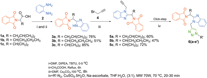
Synthesis of Pht analogues 6a–e′.
Subsequently, the alkylated products 5a–c were treated with various azides (click reactions) under microwave irradiations to acquire the final products 6a–6e′. The scope of various azides for synthesis of new Pht analogues is shown in Table S1. All the synthesized Pht analogues were characterized with various spectroscopic techniques (1H NMR, 13C NMR, HRMS and IR spectroscopy, etc.). In addition, single crystals were grown for one representative analogue 6a and subjected to single crystal X-ray diffraction. The molecular diagram of 6a is depicted in Fig. 2.
Figure 2.
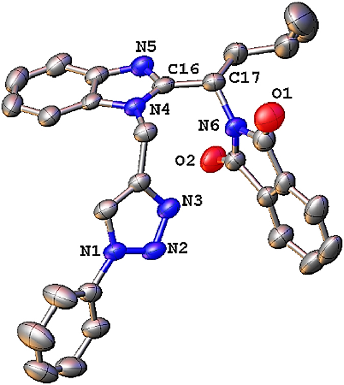
Molecular structure of 6a at 40% probability level.
Compound 6a crystallized in the monoclinic system with a C2/c space group. The details of data collection, structure solution and refinement are listed in Table S2.
Biological Studies and Structure-Activity relationships (SARs) Analysis
Antimalarial activity of all the listed Pht analogues was initially assessed on asynchronous cultures of P. falciparum 3D7 (Pf3D7) clone using the SYBR Green assay. The most active compounds were those with a mean 50% growth inhibitory concentration (IC50) < 5.14 µM, set as reference cut-off IC50 value based on the Pht (I) reference molecule mean IC50. The mean IC50 values are based on three separate experiments (Table 1). Four new analogues 6a, 6 h, 6 m and 6 u displayed antiplasmodial activity against the Pf3D7 strain with IC50 < 5.14 µM. In addition, all four compounds inhibited the growth of CQ resistant PfW2 strain with IC50 concentrations below the reference cut-off IC50; 6a IC50 = 0.7 (±0.01) µM, 6 h IC50 = 1.3 (±0.11) µM, 6 m IC50 = 3.8 (±0.34) µM and 6 u IC50 = 0.9 (±0.6) µM. Twelve out of the 31 Pht analogues lacked a dose-dependent effect on parasite growth, hence the IC50 was unattainable (no dose response, NDR). CQ and DHA were tested alongside the Phts as quality assurance and control of the assay, and SYBR Green derived IC50 values for both standard antimalarials were as recommended by literature30.
Table 1.
SAR Study of R′ Substituent: Aromatic Rings. Note: Illustration of Pht analogues, Pht (I), and chloroquine and dihydroartemisinin IC50 values.
| Entry | Compound | R | R′ | IC50 (SE) µM | IC50 (SE) µg/mL |
|---|---|---|---|---|---|
| 1 | 6a | CH2CH(CH3)2 |

|
0.9 (±0.14) | 0.7 (± 0.03) |
| 2 | 6b | CH2CH(CH3)2 |

|
NDR | NDR |
| 3 | 6c | CH2CH(CH3)2 |

|
17.3 (± 0.0) | 8.8 (± 0.0) |
| 4 | 6d | CH2CH(CH3)2 |

|
NDR | NDR |
| 5 | 6e | CH2CH(CH3)2 |

|
25 (± 1.5) | 13 (± 0.8) |
| 6 | 6f | CH(CH3)CH2CH3 |

|
NDR | NDR |
| 7 | 6g | CH(CH3)CH2CH3 |

|
NDR | NDR |
| 8 | 6h | CH(CH3)CH2CH3 |

|
0.9 (± 0.0) | 0.7 (± 0.0) |
| 9 | 6i | CH(CH3)CH2CH3 |

|
40 (± 6.2) | 22.5 (± 2.9) |
| 10 | 6j | CH(CH3)CH2CH3 |

|
73 (± 0.0) | 33.4 (± 0.0) |
| 11 | 6k | CH(CH3)2 |

|
30.86 (±0.0) | 16 (± 0.0) |
| 12 | 6l | CH(CH3)2 |

|
NDR | NDR |
| 13 | 6m | CH(CH3)2 |

|
3.5 (± 2.9) | 1.8 (± 1.3) |
| 14 | 6n | CH(CH3)2 |

|
9.6 (± 0.0) | 5.5 (±0.0) |
| 15 | 6o | CH(CH3)2 |

|
23.5 (±11.9) | 17.5 (±5.7) |
| 16 |
 I I
|
5.14 (±1.67) | 0.8 (±0.25) | ||
| 17 | CQ | 0.03 (±0.67) | 0.015 (±0.33) | ||
| 18 | DHA | 0.003 (±0.3) | 0.0017 (±0.16) |
IC50 value < 5.14 µM: active and IC50 value>5.14 µM: inactive.
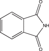
The structure-activity relationship is centred at the various substitutions of triazole ring and amino acids (Tables 1 and 2). Functionalized Phts were synthesized and screened for antimalarial activity against cultured Pf3D7 and compared with unsubstituted Pht I (IC50 = 5.14 ± 1.67 μM), CQ (IC50 = 0.03 ± 0.67 μM) and DHA (IC50 = 0.003 ± 0.3 μM). During the design of the new molecules Pht, benzimidazole scaffolds were kept constant and its flanking sides, triazoles were diversified. It is evident from the screening results (Tables 1 and 2) that the variations of R or R′ substituents play an important role in the potency of the compounds against growth of the parasite.
Table 2.
SAR of R′ Substituent Antimalarial Activity: Aliphatic and Glycoside Groups.
| Entry | Compound | R | R′ | IC50 (SE) µM | IC50 (SE) µg/mL |
|---|---|---|---|---|---|
| 1 | 6p | CH2CH(CH3)2 |

|
62 (±2.5) | 31 (±1.2) |
| 2 | 6q | CH2CH(CH3)2 |

|
8.4 (±0.5) | 4.1 (±0.3) |
| 3 | 6r | CH2CH(CH3)2 |

|
7 (±0.7) | 3.5 (±0.34) |
| 4 | 6s | CH2CH(CH3)2 |

|
No IC50 | No IC50 |
| 5 | 6t | CH2CH(CH3)2 |

|
28 (±0.7) | 14 (±0.3) |
| 6 | 6u | CH2CH(CH3)2 | H | 0.7 (±0.0) | 0.3 (±0.0) |
| 7 | 6v | CH(CH3)CH2CH3 |

|
No IC50 | No IC50 |
| 8 | 6w | CH(CH3)CH2CH3 |

|
15.6 (± 0.0) | 11.6 (± 0.0) |
| 9 | 6x | CH(CH3)CH2CH3 |

|
No IC50 | No IC50 |
| 10 | 6y | CH(CH3)CH2CH3 |

|
No IC50 | No IC50 |
| 11 | 6z | CH(CH3)CH2CH3 | H | No IC50 | No IC50 |
| 12 | 6a′ | CH(CH3)2 |

|
No IC50 | No IC50 |
| 13 | 6b′ | CH(CH3)2 |

|
7.2 (±0.0) | 3.8 (±0.0) |
| 14 | 6c′ | CH(CH3)2 |

|
No IC50 | No IC50 |
| 15 | 6d′ | CH(CH3)2 |

|
48.3 (±0.6) | 20 (±0.29) |
| 16 | 6e′ | CH(CH3)2 |

|
16.7 (±21) | 6.9 (±10.1) |
The antimalarial data demonstrates that the introduction of larger R groups (isobutyl or sec-butyl) increases the potency as noticed in case of Phts 6a, 6h and 6u whereas relatively smaller groups lower the activity (i.e. 6m). As shown in Table 1, the insertion of a phenyl substituent also influences the potency of the molecules. Pht analogues possessing 4-fluorophenyl ring on the triazoles moiety with R represented as butyl group (6h) was noticed as more active over the ananlogues containing substituted phenyl group at triazole moiety. This result, at least in part, appears to be due to the high electronegativity of fluoro group. Analogues with an unsubstituted aromatic ring also exhibited significant inhibition of the parasite growth, but only when R was replaced with an isobutyl group (e.g. 6a). In the absence of a functional group on the triazole ring with R represented as an isobutyl group, we observed the highest potency against the P. falciparum 3D7 strain (i.e. 6u).
Stage-Specificity and Effects on Parasitemia Titres
Next, we sought to determine stage-specificity of the antiplasmodial activity of the four active analogues (6a, 6h, 6m and 6u) on synchronized Pf3D7 strain cultures at 2% hematocrit and 1% parasitemia, with concentrations corresponding to individual drug IC50. To determine the effect of the compounds on both early and late ring stage parasites, the treatment was conducted on newly synchronized rings for 12 hours, and drug effect on parasite growth and morphology was monitored at 6 and 12 hours after exposure. Similarly, early trophozoites were exposed to each compound and incubated for 16 hours, and monitored at 6 and 16 hours post-exposure. The effect of the compounds on parasite morphology and development was compared in exposed and unexposed drug wells (Figs 3 and 4). Compounds 6a and 6u were active against ring-stage forms as indicated by a marked reduction in parasite density and abnormal ring stage morphology at 6 hour and 12 hour post-exposure. However, compounds 6a and 6u did not interfere with development of parasite progression from trophozoites to schizonts when they were exposed to a separate culture of trophozoites. Treatment with 6h and 6m did not arrest ring-stage maturation as indicated by the marked increase in ring-stage growth at 12 hours, like the no treatment group. However, upon examination of both 6h and 6m treatment on mature blood-stages, 6h resulted in complete destruction of trophozoites at 6 hours after drug exposure of early-stage trophozoites. Further, monitoring the effect of 6h at 16 hours showed the presence of schizonts with abnormal morphology.
Figure 3.
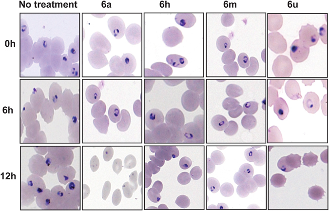
Micrographs of P. falciparum ring-stage forms treated with Pht analogues. Illustration of Pht analogues treatment effect on early erythrocytic parasite stage (rings). (All treatments were performed in parallel to a no treatment group).
Figure 4.
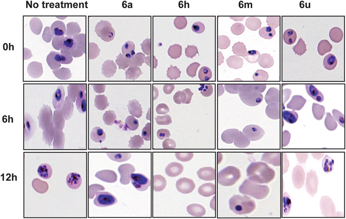
Micrographs of P. falciparum trophozoite stages treated with Pht analogues. Note: Illustration of Pht analogues treatment effect on early (rings) and mature (trophozoites and schizonts) parasite blood stages. (All treatments were performed in parallel to a no treatment group).
Although, trophozoite growth did not appear to be affected by treatment with 6m at 6 hours, the resultant schizonts appeared less granular and lacked distinguishable merozoites upon 16 hours exposure. The effect on the analogues on parasitemia counts was correlated with their stage-specificity. Analogues 6a and 6u caused a reduction in ring stage parasitemia at 6 hrs post exposure, while their effect on mature blood stage parasite titres at 16 hours was negligible (Fig. 5). Although, 6h and 6m did not affect parasite growth at 6 hours post exposure (i.e., ring-stage), both analogues caused a reduction in parasitemia at 16 hours (i.e., trophozoite stage, Fig. 5).
Figure 5.
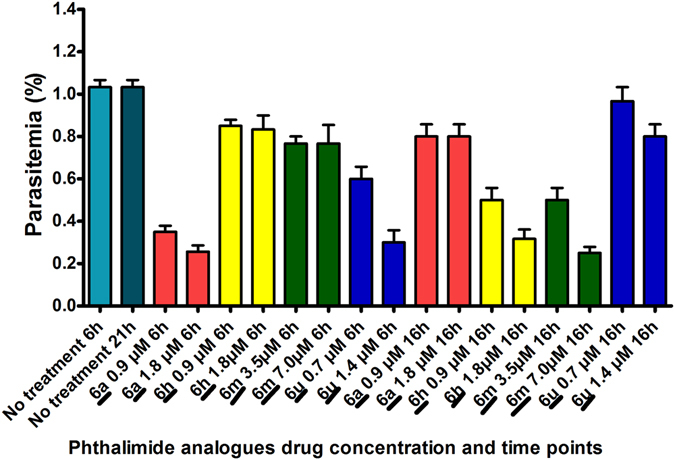
Effect of Pht analogues on parasite growth titres (Note: Graphical description of the inhibitory effect of select analogues on parasite growth titres 6 hours after incubation with ring-stages and 16 hours after drug incubation with trophozoite stage. Parasitemia percentage was derived by counting the number of infected erythrocytes from a total of 2,000 erythrocytes on Giemsa stained thin smears from each experiment. Bar diagrams represent the average of three different experiments).
Drug-Drug Interaction Assays
We then explored the synergistic drug inhibitory activities between the Pht analogues and CQ or DHA. Synergistic inhibitory activities were observed between three analogues (6a, 6h and 6u) in combination with both CQ and DHA against the 3D7 and W2 strains (Table S3). The determination of drug interactions between the analogues with CQ and DHA is necessary for identifying possible partner drugs to combat resistance to current antimalarial therapies. Recent reports on the emergence of CQ sensitive P. falciparum strains achieved in some malaria endemic regions is attributed to withdrawal of CQ pressure on parasite populations due to replacement with sulfadoxine-pyrimethamine (SP) and ACTs31, 32. CQ was an ideal antimalarial due to its pharmacokinetics, safety profile and low cost. Since P. falciparum resistance developed primarily because of administration as a monotherapy, identifying potential CQ partner drugs can beneficial in reducing development of historical parasite resistance to CQ monotherapy. Artemisinin (ART) and its derivatives are the current front line of defense against uncomplicated P. falciparum malaria and are administered as ACTs. Emergence of resistance to both ACT drug components warrants identification of ART replacement and possible combination chemotypes33–35. Notably, three Pht analogues (6a, 6h and 6u) showed synergistic activity when combined with CQ and DHA against both Pf3D7 and PfW2, suggesting their potential for use in combination therapies.
Antimalarial Effect of Pht Analogs Alone and in Combination with Artemisinin in Plasmodium berghei Infected Mice
The antimalarial effect of two active analogues, 6h and 6u (administered at 50 mg/kg of body weight), was determined in mice infected with P. berghei NK65, a strain, which results in high levels of blood-stage parasitemia. Administration of either 6h or 6u alone for four consecutive days caused suppression of the parasite load on days 5 and 8 of infection and improved survival as compared to untreated (Fig. 6A and B). However, the 6u analogue had better antimalarial efficacy than the 6h analogue (Fig. 6A). As such, we then evaluated the efficacy of 6u (50 mg/kg) in combination with artemisinin (5 mg/kg of body weight) at reduced dosage in the murine malaria model. As shown in Fig. 6, neither of the compounds delivered as monotherapy conferred clearance of parasitemia, however, co-administration of the 6u analogue with ART considerably enhanced the antimalarial efficacy by reducing the parasite load and extending survival (P < 0.05, Fig. 6A and B). The median survival times of animals treated with Pht 6u alone, as well in combination with ART were 23 and 27.5 days, respectively (P<0.001). These results demonstrate the compound 6u in combination with ART has the greatest therapeutic efficacy in the murine model of malaria.
Figure 6.
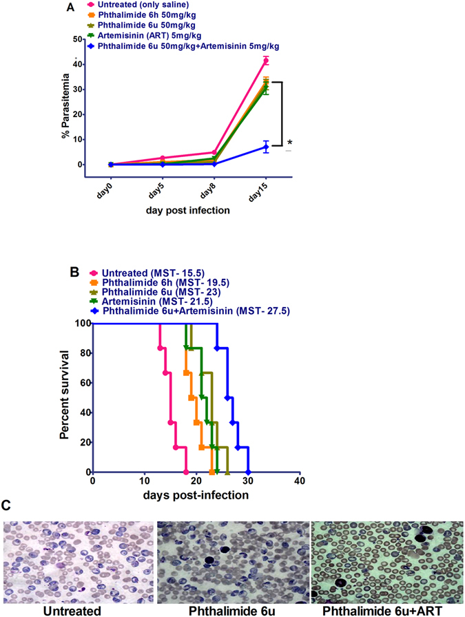
Antimalarial effect of Pht analogs (6h and 6u) alone and in combination with Artemisinin (6u and ART) on parasitemia and survival in mice infected with P. berghei NK65. Mice were injected with 1 × 10⁷ P. berghei infected RBCs. After 48 hours mice were treated with control (PBS: untreated), 6h and 6u (alone), and 6u in combination with artemisinin (all injected subcutaneously) for four consecutive days. The % of parasitemia was determined for the groups on days 5, 8 and 15 by randomly selecting 10 different optical fields on blood smears. (A) Administration of 6h and 6u (alone), and 6 u in combination with ART. Data are the mean ± SEM from six animals per treatment group. (B) Survival in the treatment groups (n = 6/group). (C) Photomicrograph of blood smears of untreated versus treatment groups at day 15 post infection (100x magnification). ART, Artemisinin; *P < 0.05.
Cytotoxicity Evaluation
As a final step in the investigational pipeline, we determined the cytotoxic effect of the four active analogues (6a, 6h, 6m and 6u) in U937 cell lines by measuring 50% cytotoxic concentrations (CC50) and calculating selectivity indices (SIs) as a measure of toxicity in human cells.
Compounds 6a and 6m displayed potent CC50 values <1 µM of 0.91 ± 0.32 µM and 0.78 ± 0.32 µM, respectively, and were considered to possess higher selectivity for U937 cell lines vs. P. falciparum (SI values of 1.01 ± 1.5 µM and 1.11 ± 1.5 µM, respectively). Analogues 6h and 6u possessed less selectivity for U937 cell lines with CC50 values of 28.82 ± 0.67 µM and 2.08 ± 1.6 µM, respectively, and SI values of 41.2 ± 1.7 µM and 2.31 ± 0.76 μM, respectively, and were, therefore, considered less toxic to human cells. As such, additional chemical modifications of 6a and 6m may be required to produce analogues with less selectivity and toxicity for human cell lines in the context of retaining their antiplasmodial activity.
Conclusion
In summary, we present simple and inexpensive chemical procedures with readily available starting materials considering the need for low-cost, novel antimalarial agents for use in malaria endemic areas. Results presented here investigated in vitro and in vivo antimalarial activity of novel Pht analogues blended with benzimidazole and triazoles, which have been synthesized by means of click reactions under microwave conditions. Amongst all active members, eight analogues displayed growth inhibition of P. falciparum in culture. The synergy of the lead compounds 6h and 6u with standard antimalarial drugs such as ART, demonstrate their suitability as combination regiments. Future studies will focus on lead optimization, pharmacokinetics and parasite target site(s) to advance Pht analogues as potential antimalarial candidates for clinical use.
Methods
Chemistry
Solvents and reagents were purchased from commercial sources and used without purification for the experiments. Homogeneity/purity of all the products was assayed by thin-layer chromatography (TLC) on alumina-coated plates (Merck). Product samples in chloroform (CHCl3) were loaded on TLC plates and developed in Ethyl acetate/Petroleum ether (1:1, v/v). When slight impurities were detected by iodine vapour/UV light visualization, compounds were further purified by chromatography on silica gel columns (100–200 mesh size, CDH). Reactions using microwave were run in a closed vial applying a dedicated CEM-Discover monomode microwave apparatus operating at a frequency of 2.45 GHz with continuous irradiation power from 0–300 W (CEM Corporation, P.O. Box 200, Matthews, NC 28106). Melting points were determined on Melting point machine M-560 (Buchi). Infrared (IR) spectra were recorded in KBr medium using a Perkin-Elmer Fourier Transform-IR spectrometer, whereas 1H and 13C nuclear magnetic resonance (NMR) spectra were recorded in CDCl3 DMSO and D2O medium on a JEOL ECX-400P NMR at 400 MHz and 100 MHz, respectively at USIC, University of Delhi, using TMS as an internal standard. Absorption frequencies (ν) are expressed in cm−1, chemical shifts in ppm (δ-scale) and coupling constants (J) in Hz. Splitting patterns are described as singlet (s), doublet (d), doublet of doublet (dd), triplet (t), quartet (q) and multiplet (m). The high-resolution mass spectral data was obtained using a Agilent Technology-6530, Accurate mass, Q-TOF LCMS spectrometer at USIC, University of Delhi. Compounds 1a–1c were prepared following literature procedures36.
General Procedure for Synthesis of Compounds 3a–3c
In first step, respective compounds 1a–1c (38 mmol) were dissolved in 250 mL of DMF and DIPEA (45 mmol) was added drop-wise at 0–5 °C. After 10 minutes the essential amount of TBTU (45 mmol) was added slowly and the reaction contents were stirred for 30 minutes at the same temperature. Thereafter, o-phenylenediamine (38 mmole) was added and the resulting mixture was stirred at 0–5 °C for 6 hours. After completion of the reaction as confirmed by TLC, the reaction mixture was quenched with ice cold water, which resulted in precipitate formation. The precipitate was filtered off, washed with excess of ice cold water and dissolved in appropriate amount of ethyl acetate. The resulting organic phase was washed with 1 N HCl followed by saturated solution of NaHCO3 and at last with water. The separated ethyl acetate layer was dried over Na2SO4 and concentrated under reduced pressure to acquire the crude product. Next, the crude product was dissolved in 150 mL glacial acetic acid and the resulting suspension was refluxed for 6 hours. After completion of the reaction as confirmed by TLC, the reaction mixture was cooled to room temperature, concentrated under reduced pressure and diluted with ice cooled water. The resulting solid was filtered off and washed thoroughly with ice cold water and saturated NaHCO3 solution to obtain the desired products. The products were purified by silica gel column chromatography eluting with 20% mixture of ethyl acetate in n-hexane and the final compounds 3a–3c were isolated.
General Procedure for Synthesis of Compounds 5a-5c
In a RB flask, respective compounds 3a–3c (15 mmol), Cs2CO3 (45 mmol) and appropriate amount of DMF were mixed and the contents were heated at 100 °C for 20 minutes and subsequently, propargyl bromide (4) (22 mmol) was added drop wise. This resulted in a turbid reaction mixture, which was stirred at 100 °C for next 8 hours. After completion of the reaction as indicated by TLC, the reaction mixture was cooled to attain room temperature and concentrated under vacuo to give residue. Thereafter, ice cooled water was added to the residue and the resulting precipitate was filtered and dried. The residue was purified by silica gel column chromatography eluting with 10% mixture of ethyl acetate in n-hexane to afford the titled compounds 5a–5c.
General Synthetic Procedure of New Analogues 6a-6e’
A microwave vial was charged with respective compounds 5a–5c (1.34 mmol) and azide (2.02 mmol) and THF:H2O (4:1, v/v, 5 mL). The required amount of CuSO4.5H2O (0.27 mmol) and sodium ascorbate (0.54 mmol) was added and the vial was sealed tightly, which was heated at 70 °C under microwave irradiation of 70 W for 20 minutes. The progress of the reaction was confirmed by TLC. After completion of the reaction, the reaction mixture was transferred into a RF flask and concentrated under vacuo to give a residue that was quenched with ammonia solution and filtered off. Thus, obtained residue was purified by silica gel column chromatography eluting with appropriate mixture (~25%) of ethyl acetate in petroleum ether to afford the titled compounds 6a–6e′. The spectral data of all new compounds is described in supporting information.
Biological Evaluation
In vitro cultivation of P. falciparum
The laboratory-adapted P. falciparum strains 3D7 (Africa; CQ sensitive) and W2 (Africa; CQ resistant), were acquired from Malaria Research and Reference Reagent Resource Center (MR4) (Manassas, VA, USA) and maintained with type O +ve erythrocytes suspended in continuous complete culture medium as described37. Complete culture media consisted of 10.43 g/litre of RPMI 1640-HEPES supplemented with 10% (vol/vol) human AB serum, 92.6 mg/litre L-glutamine, 50 mg/liter hypoxanthine, 2 g/litre sodium bicarbonate (Sigma-Aldrich, St. Louis, MO). Incubation of cultures was at 37 °C and maintained in a low oxygen atmosphere (5% O2, 5% CO2, 90% N). The levels of parasitemia in the cultures were maintained at between 2 and 10%, with 5% hematocrit. Media was changed after every 24 hours and parasitemia monitored every 48 hours. Synchronous cultures were prepared by sorbitol lysis38. Stock solutions of the Pht, Pht analogues, and DHA were prepared in absolute dimethyl sulfoxide (DMSO) while CQ was prepared in distilled water. The prepared drug stock solutions were used immediately or stored at −80 °C for not longer than one month before use. Stock solutions were further diluted in serum free RPMI 1640 media (stock solution solvent final conc. 0.05%) before performing a 2-fold serial dilution to achieve dose ranges of 0.2 µM to 100 µM (Pht and derivatives), 7.8 to 2,000 nM (CQ) and 0.17 to 87.5 nM (DHA). 25 μL of the drug diluents were aliquoted into 96 well plates and used immediately. Alternatively, the stock solutions were diluted in distilled water, 2-fold serial dilution performed on a 96 well plate and allowed to air dry in a laminar flow hood after which dried plates were stored at 4 °C until use. SYBR Green I assay technique with additional modification was used for drug susceptibility testing of the parasites. Parasite cultures of >1% parasitemia were diluted to 1% parasitemia and 2% hematocrit and 200 μL were transferred onto drug pre-dosed plates and incubated at 37 °C for 90 hours39, 40. Drug exposure was terminated by freezing the drug plates at −80 °C for 24 hours after which lysis buffer containing (per liter) 100 mM Tris-HCl, 10 mM EDTA, 0.016% Saponin, 1.6% triton X-100 and 20X SYBR Green I dye was added and the sample incubated for 2 hours in the dark. Relative fluorescence units (RFU) were read using the Perkin Elmer Wallac 1420 fluorescence plate reader, with excitation and emission wavelengths of 485 nm and 535 nm respectively. The readouts values were aligned with corresponding drug doses and analysis performed by Graph-pad Prism software. The drug concentrations (x value) were transformed using X = Log[X] and plotted against the counts (y values) and the data analysed by non-linear regression (sigmoid dose-response/variable slope equation) to yield the IC50.
Drug interaction combination assays and analysis
A modified fixed-ratio isobologram method was used to assess interaction between CQ/DHA and Pht analogues41. Briefly, a total of 5 solutions containing fixed-ratio mixtures of Pht derivatives with either CQ or DHA were prepared in the following ratios: 1:1, 2:1, 3:2, 2:3, and 1:4. The starting concentrations for ten 2 fold-serial dilutions across the microtiter plate were assigned so that the IC50 of each drug would be in the 5th serial dilution of the plate. Each drug was tested alone and at fixed ratios of its IC50. The assessment of drug interaction was based on the calculation of the fractional inhibitory concentration (FIC) of two drugs in the 4 combination ratios. FIC was calculated for each association by dividing the IC50 of the drug in the combination by the IC50 of the drug alone. The sum of these two FIC (∑FIC); (∑FIC = 1) indicates an additive effect between drug A and drug B, (∑FIC < 1), suggesting a synergistic effect and (∑FIC > 1) indicates antagonism.
Measurement of cytotoxic activity in U937 cells
Cytotoxicity of the 4 select Pht compounds on human cells was evaluated by assessing cell viability of the U937, a human acute monocyte leukemia cell lines by use of the MTT assay42. U937 was acquired from American Type Culture Collection (ATCC, Rockville, MD) and maintained in RPMI 1640 medium supplemented with 10% fetal bovine serum, and Penicillin-streptomycin (1% v/v) (Gibco, UK) at 37 °C. Robust U937 cells at a concentration of 80,000 cells/ml were plated into 96-well plates and incubated for 24 hours. Six concentrations of the Pht compounds were added in a two-fold dilution from starting concentration of 50 to 1.56 µM in triplicate and incubated with the cells for 24 hours. This was followed by addition of 10 µL MTT solution (5 mg/mL) into each well, incubated for 4 hour at 37 °C followed by addition of 50 µL DMSO to dissolve the formazan precipitate according to manufacturer’s protocol. Aliquots were drawn from each well and color intensity was measured spectrophotometrically in an ELISA plate reader (Biotek, ELx800) at 540 nm. The cell viability ratio was calculated by the following formula: % cytotoxicity = [Mean OD of test cells] − [Mean OD of control cells]/[Mean OD of control cells] × 100. Viability counts were then plotted against corresponding drug concentrations to yield cytotoxicity (CC50) through non-linear regression (sigmoid dose-response/variable slope equation). Drug CC50 values were then used to define selectivity indices, which is a measure of drug safety in human relative to parasitic cells and was calculated as the ratio of the CC50 value determined on the U937 cells (cytotoxicity) and the IC50 value determined on parasite growth inhibition 3D7 (anti-plasmodial activity). In this study, we set SI > 2 as cut-off as an indicative of low drug cytotoxicity.
In vivo experiments
All animal experiments were performed in female Swiss albino mice (4 to 5 weeks old, weighing 25 to 30 g). The animals were housed under standard controlled conditions at 25 °C with a 12-h light-dark cycle and access to sterilized food pellets and water. All experiments were carried out in accordance with the standard procedures approved by the Animal Ethics Committee of the University of Delhi South Campus, under the Control and Supervision of Experiments on Animals (CPCSEA), Ministry of Social Justice and Empowerment, Government of India. To examine the therapeutic efficacy of most potent Pht analogues 6h and 6u, a murine model of malaria was developed by intraperitoneal i.p. administration of standard inoculum of rodent strain of P. berghei Nk65 carrying 1 × 10⁷ parasitized erythrocytes per 200 µl volume to each experimental Swiss albino mouse. The antimalarial activity was carried out in accordance with a slightly modified version of the Peter’s 4-day suppressive test43. The animals were assigned to each group (n = 6). Subsequently, after 48 hour of postinfection, the parasitemia level reached 1 to 2%, and all the groups of mice were treated by subcutaneous (s.c.) injection with compounds 6h and 6u alone as well in combination with artemisinin solubilized in DMSO. One group was kept as a control and treated with physiological saline. The efficacy of the treatment was monitored by measuring the parasitemia and survival on days 5, 8 and 15 posttreatments by obtaining thin smears of blood withdrawn from the tail vein of infected mice and staining with 10% Giemsa. The level of parasitemia was determined by counting infected and noninfected erythrocytes from 10 to 15 randomly selected optical fields at 100x magnification and expressed as the number of infected erythrocytes per 100 erythrocytes. The survival of mice was recorded and observed for external symptoms, such as change in body weight, ruffled fur, lethargy and paralysis, until 30 or 40 days posttreatment. The reduction in the level of parasitemia was taken as the index for the curative activities of the drugs. The percentage of parasitemia was calculated manually with the Cell Counting Aid software44 using the formula (total no. of parasitized RBCs)/(total no. of RBCs) × 100.
Statistical analysis
For in-vivo experiments, statistical differences between two groups were determined by Student’s t test and between multiple groups using one-way analysis of variance (ANOVA), with P values of <0.05, by GraphPad Prism (version 5.01; GraphPad Software, Inc., CA). The survival of the mice was followed up to day 30 or 40 postinfection using Kaplan-Meier survival analysis, and statistical differences in animal survival were analyzed by a log rank test.
Electronic supplementary material
Acknowledgements
This work was supported by Science and Engineering Research Board (ECR/2015/000448), New Delhi, Government of India. We acknowledge University of Delhi for instrumentation facility. BR, an UGC-Raman postdoctoral fellow at MIT is highly grateful to his advisor Prof. Alexander M Klibanov, Novartis-Chair Professor, Department of Chemistry, MIT for his suggestions and encouragement. BKS acknowledges DST (INT/UKR/2012/P-10), New Delhi, India for financial assistance.
Author Contributions
Conceived and designed the experiments: Brijesh Rathi. Performed chemical synthesis and characterization: Prashant Kumar. Performed in vitro experiments: Angela O. Achieng, Prakasha, Kempaiah. Performed in vivo experiments: Vinoth Rajendran. Contributed reagents/materials/analysis tools: Brijesh Rathi, P.C. Ghosh, D.J. Perkins. All the authors helped in writing the paper and review independently.
Competing Interests
The authors declare that they have no competing interests.
Footnotes
Publisher's note: Springer Nature remains neutral with regard to jurisdictional claims in published maps and institutional affiliations.
Contributor Information
Prakasha Kempaiah, Email: Pkempaiah@salud.unm.edu.
Brijesh Rathi, Email: brijeshrathi@hrc.du.ac.in.
Electronic supplementary material
Supplementary information accompanies this paper at doi:10.1038/s41598-017-06097-z
References
- 1.World Malaria Report 2015; World Health Organization (2015).
- 2.Flannery EL, Chatterjee AK, Winzeler EA. Antimalarial drug discovery [mdash] approaches and progress towards new medicines. Nat Rev Microbiol. 2013;11:849. doi: 10.1038/nrmicro3138. [DOI] [PMC free article] [PubMed] [Google Scholar]
- 3.Bhatt S, et al. The effect of malaria control on Plasmodium falciparum in Africa between 2000 and 2015. Nature. 2015;526:207–211. doi: 10.1038/nature15535. [DOI] [PMC free article] [PubMed] [Google Scholar]
- 4.Galatas B, Bassat Q, Mayor A. Malaria Parasites in the Asymptomatic: Looking for the Hay in the Haystack. Trends Parasitol. 2016;32:296. doi: 10.1016/j.pt.2015.11.015. [DOI] [PubMed] [Google Scholar]
- 5.Bretscher MT, et al. The distribution of Plasmodium falciparum infection durations. Epidemics. 2011;3:109–118. doi: 10.1016/j.epidem.2011.03.002. [DOI] [PubMed] [Google Scholar]
- 6.Gamo FJ, et al. Thousands of chemical starting points for antimalarial lead identification. Nature. 2010;465:305–310. doi: 10.1038/nature09107. [DOI] [PubMed] [Google Scholar]
- 7.Hanboonkunupakarn B, White NJ. The threat of antimalarial drug resistance. Tropical Diseases, Travel Medicine and Vaccines. 2016;2:1–5. doi: 10.1186/s40794-016-0027-8. [DOI] [PMC free article] [PubMed] [Google Scholar]
- 8.Corey VC, et al. A broad analysis of resistance development in the malaria parasite. Nature Commun. 2016;15:11901. doi: 10.1038/ncomms11901. [DOI] [PMC free article] [PubMed] [Google Scholar]
- 9.Dondorp AM, et al. Artemisinin resistance in Plasmodium falciparum malaria. N. Engl. J. Med. 2009;361:455–467. doi: 10.1056/NEJMoa0808859. [DOI] [PMC free article] [PubMed] [Google Scholar]
- 10.Tun KM, et al. Spread of artemisinin-resistant Plasmodium falciparum in Myanmar: a cross-sectional survey of the K13 molecular marker. Lancet Infect. Dis. 2015;15:415–421. doi: 10.1016/S1473-3099(15)70032-0. [DOI] [PMC free article] [PubMed] [Google Scholar]
- 11.Phyo AP, et al. Emergence of artemisinin-resistant malaria on the western border of Thailand: a longitudinal study. Lancet. 2012;379:1960–1966. doi: 10.1016/S0140-6736(12)60484-X. [DOI] [PMC free article] [PubMed] [Google Scholar]
- 12.Takala-Harrison S, et al. Independent Emergence of Artemisinin Resistance Mutations Among Plasmodium falciparum in Southeast Asia. J. Infect. Dis. 2015;211:670–679. doi: 10.1093/infdis/jiu491. [DOI] [PMC free article] [PubMed] [Google Scholar]
- 13.Wells TN, Van Huijsduijnen RH, Van Voorhis WC. Malaria medicines: a glass half full? Nat Rev Drug Discov. 2015;14:424–442. doi: 10.1038/nrd4573. [DOI] [PubMed] [Google Scholar]
- 14.Vennerstrom JL, et al. Identification of an antimalarial synthetic trioxolane drug development candidate. Nature. 2004;430:900–904. doi: 10.1038/nature02779. [DOI] [PubMed] [Google Scholar]
- 15.Shiheido H, et al. A phthalimide derivative that inhibits centrosomal clustering is effective on multiple myeloma. PLoS ONE. 2009;7:e38878. doi: 10.1371/journal.pone.0038878. [DOI] [PMC free article] [PubMed] [Google Scholar]
- 16.Coelho LCD, et al. Novel phthalimide derivatives with TNF-α and IL-1β expression inhibitory and apoptotic inducing properties. Med. Chem. Commun. 2014;5:758–765. [Google Scholar]
- 17.Sharma U, Kumar P, Kumar N, Singh B. Recent advances in the chemistry of phthalimide analogues and their therapeutic potential. Mini Rev. Med. Chem. 2010;10:678–704. doi: 10.2174/138955710791572442. [DOI] [PubMed] [Google Scholar]
- 18.Horvat M, et al. Evaluation of antiproliferative effect of N-(alkyladamantyl)phthalimides in vitro. Chem. Biol. Drug. Des. 2012;79:497–506. doi: 10.1111/j.1747-0285.2011.01305.x. [DOI] [PubMed] [Google Scholar]
- 19.Papp K, et al. Efficacy of apremilast in the treatment of moderate to severe psoriasis: a randomised controlled trial. Lancet. 2012;380:738–46. doi: 10.1016/S0140-6736(12)60642-4. [DOI] [PubMed] [Google Scholar]
- 20.González MA, Clark J, Connelly M, Rivas F. Antimalarial activity of abietane ferruginol analogues possessing a phthalimide group. Bioorg. Med. Chem. Lett. 2014;24:5234–5237. doi: 10.1016/j.bmcl.2014.09.061. [DOI] [PubMed] [Google Scholar]
- 21.Singh AK, et al. Design, synthesis and biological evaluation of functionalized phthalimides: a new class of antimalarials and inhibitors of falcipain-2, a major hemoglobinase of malaria parasite. Bioorg. Med. Chem. 2015;23:1817–1827. doi: 10.1016/j.bmc.2015.02.029. [DOI] [PubMed] [Google Scholar]
- 22.Singh AK, et al. Hydroxyethylamine Based Phthalimides as New Class of Plasmepsin Hits: Design, Synthesis and Antimalarial Evaluation. Plos ONE. 2015;10:e0139347. doi: 10.1371/journal.pone.0139347. [DOI] [PMC free article] [PubMed] [Google Scholar]
- 23.Camacho J, et al. Synthesis and biological evaluation of benzimidazole-5-carbohydrazide derivatives as antimalarial, cytotoxic and antitubercular agents. Bioorg. Med. Chem. 2011;19:2023–2029. doi: 10.1016/j.bmc.2011.01.050. [DOI] [PubMed] [Google Scholar]
- 24.Ndakala AJ, et al. Antimalarial Pyrido[1,2-a]benzimidazoles. J. Med. Chem. 2011;54:4581–4589. doi: 10.1021/jm200227r. [DOI] [PubMed] [Google Scholar]
- 25.Patil V, et al. Antimalarial and antileishmanial activities of histone deacetylase inhibitors with triazole-linked cap group. Bioorg. Med. Chem. 2010;18:415–425. doi: 10.1016/j.bmc.2009.10.042. [DOI] [PMC free article] [PubMed] [Google Scholar]
- 26.Lentz CS, et al. In Vitro Activity of wALADin Benzimidazoles against Different Life Cycle Stages of Plasmodium Parasites. Antimicrob. Agents Chemother. 2015;59:654–658. doi: 10.1128/AAC.02383-14. [DOI] [PMC free article] [PubMed] [Google Scholar]
- 27.Magistrado PA, et al. Plasmodium falciparum Cyclic Amine Resistance Locus (PfCARL), a Resistance Mechanism for Two Distinct Compound Classes. ACS Infect. Dis. 2016;2:816–826. doi: 10.1021/acsinfecdis.6b00025. [DOI] [PMC free article] [PubMed] [Google Scholar]
- 28.Devender N, et al. Identification of β-Amino alcohol grafted 1,4,5 trisubstituted 1,2,3-triazoles as potent antimalarial agents. Eur. J. Med. Chem. 2016;109:187–198. doi: 10.1016/j.ejmech.2015.12.038. [DOI] [PubMed] [Google Scholar]
- 29.Adimulam CS, et al. Design, Synthesis and Biological Evaluation of Novel Fluorinated Heterocyclic Hybrid Molecules Based on Triazole & Quinoxaline Scaffolds Lead to Highly Potent Antimalarials and Antibacterials. Lett. Drug Des. Discov. 2015;12:393–407. [Google Scholar]
- 30.Traore K, et al. Drying anti-malarial drugs in vitro tests to outsource SYBR green assays. Malaria J. 2015;14:90. doi: 10.1186/s12936-015-0600-z. [DOI] [PMC free article] [PubMed] [Google Scholar]
- 31.Mekonnen SK, et al. Return of chloroquine-sensitive Plasmodium falciparum parasites and emergence of chloroquine-resistant Plasmodium vivax in Ethiopia. Malar. J. 2014;13:244. doi: 10.1186/1475-2875-13-244. [DOI] [PMC free article] [PubMed] [Google Scholar]
- 32.Kiarie WC, Wangai L, Agola E, Kimani FT, Hungu C. Chloroquine sensitivity: diminished prevalence of chloroquine-resistant gene marker pfcrt-76 13 years after cessation of chloroquine use in Msambweni, Kenya. Malar J. 2015;14:328. doi: 10.1186/s12936-015-0850-9. [DOI] [PMC free article] [PubMed] [Google Scholar]
- 33.Dondorp AM, et al. Artemisinin resistance: current status and scenarios for containment. Nat. Rev. Microbiol. 2010;8:272–280. doi: 10.1038/nrmicro2331. [DOI] [PubMed] [Google Scholar]
- 34.Yeung S, Socheat D, Moorthy VS, Mills AJ. Artemisinin resistance on the Thai-Cambodian border. Lancet. 2009;374:1418–1419. doi: 10.1016/S0140-6736(09)61856-0. [DOI] [PubMed] [Google Scholar]
- 35.Leang R, et al. Efficacy of Dihydroartemisinin-Piperaquine for Treatment of Uncomplicated Plasmodium falciparum and Plasmodium vivax in Cambodia, 2008 to 2010. Antimicrob Agents Chemother. 2013;57:818–826. doi: 10.1128/AAC.00686-12. [DOI] [PMC free article] [PubMed] [Google Scholar]
- 36.Furniss, B. S., Hannafold, A. J., Smith, P. W. G., Tatchell, A. R. Vogel’s Textbook of Practical Organic Chemistry, 5th ed.; Longman Scientific and Technical: London (1989).
- 37.Trager W, Jensen JB. Continuous culture of Plasmodium falciparum: its impact on malaria research. Int. J. Parasitol. 1997;27:989–1006. doi: 10.1016/s0020-7519(97)00080-5. [DOI] [PubMed] [Google Scholar]
- 38.Lambros C, Vanderberg JP. Synchronization of Plasmodium falciparum erythrocytic stages in culture. J. Parasitol. 1979;65:418–20. [PubMed] [Google Scholar]
- 39.Smilkstein M, Sriwilaijaroen N, Kelly JX, Wilairat P, Riscoe M. Simple and inexpensive fluorescence-based technique for high-throughput antimalarial drug screening. Antimicrob. Agents Chemother. 2004;48:1803–1806. doi: 10.1128/AAC.48.5.1803-1806.2004. [DOI] [PMC free article] [PubMed] [Google Scholar]
- 40.Johnson JD, et al. Assessment and continued validation of the malaria SYBR green I-based fluorescence assay for use in malaria drug screening. Antimicrob. Agents Chemother. 2007;51:1926–1933. doi: 10.1128/AAC.01607-06. [DOI] [PMC free article] [PubMed] [Google Scholar]
- 41.Fivelman QL, Adagu IS, Warhurst DC. Modified Fixed-Ratio Isobologram Method for Studying In Vitro Interactions between Atovaquone and Proguanil or Dihydroartemisinin against Drug-Resistant Strains of Plasmodium falciparum. Antimicrob. Agents Chemother. 2004;48:4097–4102. doi: 10.1128/AAC.48.11.4097-4102.2004. [DOI] [PMC free article] [PubMed] [Google Scholar]
- 42.Loosdrecht AA, Beelen RHJ, Ossenkoppele GJ, Broekhoven MG, Langenhuijsen MMAC. A tetrazolium-based colorimetric MTT assay to quantitate human monocyte mediated cytotoxicity against leukemic cells from cell lines and patients with acute myeloid leukemia. J. Immunol Methods. 1994;174:311–320. doi: 10.1016/0022-1759(94)90034-5. [DOI] [PubMed] [Google Scholar]
- 43.Peter W, Portus H, Robinson L. The four-day suppressive in vivo antimalarial test. Ann. Trop. Med. Parasitol. 1995;69:155–171. [Google Scholar]
- 44.Ma C, Harrison P, Wang L, Coppel RL. Automated estimation of parasitaemia of Plasmodium yoelii-infected mice by digital image analysis of Giemsa-stained thin blood smears. Malar J. 2010;9:348. doi: 10.1186/1475-2875-9-348. [DOI] [PMC free article] [PubMed] [Google Scholar]
Associated Data
This section collects any data citations, data availability statements, or supplementary materials included in this article.


