Abstract
Objectives
Selaginella bryopteris L. (family: Selaginaceae), is often used in traditional Indian systems of medicine for the prevention and cure of several disorders and for the treatment of patient with spermatorrhoea, venereal disease, constipation, colitis, urinary tract infections, fever, epilepsy, leucorrhoea, beri-beri and cancer. It is also used as a strength tonic. This study aimed to evaluate the mechanisms underlying the anti-inflammatory effects of topically administered aqueous, polar and non-polar methanolic fractions (10 mg/20 μL) of Selaginella bryopteris.
Methods
An acute oral toxicity study of Selaginella bryopteris at doses from 250 to 2,000 mg/kg body weight (bw) was performed. Aqueous, polar and non-polar methanolic extracts (10 mg/20 μL) applied topically for 5 days were evaluated for their anti-inflammatory effects against 12-tetra-O-decanoyl phorbol acetate (TPA)-induced inflammation by using the redness in the ear, the ear’s weight (edema), oxidative stress parameters, such as lipid-peroxide (LPO) and nitric oxide (NO), and the pro-inflammatory cytokines involved in inflammation, such as tumour necrosis factor (TNF)-α, interleukin (IL)-1β and IL-6. Indomethacine (0.5 mg/20 μL) was used for the positive control.
Results
Selaginella bryopteris produced no mortalities when administered orally at doses from 250 to 2,000 mg/kg bw. Topical treatment with the non-polar methanolic fraction (10 mg/20 αL) significantly suppressed redness (2.4 ± 0.5) and edema (30.4 ± 1) and effectively reduced the LPO level (32.3 ± 3.3). The NO level was (8.07 ± 0.55), and the TNF-α, IL-1β, and IL-6 levels were decreased to 69.6 ± 15.5, 7.7 ± 4.8 and 82.6 ± 5.9, respectively.
Conclusion
This study demonstrated for the first time the mechanisms underlying the anti-inflammatory effect of medicinal plants like Selaginella bryopteris and quantified the pharmacological interactions between them. The present study showed this herbal product to be a promising anti-inflammatory phytomedicine for the treatment of patients with inflammatory skin diseases.
Keywords: cytokines, indomethacin, sanjeevni, Selaginella bryopteris, skin inflammation
1. Introduction
Selaginella bryopteris (L.) (family: Selaginaceae) common name Sanjeevni, is a lithophytes pteridophytic plant, which is known for its remarkable recovery capabilities [1]. Selaginella bryopteris is the first known life-giving herb in India as the name of a wonder herb identified as Sanjeevni is mentioned in the well-known epic by the Hindi poet Tulsi das [2].
In India, it is used as a major ingredient in local pills for the treatment of patients with spermatorrhoea, venereal diseases, constipation, colitis, indigestion and urinary problems (diuretic). It is also used to treat patients who are unconscious, and to lower the body temperature in patient with fever [3–5].
In Madhya Pradesh, the herb is traditionally used as a strength tonic by members of the Gond tribes. The women in the tribes of the Bastar region of Chattishgarh use dried powders of this herb to treat gynecological problems like menstrual irregularities and leucorrhoea and to minimize labor pain [6]. Herb paste is used orally by the local indigenous people of Songhati, Sonbhadra UP. To cure patients with beri-beri and dysentery and for rejuvenation when given with cow’s milk [7].
Reported pharmacological effects are anti-bacterial [8] and anti-protozoal [9] activities, growth promote on [10], anti-stress cell death [10], relief from heat stroke and the burning sensation during urination [10], memory enhancement [11], anti-hyperglycemic activity [12], relief from stomach-ache [13] and anti-depression activity [14].
Traditional herbal medicines possess great significance and have a rational background hence interest in herbal medicines in terms of investigating the rationality of their use in modern scientific terms has been increasing in recent years. As a result traditional medicines all over the world are now being re-evaluated through extensive research on the base materials in plant species and on their therapeutic principles in order to determine their actual efficacies and any limitations on the safe use of these drugs and the results of such research are expected to widen the scope of their future use [15].
Skin inflammation is widely studied due to its complexity relevance to human health and the large number of people that suffer from chronic inflammation [16]. Skin damage is generally associated with inflammation due to skin burns or inflammatory illnesses such as psoriasis, contactor atopic dermatitis, vulgaris acne and skin wounds [17–21].
Thus this investigation is an attempt to authenticate scientifically the use of a methanolic extract of Selaginella bryopteris for the treatment of 12-tetra-O-decanoyl phorbol acetate (TPA)-induced inflammation in experimental rats in order to provide evidence supporting its use as a topical ointment in folk medicine.
2. Material and Methods
Whole plants of Selaginella bryopteris were collected in April, 2016, from the Andhra Pradesh and authenticated by K. Madhava Chetty, Venkateswara University, Tirupati, India (Voucher number 1098).
Dried powders were extracted by using methanol or water by adopting the process of maceration for approx 72 hours with occasional shaking. Both extracts aqueous and methanolic were filtered concentrated on a rotator evaporator and dried at 40°C in a water bath. The methanolic extract was defatted with petroleum ether to separate the polar fraction (MP) and the non-polar (MN) fractions.
In a vial 1 mg of TPA was dissolved in 1000 μL acetone (1 mg/mL) and was then labeled as stock. Then 150 μL were taken from the stock. And 2,850 μL of TPA were added to bring the volume to 3 mL to produce the required dose of 1 μg/20 μL.
For the preparation of the 10 mg/20 μL dose, 250 mg of dried powdered extracts aqueous (MN fraction) or methanolic (MP fraction) were suspended in 500 μL of distilled water (1 mg/ 2 μL) these test samples were then labeled as test doses of the respective extracts.
This study was conducted on laboratory animals at the National Botanical Research Institute (NBRI) Lucknow. Fresh healthy male Wistar rats 9 – 12 weeks old and weighing 120 – 140 g were obtained from the animal house facility at the Pharmacognosy and Ethnopharmacology Division, NBRI Lucknow. All animals were housed in poly-propylene cages at a stable room temperature (20 ± 2°C) with ad libitum access to food (standard rat/mouse pellets) and water. All experiments were performed in the morning in accordance with the current guidelines for the care of laboratory animals and the ethical guidelines for investigations involving experimental pain in conscious animals [22]. The protocol for the experiments on animals was approved by Committee for the Purpose of Control and Supervision of Experiments on Animals (CPCSEA), New Delhi, (approval number: IAEC CPCSEA/07/2014).
The animals were placed into six groups with six animals per group: The right ear of each animal in Group 1 the vehicle control group was topically treated with 20 μL of acetone for 5 days and in Group 2, the toxin control group-the right ear of each animal was topically treated with 20 μL of TPA (1 μg/20 μL) for 5 days. In Group 3, the positive control group the right ear of each rat was treated with 20 μL of the standard drug indomethacin (0.5 mg/20 μL) 30 minutes after TPA application for 5 days. The right ears of the animals in Groups 4 and 5 were treated topically with 10 mg/20 μL of the MN and the MP, respectively of the methanolic extract 30 minutes after TPA application for 5 days whereas the right ears of the animals in Group 6 were topically treated with the aqueous plant extract-at a dose of 10 mg/20 μL 30 minutes after TPA application for 5 days.
The left ear of each rat in each group was treated topically with the vehicle for 5 days. On the 5th day one hour after sample application all rats were sacrificed by using cervical dislocation and their ears were removed and kept in ice-cold phosphate buffer solution (PBS, pH 7.4) for further processing.
The redness of each ear of each rat was observed continuously for 5 days to ascertain any difference in the redness among the groups. The degree of redness was found to be a maximum in the toxin control group [23].
The weight of each ear of each rat was taken and the difference in the weights of the two ears was calculated. The swelling was assessed in terms of the mean weight increase of the ear [24].
Lipid per oxidation was assessed by measuring the malondialdehyde (MDA) levels based on the reaction of MDA with thiobarbituric acid [25].
The level of NO was estimated as a measure of the nitrite, a nitric oxide (NO) metabolite, in the control and the case groups because NO is a highly reactive free radical that is a strong oxidizer and remains stored in tissues as nitrates (NO3) or nitrite (NO2). Thus the NO concentration can be estimated by measuring the concentrations of NO3 and NO2 in combination. A simple technique for doing this is to monitoring the reductions of NO3 to NO2 by nitrate reductase or a metallic catalyst and then using the colorimetric Griess reaction to the measure NO2 levels (nitrite levels) [26].
All the pro-inflammatory cytokines in the supernatant of the ear homogenate of each rat were measured following the manufacture’s guidelines (Thermo Scientific).
The acute toxicity studies were conducted as per guideline 425 of the Organization of Economic and Cooperation Development (OECD), and a dose limit of 2,000 mg/kg of body weight (bw) was used. Swiss mice weighing 20 – 25 gm were used for the acute toxicity studies. The animals were divided into 5 groups with six animals per group. Group 1 being the normal control and Groups 2 to 5, receiving 250, 500, 1,000, 2,000, mg/kg bw respectively of the prepared extract of Selaginella bryopteris by mouth. The animals were observed continuously for the first 4 hour after dosing and daily thereafter for a total of 14 days to observe any changes in general behavior (restlessness, dullness, and agitation) or other physiological activities and any toxic symptoms. After periods of 24 and 72 hrs they were observed for lethality or death.
All the values are expressed as means ± standard errors of the mean (SEMs). The data were statistically analyzed by using the one-way analysis of variance (ANOVA) followed by Tukey’s test with the PRIZM 3.0 software. The statistical significance was defined as P < 0.05.
3. Results
In the toxicity studies, Selaginella bryopteris did not produce any mortality when administered orally at various doses from 250 to 2,000 mg/kg bw. However 2 hours post treatment reduced locomotion and dullness were observed in some animals treated with the higher dose of 2,000 mg/kg (Table 1).
Table 1.
Acute (oral) toxicity study in Swiss mice after 24 hours administration of non-polar methanolic extract of Selaginella bryopteris
| Group | Dose (mg/kg bw) | D/T | Period of observing sign (hours) | Obsereved toxicity |
|---|---|---|---|---|
| A | - | - | 24 | No toxicity observed |
| B | 250 | 0/5 | 24 | No toxicity observed |
| C | 500 | 0/5 | 24 | No toxicity observed |
| D | 1,000 | 0/5 | 24 | No toxicity observed |
| E | 2,000 | 0/5 | 24 | Slight dullness was observed in the first two hours |
Bw, body weight; D/T, ratio of the number of dead rats to the number of treated rats.
Period, mortality latency, time to death (in hour) after the oral administration of the extract; the number of rats in each dose group (n) was 12.
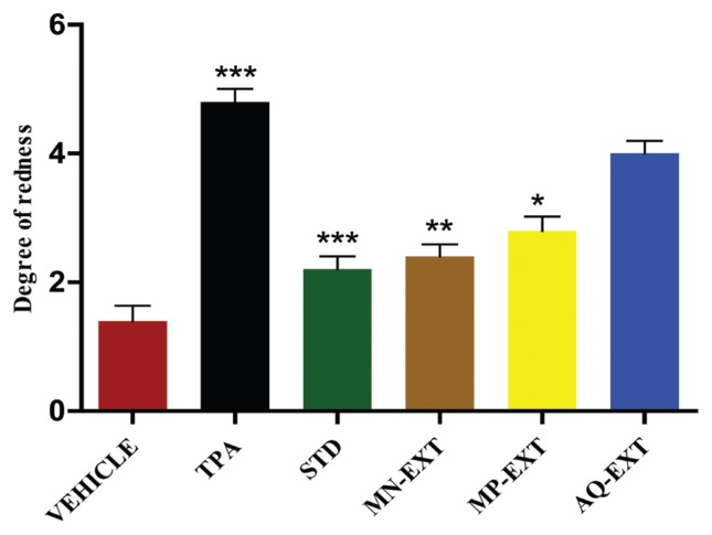
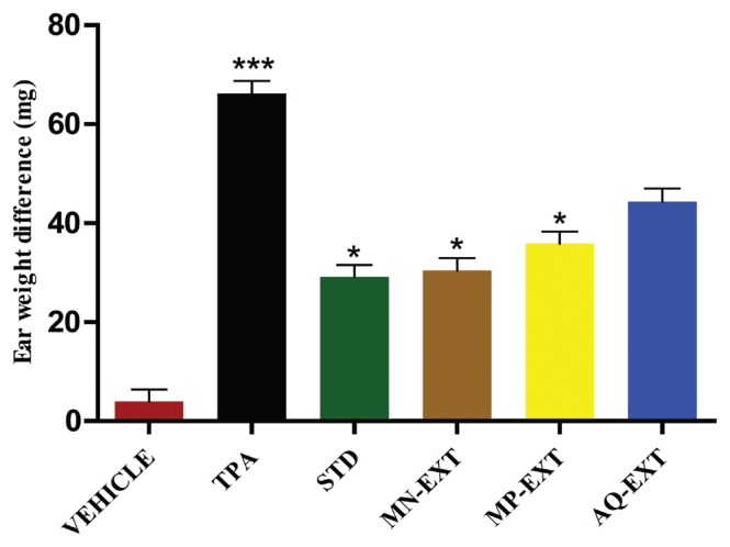
Phorbol ester in the rat’s ear induced an edemetogenic response as evidenced by a marked increase in ear redness and weight. Topical applications of the MN and the polar methanolic extracts and the aqueous extract of Selaginella bryopteris caused significant suppression of the TPA-induced skin inflammation i.e. degrees of redness of 2.4 ± 0.5 (P < 0.01), 2.8 ± 0.5 (P < 0.05), 4.0 ± 0.3 for the MN, the polar and the aqueous extract of Selaginella bryopteris respectively compared to 4.8 ± 0.2 for the toxin control group. Furthermore the edema (difference in the weights of the ears) was significantly reduced being- 30.4 ± 1.2 (P < 0.05), 35.9 ± 0.6 (P < 0.05), and 44.4 ± 2.4 for the MN the polar and the aqueous extract of Selaginella bryopteris, respectively, compared to 66.2 ± 0.8 for the TPA group (Figs. 1, 2).
Figure 1.
Degree of redness in the right ear of the rat (P < 0.05).
TPA, tetra-O-decanoyl phorbol acetate; MN, non-polar; MP, polar fraction, *P < 0.05, **P < 0.01, and ***P < 0.001.
When compared to the toxin control group. Marking from 1 to 5 are arbitrary and show the degree of redness in increasing order: 1-very low, 2-low, 3-mild 4-high and 5-very high.
Figure 2.
Difference in the weights of the right and the left ears of the rats (mg).
TPA, tetra-O-decanoyl phorbol acetate; MN, non-polar; MP, polar fraction, *P < 0.05, **P < 0.01, and ***P < 0.001.
When compared to the toxin control group. Marking from 1 to 5 are arbitrary and show the degree of redness in increasing order: 1-very low, 2-low, 3-mild 4-high and 5-very high.
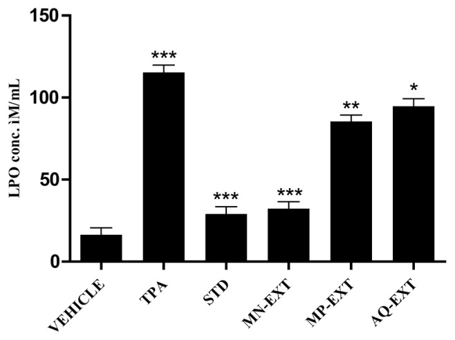
The molecular formation of MDA (i.e. lipid per-oxidation, LPO) in ear homogenate was found to be markedly reduced in the test groups. Groups 4 – 6 in comparison to the toxin control group. Group 2 (115.3 ± 5.4). Estimated levels of LPO were found to be 32.3 ± 3.3 (P < 0.001), 85.4 ± 7.4 (P < 0.01), and 94.7 ± 3.5 (P < 0.05) μM/mL for the groups of receiving the MN fraction of the methanolic extract (Group 4, MN), the polar fraction of the methanolic extract (Group 5 MP), and the aqueous extract (Groups 6 AQ) of Selaginella bryopteris, respectively. Clearly from the data, the MN fraction of the methanolic extract (MN) was more effective in reducing the LPO level than either the polar fraction of the methanolic extract (MP) or the aqueous extract (Fig. 3).
Figure 3.
Level of LPO (MDA) in ear tissue (mM/mL) (P < 0.05).
LPO, lipid-peroxide; MDA, malondialdehyde; TPA, tetra-O-decanoyl phorbol acetate; MN, non-polar, *P < 0.05, **P < 0.01, and ***P < 0.001.
When compared to the toxin control group.
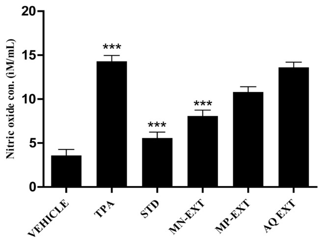
The production of NO was estimated based on the accumulation of nitrite, which is a stable product of NO metabolism, in the ear homogenates by using the Griss reagent. The levels of NO were found to be 8.07 ± 0.55 (P < 0.001), 10.8 ± 1.8, and 13.6 ± 0.92 μM/mL for the groups receiving the MN fraction of the methanolic extract (MN, Group 4), the polar fraction of the methanolic extract (MP, group 5) and the aqueous extract (AQ, Group 6) of Selaginella bryopteris respectively. Thus the reductions in the NO levels in plant-treated groups were significant compared to the level of 14.3 ± 0.4 for the toxin control group (TPA, Group 2) (Fig. 4).
Figure 4.
Level of NO in ear tissue (P < 0.05).
NO, nitric oxide; TPA, tetra-O-decanoyl phorbol acetate; MN, non-polar; MP, polar fraction, *P < 0.05, **P < 0.01, and ***P< 0.001.
When compared to the toxin control group.
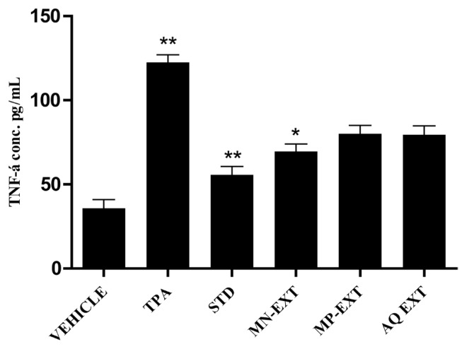
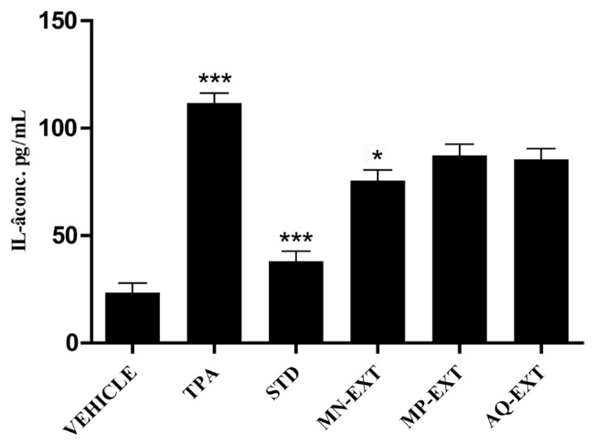
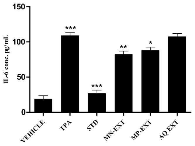
Having shown that the methanolic extracts both the MN (more effective) and the polar (less effective than MN) fractions of the plant markedly inhibited the ear’s inflammatory responses caused by the topical use of TPA, We, then investigated whether or not these inhibitions involved reductions in the levels of pro-inflammatory cytokines for tumour necrosis factor (TNF)-α: 69.6 ± 15.5 (MN Group 4, P < 0.01), 80.1 ± 3.1 (MP Group 5, P < 0.01 ), and 79.5 ± 5.3 (AQ Group 6, P < 0.01), as compared to 122.5 ± 4.5 (TPA, Group 2); for interleukin (IL)-1β: 75.7 ± 4.8 (MN Group 4, P < 0.05), 87.3 ± 5.3 (MP Group 5, P < 0.01), and 85.4 ± 1.2 (AQ Group 6, P < 0.01), as compared to 111.7 ± 5.4 (TPA, Group 2); and for IL-6- 82.6 ± 5.9 (MN Group 4, P < 0.05), 88.13 ± 4.4 (MP Group 5, P < 0.01), 107.8 ± 3.6 (AQ Group 6, P < 0.01), as compared to 109 ± 2.5 (TPA, Group 2). Topical application of TPA to the ear caused increases in the tissue levels of pro-inflammatory cytokines, which were significantly reduced after application of the MN fraction of the methanolic extract of the plant (Figs. 5, 6, 7).
Figure 5.
Level of TNF-α in ear tissue (P < 0.05).
TNF, tumour necrosis factor; TPA, tetra-O-decanoyl phorbol acetate; MN, non-polar; MP, polar fraction, *P < 0.05, **P < 0.01, and ***P < 0.001.
When compared to the toxin control group.
Figure 6.
Level of IL-1β in ear tissue (P < 0.05).
IL, interleukin; TPA, tetra-O-decanoyl phorbol acetate; MN, non-polar; MP, polar fraction, *P < 0.05, **P < 0.01, and ***P< 0.001.
When compared to the toxin control group.
Figure 7.
Level of IL-6 in ear tissue (P < 0.05).
IL, interleukin; TPA, tetra-O-decanoyl phorbol acetate; MN, non-polar; MP, polar fraction, *P < 0.05, **P < 0.01, and ***P < 0.001.
When compared to the toxin control group.
4. Discussion
The extracts of Selaginella bryopteris used in the acute toxicity study produced no deaths among the mice within 24 hours of treatment via an oral route. Thus the oral minimum lethal dose (LD50) of the methanolic extract of Selaginella bryopteris was estimated to be > 2,000 mg/kg, implying the relative safety of the plant extract when administered acutely. The major signs of toxicity noticed within the first 2 hours included slight dullness in some of the animals at the higher dose of 2,000 mg/kg administered by the oral route.
The results of this investigation provides evidence that the MN fraction of methanolic extract of Selaginella bryopteris is topically active in the attenuation of inflammation induced by TPA. The MN fraction of the extract of the plant resulted in marked inhibitions of important events related to the skin inflammatory response induced by TPA namely ear redness, ear edema, molecular formation of MDA, accumulation of NO, and increases in the tissue levels of pro-inflammatory cytokines, in a manner similar to that of Indomethacine, the reference anti-inflammatory drug. The non-polar fraction of the extract also affects the responses of pro-inflammatory cytokines by inhibiting the decrease in the levels of anti-inflammatory cytokine.
Although inflammation is a defense mechanism that helps the body to protect itself against infections, burns, toxic chemicals, allergens and other noxious stimuli, the complex events and mediators involved in the inflammatory reaction can induce, maintain or aggravate many diseases [27]. Edema, or swelling, one of the cardinal signs of acute inflammation, is an important parameter to be considered when evaluating compounds with a potential anti-inflammatory activity [28]. TPA causes increases in ear redness and weight due to hyperemia, cellular infiltration and accumulation of exuded serum [29]. The applications of the extracts (non-polar, polar, and aqueous) of the plant produced significant inhibitions of ear redness and ear weight (P < 0.05) in comparison to the control groups.
The primary action of the phorbolesteror TPA is on biological membranes. The initial membrane effects include modifications in the activities of cell receptors, altered cell adhesion, induction of arachidonic acid release, prostaglandin synthesis, inhibition of the binding of epidermal growth factor to cell surface receptors, and altered lipid metabolism. Phorbol produces a generalized change in the physical properties of the lipid phase of the membranes, and in cellular adhesion [30]. Thus the lipid per-oxidation is an important aspect of the inflammatory response. Lipid per-oxidation is a free-radical-mediated process and a potential marker of early and irreversible tissue damage. Lipid per-oxidation in-vivo destroys biological membranes leading to changes in fluidity and permeability [31]. Because free radicals are important mediators that provoke inflammatory processes their neutralizations by antioxidants and by radical scavengers can attenuate inflammation [32, 33]. In this study treatment with the MN fraction of the methanolic extract resulted in a significant fall in the level of MDA (P < 0.05) when compared to the value for the toxin control group.
NO is another important mediator in the inflammatory process and is produced at inflamed sites by inducible nitric-oxide synthase (iNOS). High levels of NO have been reported in a variety of pathological processes including various forms of inflammation, circulatory shock, and carcinogenesis [34]. Therefore, an inhibitor of (NOS) might be effective as a therapeutic agent for treating patients with inflammatory disease [35]. In this study topical application of the MN fraction of the methanolic extract (MN) of the plant reduced the levels of NO to a significant extent when compared to the level in the toxin control group.
The literature has provided evidence showing that a vast array of inflammatory mediators including prostaglandins (PGs), kinins, platelet-activating factor, leukotrienes (LTs), amines, purine, cytokines, adhesion molecules and chemokines act on specific sites (e.g. the microvasculature), leading to changes in vascular tonus and blood flow and to the local activation of leukocytes and endothelial cells [36]. In particular, cytokines initiate an inflammatory response. In this study topical applications of the plant extracts suppressed the increased levels of the proinflammatory cytokines TNF-α, IL-1β, and IL-6 (P < 0.05). The MN fraction of the methanolic extract had a much more pronounced effect against TPA-induced proinflammatory cytokines than either the polar fraction of the methanolic extract or the aqueous extract.
5. Conculsion
In this report we have provided evidence that the MN fraction of the methanolic extract of Selaginella bryopteris has topical anti-inflammatory activities by assessing its protective effects against TPA-induced skin inflammation. The anti-inflammatory effects of Selaginella bryopteris depend on down-regulation of TPA-induced expression of pro-inflammatory cytokines such as TNF-α, IL-1β and IL-6, down regulation of the expression of NO and the inhibition of TPA-induced LPO.
In conclusion, in the light of the present work, we can suggest that the non-polar fraction of the methanolic extract (MN) of Selaginella bryopteris shows topical anti-inflammatory activity against TPA-induced animal-ear inflammation. Furthermore, the present study scientifically validates its use in folk medicine.
Acknowledgment
We are highly grateful to our Honourable Director CSIR-NBRI Lucknow for the facilities provided and to the University Grant Commission (UGC-RGNSRF) for providing the fellowship. We are very much thankful to our staff especially Mr. Lalu Prasad for their constant support and encouragement throughout the study.
Footnotes
This paper meets the requirements of KS X ISO 9706, ISO 9706-1994 and ANSI/NISO Z39.48-1992 (Permanence of Paper).
Conflict of interest
The authors declare that there are no conflicts of interest.
References
- 1.Ganeshaiah KN, Vasudeva R, Uma Shaanker R. In search of Sanjeevani. Curr Sci. 2009;97(4):484–9. [Google Scholar]
- 2.Srimad Valmiki Ramayana. Yuddakanda. Slokas. 74th chapter:29–34. [Google Scholar]
- 3.Singh S, Singh R. Ethnomedicinal use of pteridophytes in reproductive health of tribal women of pachmarhi biosphere reserve, madhya pradesh, India. IJPSR. 2012;3(12):4780–90. [Google Scholar]
- 4.Singh S, Singh R. Utilization of pteridophytes of achanakmar-amarkantak biosphere reserve, central India in women’s health and beauty care practices. IRJP. 2013;4(1):235–40. [Google Scholar]
- 5.Shweta S, Singh R, Sahu TR. Floral Diversity and their conservation. Publisher Biotech Book; 2013. Ethnomedicinal uses of Pachmarhi Hills, Madhya Pradesh, India; pp. 267–90. [Google Scholar]
- 6.Shweta Singh, Singh Rita. Ethnobotany of India. Deep Publication Published; 2014. Ethnobotany of Pteridophytes in Bastar region of Chhathisgarh State. [Google Scholar]
- 7.Singh AK, Raghubanshi AS, Singh JS. Medical ethnobotany of the tribals of Sonaghati of Sonbhadra district, Uttar Pradesh, India. J Ethnopharmacol. 2002;81(1):31–41. doi: 10.1016/S0378-8741(02)00028-4. [DOI] [PubMed] [Google Scholar]
- 8.Agarwal SS, Singh VK. Immunomodulators: a review of studies on Indian medicinal plants and synthetic peptides. part I: medicinal plants. Pinsa. 1999;65(3–4):179–204. [Google Scholar]
- 9.Miki K, Nagai T, Nakamura T, Tuji M, Koyama K, Kinoshita K, et al. Synthesis and evaluation of influenza virus sialidase inhibitory activity of hinokiflavone-sialicacid conjugates. Heterocycles. 2008;75(4):879–86. doi: 10.3987/COM-07-11285. [DOI] [Google Scholar]
- 10.Sah P. Does the magical himalayan herb “Sanjeevani Booti” really exist in nature? J Am Sci. 2008;4(3):65–7. [Google Scholar]
- 11.Garg NK. Pharma Tutor. Pharmacy Infopedia; 2011. Memory enhancing activity of methanolic extract of Selaginella bryopteris in Swiss albino mice [internet] [cited 2017]. Available from: http://www.pharmatutor.org/articles/memory-enhancing-activity-of-methanolic-extract-of-selaginella-bryopteris-in-swiss-albino-mice. [Google Scholar]
- 12.Kumari R, Singh JK, Kumari P, Jha AM. Impact of Selaginella bryopteris on biochemical analysis of diabetic swiss albino mice caused induced by alloxan. Int J Basic Appli Sci Res. 2014;1(2):95–9. [Google Scholar]
- 13.Rupa P, Bhavani NL. Preliminary phytochemical screening of desiccated Fronds of Selaginella bryopteris (L.) baker (pittakalu) World J Pharm Sci. 2014;3(9):1370–8. [Google Scholar]
- 14.Chandrakant J, Mathad P, Mety S, Ahmed L, Sanaullah MD. Phytochemical and antidepressant activities of Selaginella bryopteris (L.) baker on albino mice. IJABPT. 2015;6(4):14–9. [Google Scholar]
- 15.Shah SK, Shah RP, Xu HL, Aryal UK. Biofertilizers: an alternative source of nutrients for sustainable production of tree crops. J Sustainable Agric. 2006;29(2):85–95. doi: 10.1300/J064v29n02_07. [DOI] [Google Scholar]
- 16.Alam G, Singh MP, Singh A. Wound healing potential of some medicinal plants. IJPSRR. 2011;9(1):136–45. [Google Scholar]
- 17.Gittler JK, Krueger JG, Guttman-Yassky E. Atopic dermatitis results in intrinsic barrier and immune abnormalities: implications for contact dermatitis. J Allergy Clin Immunol. 2013;131(2):300–13. doi: 10.1016/j.jaci.2012.06.048. [DOI] [PMC free article] [PubMed] [Google Scholar]
- 18.Haertel E, Werner S, Schafer M. Transcriptional regulation of wound inflammation. Semin Immunol. 2014;26(4):321–8. doi: 10.1016/j.smim.2014.01.005. [DOI] [PubMed] [Google Scholar]
- 19.Kendall A, Nicolaou A. Bioactive lipid mediators in skin inflammation and immunity. Prog Lipid Res. 2013;52(1):141–64. doi: 10.1016/j.plipres.2012.10.003. [DOI] [PubMed] [Google Scholar]
- 20.Nicolaou A, Pilkington SM, Rhodes LE. Ultraviolet-radiation induced skin inflammation: dissecting the role of bioactive lipids. Chem Phys Lipids. 2011;164(6):535–43. doi: 10.1016/j.chemphyslip.2011.04.005. [DOI] [PubMed] [Google Scholar]
- 21.Wagener F, Carels CE, Lundvig DMS. Targeting the redox balance in inflammatory skin conditions. Int J Mol Sci. 2013;14(5):9126–67. doi: 10.3390/ijms14059126. [DOI] [PMC free article] [PubMed] [Google Scholar]
- 22.Zimmerman M. Ethical guidelines for investigation of experimental pain in conscious animals. Pain. 1983;16(2):109–10. doi: 10.1016/0304-3959(83)90201-4. [DOI] [PubMed] [Google Scholar]
- 23.Chang J, Blazek E, Skowronek M, Marinari L, Carlson RP. The anti-inflammatory action of guanabenz is mediated through 5-lipoxygenase and cyclo-oxygenase inhibition. Eur J Pharmacol. 1987;142(2):197–205. doi: 10.1016/0014-2999(87)90108-7. [DOI] [PubMed] [Google Scholar]
- 24.Rasadah MA, Khozirah S, Aznie AA, Nik MM. Anti-inflammatory agents from Sandoricum koetjape Merr. Phytomedicine. 2005;11(2–3):261–3. doi: 10.1078/0944-7113-00339. [DOI] [PubMed] [Google Scholar]
- 25.Shafiq-ur-Rehman, Rehman S, Chandra O, Abdulla M. Evaluation of malonaldehyde as an index of lead damage in rat brain homogenates. Biometals. 1995;8(4):275–9. doi: 10.1007/BF00141599. [DOI] [PubMed] [Google Scholar]
- 26.Menaka KB, Ramesh A, Thomas B, Kumari NS. Estimation of nitric oxide as an inflammatory marker in periodontitis. J Indian Soc Periodontol. 2009;13(2):75–8. doi: 10.4103/0972-124X.55842. [DOI] [PMC free article] [PubMed] [Google Scholar]
- 27.Sosa S, Balick MJ, Arvigo R, Esposito RG, Pizza C, Altinier G, et al. Screening of the topical anti-inflammatory activity of some Central American plants. J Ethnopharmacol. 2002;81(2):211–5. doi: 10.1016/S0378-8741(02)00080-6. [DOI] [PubMed] [Google Scholar]
- 28.Morris CJ. Carragenan-induced paw edema in rat and mouse. Methods Mol Biol. 2003;225:115–21. doi: 10.1385/1-59259-374-7:115. [DOI] [PubMed] [Google Scholar]
- 29.Ferrandez-Arche A, Saenz MT, Arroyo M, de la Puerta R, Garcia MD. Topical anti-inflammatory effect of tirucallol a triterpene isolated from Euphorbia lactea latex. Phytomedicine. 2010;17(2):146–48. doi: 10.1016/j.phymed.2009.05.009. [DOI] [PubMed] [Google Scholar]
- 30.Weinstein IB, Lee LS, Fisher PB, Mufson A, Yamasaki H. Action of phorbol esters in cell culture. mimicry of transformation altered differentiation and effects on cell membranes. J Supramol Struct. 1979;12(2):195–208. doi: 10.1002/jss.400120206. [DOI] [PubMed] [Google Scholar]
- 31.Torres RL, Torres IL, Gamaro GD, Foretell FU, Silveira PP, Moreina JS, et al. Lipid peroxidation and total redical-trapping potential of the lungs of the rats submitted to chronic and sub-chronic stress. Braz J Med Biol Res. 2004;37(2):185–92. doi: 10.1590/S0100-879X2004000200004. [DOI] [PubMed] [Google Scholar]
- 32.Delporte C, Backhousea N, Erazoa S, Negretea R, Vidala P, Silvab X, et al. Analgesic-antiinflammatory properties of Proustia pyrifolia. J Ethnopharmacol. 2005;99(1):119–24. doi: 10.1016/j.jep.2005.02.012. [DOI] [PubMed] [Google Scholar]
- 33.Geronikaki AA, Gavals AM. Antioxidant and inflammatory diseases: synthetic and natural anti-oxidant with anti-inflammatory activity. Comb Chem High Thoughput Screen. 2006;9(6):425–42. doi: 10.2174/138620706777698481. [DOI] [PubMed] [Google Scholar]
- 34.Ohshima H, Bartsh H. Chronic infection and inflammatory processes as cancer risk factors: possible role of nitric oxide in carcinogenesis. Mutat Res. 1994;305(2):253–64. doi: 10.1016/0027-5107(94)90245-3. [DOI] [PubMed] [Google Scholar]
- 35.Koo HJ, Song YS, Kim HJ, Lee YH, Hong SM, Kim SJ, et al. Anti-inflammatory effects of genipin, an active principle of gardenia. Eur J Pharmacol. 2004;495(2–3):201–8. doi: 10.1016/j.ejphar.2004.05.031. [DOI] [PubMed] [Google Scholar]
- 36.Calixo JB, Otukai MF, Santos AR. Anti-inflammatory compounds of plant origin part I action on arachidonic acid pathway, nitric oxide and nuclear factor kappa, B (NF kappa B) Planta Med. 2003;69(11):973–83. doi: 10.1055/s-2003-45141. [DOI] [PubMed] [Google Scholar]


