



Abstract
Objective
To provide descriptive data on ambulatory ability and muscle strength in a large cohort of individuals with spina bifida enrolled in a National Spina Bifida Patient Registry (NSBPR) and to investigate factors associated with ambulatory status.
Design
Cross-sectional analysis of data from a multi-site patient registry
Results
Descriptive analysis of mobility variables for 2604 individuals with spina bifida age 5 and above are presented from 19 sites in the United States. Analysis of a subset of NSBPR data from 380 individuals from three sites accompanied by data from a specialized spina bifida electronic medical record revealed that those with no history of a shunt, lower motor level, and no history of hip or knee contracture release surgery were more likely to be ambulatory at the community level than at the household or wheelchair level.
Conclusion
This study is the first to examine factors associated with ambulatory status in a large sample of individuals with myelomeningocele and non-myelomeningocele subtypes of SB. Results of this study delineate the breadth of strength and functional abilities within the different age groups and subtypes of SB. The results may inform clinicians of the characteristics of those with varying ambulatory abilities.
Keywords: rehabilitation, mobility, spina bifida, spinal dysraphism, walking, wheelchair
Introduction
Spina bifida (SB) is caused by incomplete closure of the neural tube of the spine and is the most common congenital condition that results in physical disability.1 Approximately 166,000 Americans are living with the more severe types of SB.1 The physical manifestations of SB may include partial or complete paralysis of the lower limbs and/or trunk muscles and orthopedic deformities of the spine and limbs.2 Individuals with SB have a wide spectrum of functional abilities ranging from ambulation in the community to achieving mobility through an attendant propelled or power chair.3
Several single site studies4–12 conducted in the last decade have identified negative associations between ambulatory ability and factors such as level of spinal lesion, shunt status, and spasticity in those with upper motor neuron complications like tethered cord syndrome. These studies utilized different classification systems rather than using one common system to define the severity of the spinal lesion (i.e., anatomic level of the lesion versus level based on motor and/or sensory findings). Bartonek, et al.13 compared six common classification systems14–19 and recommended use of manual muscle testing of specific muscle groups as the best approach.
This study had two aims; the first was to describe ambulatory ability and muscle strength in a large cohort of individuals with SB enrolled in the National Spina Bifida Patient Registry (NSBPR),20 a project formed through a collaborative partnership between the Centers for Disease Control and Prevention (CDC) and the Spina Bifida Association. The NSBPR includes detailed data on individuals with SB from 19 sites in the U.S. starting in 2009. A second aim was to investigate factors associated with different levels of ambulatory status in individuals with SB. We hypothesized that a significant association exists between ambulatory status and the following variables: SB subtype, motor level, history of shunted hydrocephalus, and prior history of tethered cord release.
Methods
The NSBPR is a secure database comprised of several required key variables collected at each participating site. Examples of data in the registry are socio-demographic information, type of SB, shunting for hydrocephalus, bowel and bladder management strategies, urologic surgeries, motor level, and ambulatory status.20 The NSBPR is linked to an electronic medical record (EMR) designed specifically for individuals with SB. This EMR allows for collection of additional SB specific variables not included in the NSBPR such as surgical release of tethered cord or hip, knee, foot and ankle orthopedic surgeries.
All data in this study were collected under each participating institution’s Institutional Review Board (IRB) approved protocols. A multi-site IRB was not required. Inclusion criteria were diagnosis of myelomeningocele, meningocele, lipomyelomeningocele or fatty filum/tethered cord; adult participants who were their own medical power of attorney must have been able to give written informed consent; and a parent, guardian, or medical power of attorney must have been able to give written informed consent by proxy if the subject was a child or was unable to make his or her own medical decisions. Exclusion criterion was any other type of spinal dysraphism such as split cord malformation or myelocystocele. Data from individuals ages 5 and up were analyzed since motor function prior to this age is often unreliable.21 Data collection occurred at the initial enrollment of each participant. Trained investigators at each of the 19 participating sites interviewed participants and also conducted a review of medical records. This study design utilized two data sets to explore what factors are associated with ambulation status.
Phase I of the study extracted data only from the NSBPR. These variables were collected from 19 sites participating in the registry from 2009–2012 and are shown in Figure 1. Motor level was based on manual muscle testing, which included both left and right side. The overall level assigned was that of the more impaired side. The Hoffer classification16 was used to define ambulatory status based on the historic four categories (see Figure 1).
Figure 1.
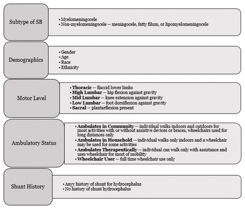
Phase I NSBPR variables
Phase II of the study utilized data from a subset of three of the national sites (University of Pittsburgh Medical Center and Children’s Hospital of Pittsburgh, Wayne State University/Detroit Medical Center and Children’s Hospital of Michigan, and Children’s Hospital Colorado). These sites had collected additional data in the EMR beyond those in the NSBPR in order to create a more robust dataset. A more extensive neurosurgical and orthopedic history was therefore available for analysis (see Figure 2).
Figure 2.
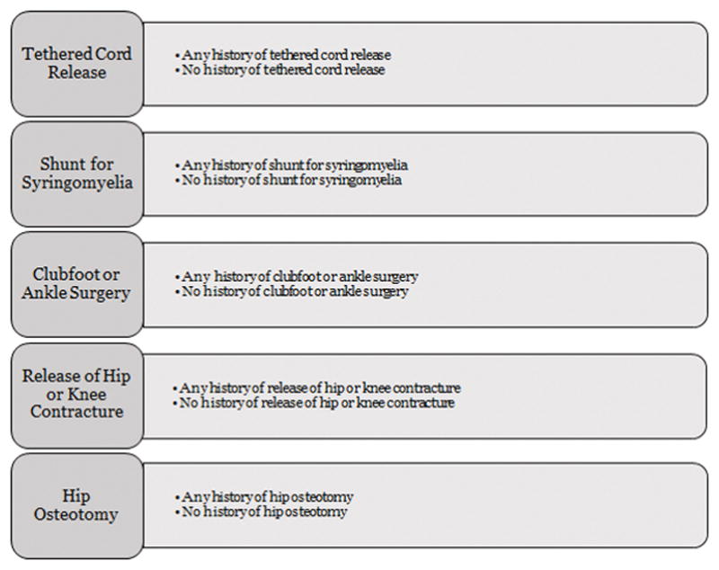
Phase II Additional EMR Variables
Analysis
Statistical Analysis Software 9.4 and IBM SPSS version 21 were used for all data analyses. Univariate analyses were used to describe distributions of characteristics of participants in each of the two Phases. To determine if participants in Phase II were similar to those in Phase I, the groups were divided into myelomeningocele and non-myelomeningocele (meningocele, fatty filum, and lipomyelomeningocele) subtypes and then compared with respect to age (categorized as 5–10yrs, 11–15yrs, 16–20yrs, 21–35yrs, 36–50yrs, and 51+), gender, ethnicity, race, shunt history, ambulation status and motor level (categorized as thoracic/high lumbar, mid-lumbar, and low lumbar/sacral) using bivariate analyses. Spearman’s correlations were run to determine whether age was related to either ambulatory status or motor level.
For Phase II, bivariate analyses were used to determine the association of independent variables in Figs. 1 and 2 with ambulation status, as well as to determine if interactions existed among independent variables. Variables that were associated with ambulation status were included, and collinear variables were excluded. Then, an ordered logistic regression model was built with ambulatory status as the dependent variable (4 levels). This model was chosen because all assumptions of the model were met. Ordinal regression was ruled out because of violation of the proportional odds ratio. “Ambulates in community” was used as the reference category. Some of the independent variables were collapsed further into categories due to insufficient numbers of participants in some of the ambulation status categories. Independent variable categories were as follows: age category (0–15 years, and 16+ years), gender, race (Caucasian/White or other), subtype (myelomeningocele or non-myelomeningocele) using myelomeningocele as the reference category, motor level (thoracic/high lumbar, mid-lumbar, and low lumbar/sacral) with thoracic/high lumbar as the reference category, shunt history (yes or no) with history of shunt as the reference category, and history of hip or knee contracture release (yes or no) with history of release as the reference category. Significance was defined a priori as a p-value less than or equal to 0.05.
Results
Phase I
The Phase I dataset contained a total of 3738 unique participants from 19 sites that contributed data from the beginning of the project in 2009 until December 31, 2012. Of those participants, 2604 (69.7%) individuals were age 5 or above at enrollment. One participant was missing motor level data and was excluded from statistical analyses (n=2603). General characteristics of these participants are displayed in Table 1. A total of 888 additional individuals were deemed eligible but did not participate in the NSBPR.
Table 1.
General characteristics
General Characteristics of Phase I and II Participants
| Phase I n=2604 |
Phase II n=381 |
p value | |
|---|---|---|---|
| General Demographics | |||
| Mean Age (SD) [range] years | 14.6 (8.4) [5–73] | 15.0 (8.4) [5–57] | NS |
| Female n (%) | 1373 (52.7%) | 188 (49.3%) | NS |
| Race n (%) | |||
| White/Caucasian | 2198 (84.4%) | 319 (83.7%) | NS |
| Black/African American | 231 (8.9%) | 44 (11.5%) | |
| Asian | 80 (3.1%) | 9 (2.4%) | |
| American Indian/Alaska Native | 11 (0.4%) | 0 (0%) | |
| Native Hawaiian/Pacific Islander | 8 (0.3%) | 0 (0%) | |
| Refused to provide | 4 (0.2%) | 0 (0%) | |
| Missing | 25 (1.0%) | 0 (0%) | |
| Other/more than one race | 47 (1.8%) | 9 (2.4%) | |
| Ethnicity n (%) | |||
| Hispanic or Latino | 536 (20.6%) | 66 (17.3%) | NS |
| Not Hispanic or Latino | 2057 (79.0%) | 315 (82.7%) | |
| Did not provide | 11 (0.4%) | 0 (0%) | |
| Subtypes and shunting n (%) | |||
| missing subtype | 1 (0.0%) | 1 (0.3%) | NS |
| myelomeningocele | 2156 (82.8%) | 323 (84.8%) | |
| non-myelomeningocele | 447 (17.2%) | 57 (15.0%) | |
| >lipomyelomeningocele | 349 (13.4%) | 45 (11.8%) | |
| >meningocele | 43(1.7%) | 7 (1.8%) | |
| >fatty filum/tethered cord | 55 (2.1%) | 5 (1.3%) | |
| Shunting | |||
| myelomeningocele and shunted | 1723 (79.9%) | 279 (86.4%) | p=0.002 |
| non-myelomeningocele and shunted | 20 (4.5%) | 4 (7.0%) | NS |
SD=standard deviation, NS=not statistically significant
Table 2 shows the percentage of Phase I participants with myelomeningocele and non-myelomeningocele subtypes stratified by ambulatory status. Figures 3a and 3b show subtypes and ambulatory status further stratified by motor level while Figures 4a and 4b show subtypes and ambulatory status further stratified by age category.
Table 2.
Ambulatory Status of Phase I Participants
Ambulatory Status of Phase I and II Participants
| Subtype | n | Percent of Subtype | Percent of Total | ||
|---|---|---|---|---|---|
| Phase I | Myelomeningocele | Community | 988 | 45.8 | 38.0 |
| Household | 197 | 9.1 | 7.6 | ||
| Therapeutic | 165 | 7.7 | 6.3 | ||
| Wheelchair | 806 | 37.4 | 31.0 | ||
| Total | 2156 | 100.0 | 82.8 | ||
| Non-myelomeningocele | Community | 413 | 92.4 | 15.9 | |
| Household | 11 | 2.5 | 0.4 | ||
| Therapeutic | 8 | 1.8 | 0.3 | ||
| Wheelchair | 15 | 3.4 | 0.6 | ||
| Total | 447 | 100.0 | 17.2 | ||
| Phase II | Myelomeningocele | Community | 140 | 43.3 | 36.8 |
| Household | 34 | 10.5 | 8.9 | ||
| Therapeutic | 29 | 9.0 | 7.6 | ||
| Wheelchair | 120 | 37.2 | 31.6 | ||
| Total | 323 | 100.0 | 85.0 | ||
| Non-myelomeningocele | Community | 51 | 89.5 | 13.4 | |
| Household | 3 | 5.3 | 0.8 | ||
| Therapeutic | 1 | 1.8 | 0.3 | ||
| Wheelchair | 2 | 3.5 | 0.5 | ||
| Total | 57 | 100.0 | 15.0 | ||
Figure 3.
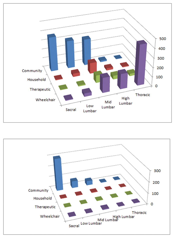
Figure 3a. Number of Phase I participants with myelomeningocele subtype (y axis) sorted by ambulatory status and motor level
Figure 3b. Number of Phase I participants with non-myelomeningocele subtypes (y axis) sorted by ambulatory status and motor level
Figure 4.
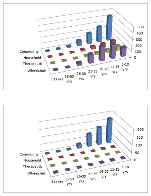
Figure 4a. Number of Phase I participants with myelomeningocele subtype (y axis) sorted by ambulatory status and age category
Figure 4b. Number of Phase I participants with non-myelomeningocele subtypes (y axis) sorted by ambulatory status and age category
The Spearman correlations between age and both ambulatory status and motor level was of low strength in the subtypes of myelomeningocele (p<0.001, rs=−0.23; p<0.001, rs=+0.22,) and non-myelomeningocele (p=0.025, rs=−0.10; p=0.014, rs=+0.09).
Phase II
Three sites collected additional EMR data beyond what is included in the NSBPR-contributed data from a subset of 381 participants aged 5 years and older. Two individuals had missing motor level data and were excluded from the statistical analyses (n=379). Table 1 shows a comparison between the general characteristics of Phase II participants compared to those of Phase I. A significant difference was seen within the myelomeningocele group in terms of history of shunting (p=0.002); a higher percentage of Phase II participants had been shunted, as compared to those in Phase I.
Table 2 shows the percentage of Phase II participants with myelomeningocele and non-myelomeningocele subtypes stratified by ambulatory status. Graphs of subtypes and ambulatory status further stratified by motor level are not shown, but followed the same trends as that seen in Phase I with one exception. Phase II participants with non-myelomeningocele subtypes were more likely to be in the mid lumbar category (28.1% vs. 12.8%) and less likely to be in the low lumbar category (5.3% vs. 16.1%) compared to those in Phase I (p<0.001). Graphs of subtypes and ambulatory status further stratified by age category are not shown, but displayed similar trends as those of Phase I participants. Phase II participants did not differ from Phase I in terms of age category, gender, ethnicity, race, or ambulation status. Table 3 shows subtypes of Phase II participants stratified by surgical history.
Table 3.
Subtypes of Phase II participants stratified by surgical history
| shunt for hydro-cephalus | tethered cord release | shunt for syringomelia | clubfoot or ankle surgery | Hip or knee contracture release | hip osteotomy | ||||||||
|---|---|---|---|---|---|---|---|---|---|---|---|---|---|
| n | % of subtype | n | % of subtype | n | % of subtype | n | % of subtype | n | % of subtype | n | % of subtype | ||
| Myelomeningocele | history of | 279 | 86.4 | 107 | 33.1 | 10 | 3.1 | 113 | 35.0 | 76 | 23.5 | 14 | 4.3 |
| no history of | 44 | 13.6 | 216 | 66.9 | 313 | 96.9 | 210 | 65.0 | 247 | 76.5 | 309 | 95.7 | |
| Non-myelomeningocele | history of | 4 | 7.0 | 35 | 61.4 | 4 | 7.0 | 13 | 22.8 | 7 | 12.3 | 1 | 1.8 |
| no history of | 53 | 93.0 | 22 | 38.6 | 53 | 93.0 | 44 | 77.2 | 50 | 87.7 | 56 | 98.2 | |
Regression results are shown in Tables 4 and 5. The percent concordance of the model was 81.8%. Independent variables that were significantly and inversely associated with ambulation status (overall analysis of effects) were motor level (p <.0001), shunt history (p = 0.0135), and history of hip or knee contracture release surgery (p = 0.0170). The remaining independent variables were not significantly associated with ambulatory status.
Table 4.
Odds Ratio Estimates
Odds Ratio Estimates from Phase II Regression Model
| effect | ambulatory status | odds ratio | 95% Wald Confidence Limits | |
|---|---|---|---|---|
| NonMMC vs MMC | therapeutic | 0.071 | 0.006 | 0.85 |
| NonMMC vs MMC | wheelchair | 0.405 | 0.096 | 1.711 |
| NonMMC vs MMC | household | 0.343 | 0.102 | 1.149 |
| Low Lumbar+Sacral vs High Lumbar+Thoracic | therapeutic | 0.534 | 0.174 | 1.638 |
| Low Lumbar+Sacral vs High Lumbar+Thoracic | wheelchair | 0.013 | 0.005 | 0.031 |
| Low Lumbar+Sacral vs High Lumbar+Thoracic | household | 0.02 | 0.008 | 0.046 |
| Mid Lumbar vs High Lumbar+Thoracic | therapeutic | 1.12 | 0.467 | 2.689 |
| Mid Lumbar vs High Lumbar+Thoracic | wheelchair | 0.063 | 0.031 | 0.13 |
| Mid Lumbar vs High Lumbar+Thoracic | household | 0.09 | 0.041 | 0.198 |
| No Shunt vs Shunt Present | therapeutic | 3.119 | 0.621 | 15.673 |
| No Shunt vs Shunt Present | wheelchair | 0.271 | 0.093 | 0.787 |
| No Shunt vs Shunt Present | household | 0.398 | 0.161 | 0.985 |
| Hip or Knee Contracture Surgery vs no surgery | therapeutic | 0.544 | 0.239 | 1.236 |
| Hip or Knee Contracture Surgery vs no surgery | wheelchair | 0.361 | 0.188 | 0.692 |
| Hip or Knee Contracture Surgery vs no surgery | household | 0.423 | 0.219 | 0.818 |
MMC = myelomeningocele
Table 5.
Analysis of Maximum Likelihood Estimates
Analysis of Maximum Likelihood Estimates from Phase II Regression Model
| Independent Variable | Ambulatory Status | Beta | Standard Error | p value |
|---|---|---|---|---|
| Low lumbar/sacral level | wheelchair | 0.6274 | 0.5719 | 0.2726 |
| Low lumbar/sacral level | therapeutic | −4.3663 | 0.4619 | <0.0001* |
| Low lumbar/sacral level | household | −3.9327 | 0.4338 | <0.0001* |
| Mid-lumbar level | wheelchair | 0.1135 | 0.4468 | 0.7995 |
| Mid-lumbar level | therapeutic | −2.7653 | 0.3692 | <0.0001* |
| Mid-lumbar level | household | −2.4098 | 0.4030 | <0.0001* |
| No shunt | wheelchair | 1.1375 | 0.8237 | 0.1673 |
| No shunt | therapeutic | −1.3059 | 0.5439 | 0.0164* |
| No shunt | household | −0.9201 | 0.4616 | 0.0462* |
| No hip or knee contracture surgery | wheelchair | −0.6092 | 0.4191 | 0.1460 |
| No hip or knee contracture surgery | therapeutic | −1.0187 | 0.3318 | 0.0021* |
| No hip or knee contracture surgery | household | −0.8595 | 0.3358 | 0.0105* |
indicates significance at 0.05 level
Discussion
To our knowledge, this is the first study to present detailed analyses of factors related to ambulatory status in individuals with SB in a large, national sample. Collection of additional EMR variables at 3 sites provided data for further analysis than what was possible with variables collected in the NSBPR at the time of this study. However, the NSBPR has recently been expanded and more surgical history variables are being collected at participating sites. Although the general demographics and ambulatory status of Phase II participants were generally representative of those in Phase I, the smaller cohort differed from the larger in two ways: more individuals in Phase II with myelomeningocele had a history of a shunt, and more individuals in Phase II with non-myelomeningocele subtypes had a higher motor level. These differences suggest that the Phase II participants may have been slightly more impaired than the larger cohort.
Spina bifida subtype was not significantly associated with ambulatory status in Phase II participants. Although approximately 92% of those with the non-myelomeningocele subtype were ambulatory at the community level, a moderate percentage of those with myelomeningocele (43–46%) were also able to ambulate in the community. It is important to note that the Hoffer classification of ambulation does not distinguish how ambulation is achieved. For example, all individuals who use assistive devices or orthoses for community ambulation are classified with individuals who do not require any assistive devices or orthoses. As such, it was not possible to determine how much assistance from these devices was needed to achieve functional ambulation in either subtype. The latest version of NSBPR now includes information about assistive device and orthoses which should help drive future research related to ambulation. A recent systematic review22 revealed that crutches and ankle foot orthoses do provide some benefits for gait pattern, stride, and oxygen cost but research on the functional benefits of orthoses and assistive devices in SB is quite limited.
History of shunting for hydrocephalus was inversely associated with ambulatory status. Compared to those who had shunting for hydrocephalus, those with no history of a shunt were more likely to be ambulatory at the community level than at the household or wheelchair level. Damage to the corticospinal tract from white matter or hindbrain abnormalities, regardless of the contributions of hydrocephalus, is known to impair walking ability because the neural signal cannot travel to the lower limbs.23 Therefore, it is possible that having a history of hydrocephalus necessitating shunting in this study was a proxy measure of upper motor neuron damage which in itself can impair walking ability. Another, but not mutually exclusive, explanation is that, although SB subtype was not independently associated with ambulatory status, those with myelomeningocele are more likely to be shunted. In addition they are likely to have more comorbid and secondary conditions, including neurological and orthopedic sequelae that affect ambulation. Thus, shunting may be a proxy for severity or complexity of the condition, which is contributing to mobility impairments.
Higher motor level was inversely associated with ambulatory status, which was expected, based on the known association of these variables from smaller studies.14–19 Compared to those with thoracic or high lumbar motor levels, those with mid lumbar (knee extension) or low lumbar/sacral motor levels (dorsiflexion/plantarflexion) were more likely to be ambulatory at the community level than at the household or wheelchair level. Bartonek, et al.13 identified knee extensors and plantarflexors as muscle groups important for ambulation and recommended they be included in classification systems of muscle strength. Our study results reinforce the importance of testing these key muscle groups along with the dorsiflexors. Additional research4,7 has suggested that hip abduction may also be a predictor of ambulation ability.
A history of hip or knee contracture release was inversely associated with ambulatory status. Hip or knee contracture releases are surgical procedures often needed to preserve lower limb function, prevent pain, improve wheelchair positioning, or prevent progressive loss of range of motion. Our results likely indicate that tendon release surgery is a proxy measure for contracture severity, i.e. those who did undergo surgery were more likely to have orthopedic limitations to ambulation, even before having surgery. Lower limb contractures have been found to contribute to impairments in function in children with myelomeningocele.21
Age in our study was treated as a covariate in the regression model, but when evaluated in bivariate analyses, had only a weak negative association with ambulatory status and motor level. This finding is consistent with previous, smaller single cohort studies11,24,25 that suggest an age-related decline in ambulatory ability may occur. This decline may be confounded in part by obesity and progressive orthopedic complications. Because the cohorts in this study were relatively young (mean age around 15 years), and because relatively fewer individuals were ambulatory at the household or therapeutic levels, compared to being ambulatory in the community or being a wheelchair user, additional research will be needed to determine how aging and secondary conditions affect ambulatory ability for each of the two subtypes independently over time.
In summary, mobility outcomes from a large cohort of individuals with SB were described, and history of shunting for hydrocephalus, higher motor level, and history of surgical release of hip or knee contractures were inversely associated with ambulatory status. These findings may help investigators design future studies aimed at determining reasons for these associations and interventions aimed at improving mobility outcomes.
Limitations and future work
One clear limitation of this study is that it did not investigate the relationship between obesity and ambulatory status. Some measures of obesity are associated with impairments in ambulation and could have been an important variable in our analysis. Unfortunately, obesity in the SB population is difficult to measure for several reasons.26–28 The legs are often proportionally shorter than the arms, which creates a potential in biased body mass index (BMI) calculations. Although suggestions have been made for ways to calculate a modified BMI taking into account anthropometric data, this practice has not been adopted by all clinics. Moreover, no standardized methods to collect height or weight have been adopted nationally for this population or those with disabilities in general. For example, it is often recommended to obtain segmental measurements in those with contractures, or to remove clothing and orthoses when obtaining weight, but this can be impractical in some clinical settings. For these reasons, standardization of data collection protocols and comparisons of formulas for calculating BMI are important topics for future research.
Another limitation of this study is the low prevalence of therapeutic ambulators in our cohort, limiting our ability to detect any association between that category of ambulation and either shunt history or motor level. Ambulation purely for therapeutic purposes and not for independent mobility often requires assistance or oversight from a caregiver or therapist, use of assistive devices and orthoses, and/or the motivation, funding, and access for therapy services. For these reasons, not as many individuals fall into this category, and as a result, this subpopulation is understudied. We did evaluate whether collapsing ambulatory status into two categories (community and household ambulators versus therapeutic ambulators and wheelchair users) would change the results of the regression model but similar results were seen.
Several additional limitations deserve discussion. First, sampling bias may have occurred for various reasons. The participants in this study were recruited from large institutions that serve individuals with SB, and data are not necessarily representative of individuals who receive care at locations not participating in the NSBPR. In fact, many adults with SB do not have access to care in formalized SB clinics. Additionally, some subtypes of SB, such as split cord malformation or myelocystocele, although not as common, were not included in this study because eligibility for enrollment in the NSBPR at the time of this study was limited to the four diagnoses listed in the inclusion criteria. The NSBPR has since been expanded to include these two diagnoses, and is also now collecting demographic information on those who are eligible but do not enroll. These variables will allow better delineation of external validity in future work. Also, survivor bias may have prevented those with more severe comorbidities or secondary conditions to be under-represented in the sample. Our study sample was in large part comprised of younger participants. This issue is being addressed currently as more sites that treat adults are being added to the NSBPR registry.
Several future opportunities for use of the NSBPR to analyze ambulation status and other mobility-related outcomes exist. Currently, additional variables that may help inform work on mobility outcomes are being collected in the NSBPR (version 2.0). Concurrent work is also being conducted to refine variables in a future database (version 3.0) for more robust analyses. Clearly, longitudinal studies, and those that include more adults, are greatly needed and will be possible with additional funding. More work is also needed to develop and define motor impairment scales similar to those used in spinal cord injury29,30 for use in research in this population.
Conclusions
This is the first study to examine ambulatory status in a large sample of individuals with SB of varying subtypes. Studies to date on ambulatory outcomes in this population have been on small cohorts of individuals. This study found that history of shunting, higher motor level and history of hip or knee contracture release surgery were inversely associated with ambulatory status. Results of this study also help to provide an initial delineation of the breadth of functional abilities within the different age groups and subtypes of SB, and also inform clinicians of the characteristics of those with varying ambulatory ability. Promoting the use of standardized rating scales for ambulation and motor function such as those in this study will also allow results to be compared across studies and to compare effectiveness of medical and rehabilitation interventions for individuals with SB.
Acknowledgments
We would like to thank the many individuals with SB and their family members who participated in this research, without whom the NSBPR would not be possible. The NSBPR has also been successful due to the contributions of the Centers for Disease Control and Prevention, the Spina Bifida Association, and all members of the NSBPR Coordinating Committee. Members of this Committee during the collection of the data reported here were William Walker, Seattle Children’s Hospital; Kathryn Smith, Children’s Hospital, Los Angeles; Kurt Freeman, Oregon Health and Science University; Pamela Wilson, Children’s Hospital Colorado; Kathleen Sawin, Children’s Hospital of Wisconsin; Jeffrey Thomson, Connecticut Children’s Medical Center and Shriners Hospital for Children, Massachusetts; Heidi Castillo, Cincinnati Children’s Hospital Medical Center; Timothy Brei, Riley Hospital for Children; David Joseph, Children’s Hospital of Alabama; Mark Dias, Hershey Medical Center; Karen Ratliff-Schaub, Nationwide Children’s Hospital; James Chinarian, Children’s Hospital of Michigan; John Wiener, Duke University Medical Center; Brad Dicianno, University of Pittsburgh Medical Center; Jacob Neufeld, Children’s Hospital and Research Center at Oakland and UC San Francisco; Robin Bowman, Ann and Robert H. Lurie Children’s Hospital of Chicago; and Paula Peterson, Primary Children’s Medical Center. We would also like to thank Georgia Wiltsie-Tilford, Renee Bischoff, Carlin Collins, Marianne Pan, Amanda Garver and Shanon Safi for assistance with data management.
Footnotes
Author Disclosures: This manuscript has not been published and is not under consideration for publication elsewhere. Data from this manuscript were accepted as part of an abstract and was presented at the AAP 2014 Annual Assembly. This project was funded by the National Center on Birth Defects and Developmental Disabilities, Centers for Disease Control and Prevention, Atlanta, Georgia, grant #1UO1DDD000744.01 and by the Center for Rehabilitation Research using Large Datasets (CRRLD), Eunice Kennedy Shriver National Institute of Child Health and Human Development of the National Institutes of Health (R24HD065702-03). The authors have no conflicts of interest to report. The findings and conclusions in this report are those of the authors and do not necessarily represent the official position of the Centers for Disease Control and Prevention or the National Institutes of Health.
Contributor Information
Brad E. Dicianno, Associate Professor, Dept. of Physical Medicine and Rehabilitation, University of Pittsburgh Medical Center (UPMC), Pittsburgh, PA.
Amol Karmarkar, Assistant Professor, Division of Rehabilitation Sciences, University of Texas Medical Branch, Galveston, TX.
Amy Houtrow, Associate Professor, Depts. of Physical Medicine and Rehabilitation and Pediatrics, UPMC, Pittsburgh, PA.
Theresa M. Crytzer, Assistant Professor, Department of Rehabilitation Science and Technology, University of Pittsburgh, Pittsburgh, PA.
Katelyn M. Cushanick, medical student researcher, Dept. of Physical Medicine and Rehabilitation, University of Pittsburgh Medical Center, Pittsburgh, PA
Andrew McCoy, medical student researcher, Dept. of Physical Medicine and Rehabilitation, University of Pittsburgh Medical Center, Pittsburgh, PA.
Pamela Wilson, Associate Professor, Department of Physical Medicine and Rehabilitation, Children’s Hospital Colorado, Aurora, CO.
James Chinarian, Assistant Professor, Departments of Pediatrics and Physical Medicine and Rehabilitation, Wayne State University School of Medicine, Detroit, MI.
Jacob Neufeld, Associate Professor, Department of Physical Medicine and Rehabilitation, St. Luke’s Children’s Hospital, Boise, ID.
Kathryn Smith, Associate Director for Administration, USC University Center for Excellence in Developmental Disabilities, Children’s Hospital Los Angeles, Los Angeles, CA.
Diane M. Collins, Assistant Professor, Department of Occupational Therapy, University of Texas Medical Branch, Galveston, TX.
References
- 1.Spina Bifida Association. [Accessed February 5, 2015]; http://www.spinabifidaassociation.org/site/c.knKLINNkEiG/b.5712305/k.3010/The_National_Spina_Bifida_Program.htm.
- 2.Dicianno BE, Kurowski BG, Yang JM, et al. Rehabilitation and medical management of the adult with spina bifida. American Journal of Physical Medicine & rRehabilitation/Association of Academic Physiatrists. 2008;87(12):1027–1050. doi: 10.1097/PHM.0b013e31818de070. [DOI] [PubMed] [Google Scholar]
- 3.Dicianno BE, Gaines A, Collins DM, Lee S. Mobility, assistive technology use, and social integration among adults with spina bifida. AmericanJournal of Physical Medicine & Rehabilitation/Association of Academic Physiatrists. 2009;88(7):533–541. doi: 10.1097/PHM.0b013e3181aa41d4. [DOI] [PubMed] [Google Scholar]
- 4.Schoenmakers MA, Gulmans VA, Gooskens RH, Helders PJ. Spina bifida at the sacral level: More than minor gait disturbances. Clin Rehabil. 2004;18(2):178–185. doi: 10.1191/0269215504cr718oa. [DOI] [PubMed] [Google Scholar]
- 5.Verhoef M, Barf HA, Post MW, van Asbeck FW, Gooskens RH, Prevo AJ. Secondary impairments in young adults with spina bifida. Developmental Medicine and Child Neurology. 2004;46(6):420–427. doi: 10.1017/s0012162204000684. [DOI] [PubMed] [Google Scholar]
- 6.Bartonek A, Gutierrez EM, Haglund-Akerlind Y, Saraste H. The influence of spasticity in the lower limb muscles on gait pattern in children with sacral to mid-lumbar myelomeningocele: A gait analysis study. Gait & Posture. 2005;22(1):10–25. doi: 10.1016/j.gaitpost.2004.06.003. [DOI] [PubMed] [Google Scholar]
- 7.Gutierrez EM, Bartonek A, Haglund-Akerlind Y, Saraste H. Kinetics of compensatory gait in persons with myelomeningocele. Gait & Posture. 2005;21(1):12–23. doi: 10.1016/j.gaitpost.2003.11.002. [DOI] [PubMed] [Google Scholar]
- 8.Danielsson AJ, Bartonek A, Levey E, McHale K, Sponseller P, Saraste H. Associations between orthopaedic findings, ambulation and health-related quality of life in children with myelomeningocele. Journal of Children’s Orthopaedics. 2008;2(1):45–54. doi: 10.1007/s11832-007-0069-6. [DOI] [PMC free article] [PubMed] [Google Scholar]
- 9.Bartonek A. Motor development toward ambulation in preschool children with myelomeningocele--a prospective study. Pediatric Physical Therapy : The official publication of the Section on Pediatrics of the American Physical Therapy Association. 2010;22(1):52–60. doi: 10.1097/PEP.0b013e3181cc132b. [DOI] [PubMed] [Google Scholar]
- 10.Oakeshott P, Hunt GM, Poulton A, Reid F. Open spina bifida: Birth findings predict long-term outcome. Archives of Disease in Childhood. 2012;97(5):474–476. doi: 10.1136/archdischild-2011-300624. [DOI] [PubMed] [Google Scholar]
- 11.Pauly M, Cremer R. Levels of mobility in children and adolescents with spina bifida-clinical parameters predicting mobility and maintenance of these skills. European journal of pediatric surgery : official journal of Austrian Association of Pediatric Surgery ... [et al] = Zeitschrift fur Kinderchirurgie. 2013;23(2):110–114. doi: 10.1055/s-0032-1324689. [DOI] [PubMed] [Google Scholar]
- 12.Sibinski M, Synder M, Higgs ZC, Kujawa J, Grzegorzewski A. Quality of life and functional disability in skeletally mature patients with myelomeningocele-related spinal deformity. Journal of Pediatric Orthopedics. Part B. 2013;22(2):106–109. doi: 10.1097/BPB.0b013e32835c2a65. [DOI] [PubMed] [Google Scholar]
- 13.Bartonek A, Saraste H, Knutson LM. Comparison of different systems to classify the neurological level of lesion in patients with myelomeningocele. Developmental Medicine and Child Neurology. 1999;41(12):796–805. doi: 10.1017/s0012162299001607. [DOI] [PubMed] [Google Scholar]
- 14.Broughton NS, Menelaus MB, Cole WG, Shurtleff DB. The natural history of hip deformity in myelomeningocele. J Bone Joint Surg Br. 1993;75(5):760–763. doi: 10.1302/0301-620X.75B5.8376434. [DOI] [PubMed] [Google Scholar]
- 15.Ferrari A, Boccardi S, Licari V. La stazione eretta ed il cammino nella spina bifida. Proceedings from the 14 meetings of the Italian Society of Physical Medicine and Rehabilitation; 1985. pp. 167–205. [Google Scholar]
- 16.Hoffer MM, Feiwell E, Perry R, Perry J, Bonnett C. Functional ambulation in patients with myelomeningocele. The Journal of Bone and Joint Surgery. American volume. 1973;55(1):137–148. [PubMed] [Google Scholar]
- 17.Lindseth RE. Treatment of the lower extremity in children paralyzed by myelomeningocele (birth to 18 months) American Academy of Orthopaedic Surgeons Instructional Course Lectures. 1976;25:76–82. [Google Scholar]
- 18.McDonald CM, Jaffe KM, Shurtleff DB. Assessment of muscle strength in children with meningomyelocele: accuracy and stability of measurements over time. Arch Phys Med Rehabil. 1986;67(12):855–861. [PubMed] [Google Scholar]
- 19.Sharrard WJ. The segmental innervation of the lower limb muscles in man. Ann R Coll Surg Engl. 1964;35:106–122. [PMC free article] [PubMed] [Google Scholar]
- 20.Thibadeau JK, Ward EA, Soe MM, et al. Testing the feasibility of a National Spina Bifida Patient Registry. Birth Defects Research. Part A, Clinical and molecular teratology. 2013;97(1):36–41. doi: 10.1002/bdra.23094. [DOI] [PMC free article] [PubMed] [Google Scholar]
- 21.Schoenmakers MA, Uiterwaal CS, Gulmans VA, Gooskens RH, Helders PJ. Determinants of functional independence and quality of life in children with spina bifida. Clin Rehabil. 2005;19(6):677–685. doi: 10.1191/0269215505cr865oa. [DOI] [PubMed] [Google Scholar]
- 22.Ivanyi B, Schoenmakers M, van Veen N, Maathuis K, Nollet F, Nederhand M. The effects of orthoses, footwear, and walking aids on the walking ability of children and adolescents with spina bifida: A systematic review using International Classification of Functioning, Disability and Health for Children and Youth (ICF-CY) as a reference framework. Prosthetics and Orthotics International. 2014 doi: 10.1177/0309364614543550. 0309364614543550. [DOI] [PubMed] [Google Scholar]
- 23.Geerdink N, Cuppen I, Rotteveel J, Mullaart R, Roeleveld N, Pasman J. Contribution of the corticospinal tract to motor impairment in spina bifida. Pediatric Neurology. 2012;47(4):270–278. doi: 10.1016/j.pediatrneurol.2012.06.010. [DOI] [PubMed] [Google Scholar]
- 24.Asher M, Olson J. Factors affecting the ambulatory status of patients with spina bifida cystica. The Journal of Bone and Joint Surgery. American Volume. 1983;65(3):350–356. [PubMed] [Google Scholar]
- 25.De Souza LJ, Carroll N. Ambulation of the braced myelomeningocele patient. The Journal of Bone and Joint Surgery. American Volume. 1976;58(8):1112–1118. [PubMed] [Google Scholar]
- 26.McPherson AC, Swift JA, Yung E, Lyons J, Church P. The assessment of weight status in children and young people attending a spina bifida outpatient clinic: A retrospective medical record review. Disability and Rehabilitation. 2013;35(25):2123–2131. doi: 10.3109/09638288.2013.771705. [DOI] [PMC free article] [PubMed] [Google Scholar]
- 27.Mita K, Akataki K, Itoh K, Ono Y, Ishida N, Oki T. Assessment of obesity of children with spina bifida. Developmental Medicine and Child Neurology. 1993;35(4):305–311. doi: 10.1111/j.1469-8749.1993.tb11642.x. [DOI] [PubMed] [Google Scholar]
- 28.Dosa NP, Foley JT, Eckrich M, Woodall-Ruff D, Liptak GS. Obesity across the lifespan among persons with spina bifida. Disability and Rehabilitation. 2009;31(11):914–920. doi: 10.1080/09638280802356476. [DOI] [PubMed] [Google Scholar]
- 29.Marino R, Barros T, Biering-Sorensen F, et al. International standards for neurological classification of spinal cord injury. The Journal of Spinal Cord Medicine. 2003;26:S50. doi: 10.1080/10790268.2003.11754575. [DOI] [PubMed] [Google Scholar]
- 30. [Accessed February 5, 2015];American Spinal Injury Association exam sheet. http://www.asia-spinalinjury.org/elearning/isncsci_exam_sheet_r4.pdf.