Abstract
Purpose
The diagnostic criteria for disseminated intravascular coagulation (DIC) established by the Japanese Association for Acute Medicine (JAAM) is able to diagnose DIC accurately and promptly. The aim of this retrospective study is to evaluate the degree of association between each parameter of JAAM DIC criteria and the diagnosis of trauma induced DIC (T-DIC) utilizing thromboelastometry (ROTEM).
Methods
Trauma patients transported to our hospital with ROTEM performed in the emergency department between January 2013 and December 2015 were enrolled in this study. We evaluated (1) the characteristics of T-DIC, (2) the relationships between T-DIC and each parameter of the JAAM DIC criteria and (3) the diagnostic accuracies of each parameter for T-DIC by statistical measurement.
Results
All 72 patients (21 T-DIC and 51 control) were included in primary analysis. T-DIC was significantly related to younger age, more severe trauma scores, more cases of massive transfusions, and remarkable coagulation abnormality detected by standard coagulation tests. In the cases of T-DIC, ROTEM showed longer clotting time, lower acceleration, lower clot firmness, and inhibited fibrinolysis in EXTEM/INTEM. Within the JAAM DIC score, PT-INR ≥1.2 was the most accurate factor for T-DIC diagnosis; sensitivity 60.0%, specificity 100.0%, and accuracy 88.7%. PT-INR ≥1.2 was statistically correlated with the JAAM DIC score (p < 0.001, r = 0.709). The univariate analysis based on 1.2 of PT-INR indicated statistical differences in most categories of ROTEM, which is similar to analysis performed for the presence and absence of T-DIC.
Conclusions
Among JAAM DIC criteria, the PT-INR ≥1.2 was the most accurate factor for both the diagnosis of T-DIC and the evaluation of its severity.
Keywords: Trauma, DIC, JAAM DIC score, PT-INR, Thromboelastometry
Introduction
Trauma is a leading cause of death and disability worldwide [1, 2]. The pathophysiological features of trauma are affected by various factors including type of injury source, underlying medical conditions, demographics of the victim, quality of the initial trauma care at the scene, and the strategy of advanced trauma resuscitation in a trauma center [3–8].
Bleeding is a primary cause of trauma death, and coagulopathy is strongly associated with an increased requirement for blood transfusions and poor clinical outcome [9–11]. Recent articles report that disseminated intravascular coagulation (DIC) plays a pivotal role in the pathogenesis of post-traumatic organ dysfunction in severe trauma patients [12]. However, it is necessary to determine the hematological status promptly and correctly to initiate effective hematological resuscitations in the emergency department (ED). Scoring systems utilizing several biomarkers were developed by the Japanese Ministry of Health and Welfare (JMHW) in 1987 and the International Society of Thrombosis and Homeostasis (JSTH) in 2001 [13, 14]. Although these criteria had potential to accurately diagnose DIC in critically ill patients, some practical limitations were stated [15, 16]. Thus, new diagnostic criteria were established by the Japanese Association for Acute Medicine (JAAM) in 2006 [17]. These criteria were proven to be able to diagnose DIC more accurately and select patients were able to receive resuscitation at the early phase of DIC compared with former criteria [17, 18].
Rotational thromboelastometry (ROTEM; TEM International, GmbH, Munich, Germany) is known as a point-of-care viscoelastic test using a citrated whole-blood sample. This test can detect coagulation and fibrinolytic abnormalities more rapidly and pathophysiologically than standard coagulation tests [19]. To date, more than 1000 articles about ROTEM have been published in various fields including cardiovascular surgery, liver transplantation, and trauma surgery [20–22].
The JAAM DIC criteria, which were established based upon all possible causes of critically ill patients, consist of widely available biomarkers and vital signs that can be measured in the emergency room [17]. However, few studies have reported about the level of contribution of each parameter within these criteria to the diagnosis of DIC. Further, T-DIC should be differentiated from non-trauma related DIC, because each DIC patient with different etiology tends to have a wide variety of clinical manifestations and mechanisms of coagulopathy.
The aim of this retrospective study is to elucidate the most meaningful and relevant parameter of the JAAM DIC criteria to the diagnosis of trauma induced DIC (T-DIC).
Methods
Patients and study design
This retrospective study has been approved by the institutional review board of Saga University Hospital (Protocol Identification Number: 2014-09-08). Trauma patients who were transported to our hospital and had ROTEM performed in the emergency department (ED) between January 2013 and December 2015 were enrolled. Patients with out-of-hospital cardiac arrest, burn injury, electrical injury, less than 18 years of age, or shorter length of hospital stay (<2 days) were excluded. The enrolled patients were divided into two groups based on the presence or absence of DIC. Univariate analysis was performed to evaluate the hematological characteristics of T-DIC, the relationships between T-DIC and each parameter of the JAAM DIC criteria, and the diagnostic accuracies of each parameter for diagnosing T-DIC. We further evaluated the differences in ROTEM data in accordance with each factor of the JAAM criteria. The correlation between the JAAM DIC score and the most accurate parameter in the criteria was also evaluated.
Diagnosis of DIC and other clinical parameters
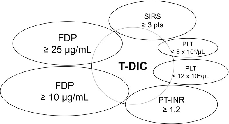
All trauma patients were diagnosed with DIC by the JAAM DIC criteria on their admission to the ED [17]. DIC was defined when the total score was 4 points or greater (range 0–8) by the criteria. We utilized the parameters of the JAAM DIC criteria for subsequent analyses including (1) systemic inflammatory response syndrome (SIRS) ≥3 pts, (2) platelet (PLT) <8 × 10⁴/µL or (3) PLT <12 × 104/µL, (4) international normalized ratio of prothrombin time (PT-INR) ≥1.2, (5) fibrinogen and fibrin degradation products (FDP) ≥25 µg/mL or (6) FDP ≥10 µg/mL (Fig. 1). Patient characteristics and past medical histories were evaluated. The definition of shock was a systolic blood pressure of less than 90 mmHg on admission. Various trauma scores including the injury severity score (ISS), the revised trauma score (RTS) and the probability of survival (Ps) were evaluated from the medical records, retrospectively. Massive transfusion was defined to be more than 10 units of red blood cell (RCC-LR) transfusion required within the first 24 h of admission to the ED. Emergency surgery was performed for hemostasis within the first 24 h.
Fig. 1.
Scheme of this study
Laboratory tests
Blood samples were collected by an emergency physician immediately after admission to the ED. Laboratory data [white blood cell (WBC), hemoglobin (Hb), PLT, PT-INR, activated partial thromboplastin time (APTT), fibrinogen, FDP, d-dimer (DD) and lactate] were measured in all trauma patients.
ROTEM analysis
To determine the coagulation and fibrinolytic status, ROTEM was often performed for trauma patients in our department. Our thromboelastometric analysis was focused on four assays: extrinsic coagulation cascade (EXTEM), intrinsic coagulation cascade (INTEM), function of fibrinogen (FIBTEM) and anti-fibrinolytic cascade using aprotinin (APTEM). ROTEM parameters analyzed in this study included the clotting time (CT), the clot formation time (CFT), the alpha angle (α), the amplitude at 10 min (A10), 20 min (A20), and 30 min (A30), the maximum clot firmness (MCF), the lysis index at 30 min (LI30), and maximum lysis (ML). These were classified into four main categories according to their features: (1) initiation (CT), (2) acceleration (CFT, α), (3) clot firmness (A10, A20, A30, MCF), and (4) fibrinolysis (LI30, ML). Hyperfibrinolysis was defined by 20% improvement of fibrinolysis in the APTEM test compared with the EXTEM test, when the ML of EXTEM was 15% or more. Physician in charge selected patients who needed to take ROTEM analysis. All tests were started less than 1 h after admission to the ED and ran more than 60 min at 37 °C.
Statistical analysis
All continuous variables are represented as median [interquartile range (IQR); Q1–Q3] and categorical variables as numbers (percentages). The p values were calculated from the Mann–Whitney U test for continuous variables, and Fisher’s exact test and Chi-square tests were used for categorical variables. Spearman correlation analysis and curve fitting were used to evaluate the relationship between the JAAM DIC score and a statistically significant parameter. Values of p < 0.05 were considered to be significant. Statistical analyses were performed by IBM SPSS Statistics version 23 (IBM Corp., Armonk, NY, USA).
Results
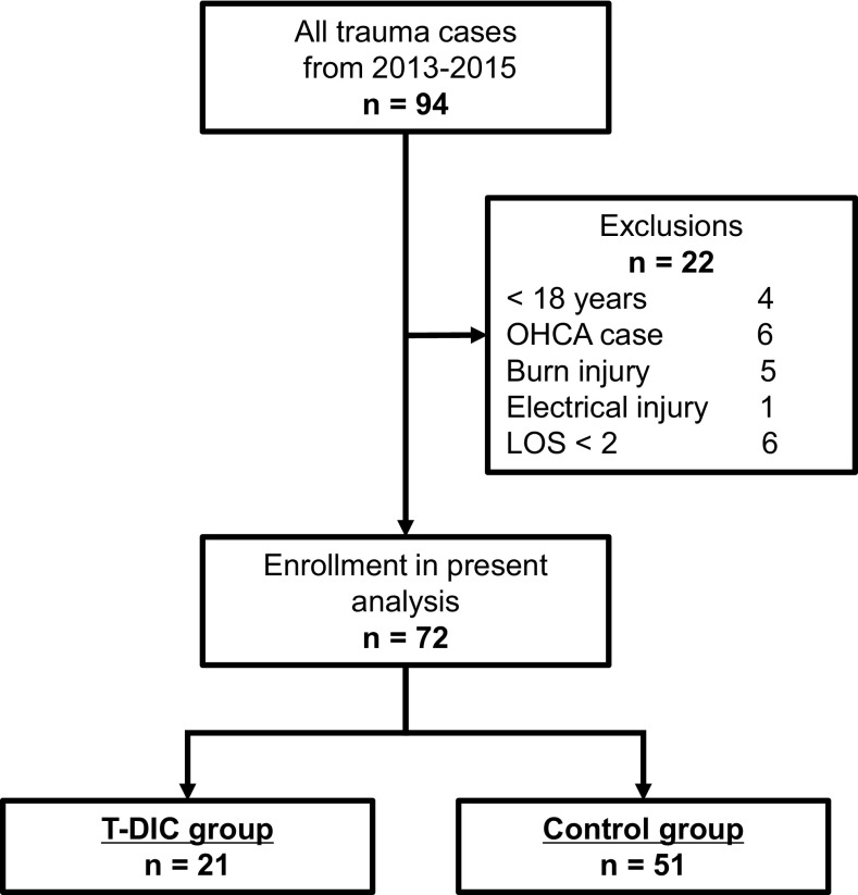
Ninety-four patients matched the inclusion criteria in this study (Fig. 2). However, 22 of them were eventually excluded: 4 for being less than 18 years old, 6 for out-of-hospital cardiac arrest, 5 for burn injury, 1 for electrical injury and 6 for less than 2 days of length of hospital stay. The 72 remaining patients were assigned to primary analysis. They were divided into two groups based on the presence of DIC on admission: traumatic-DIC (T-DIC) group (n = 21) and Control group (n = 51), respectively.
Fig. 2.
Study design
Univariate analyses between the T-DIC group and the Control group
Table 1 shows patients’ characteristics, treatment, and clinical outcome. All were blunt trauma cases. The T-DIC group was significantly younger than the Control group (71 vs. 57 years; p = 0.034). Significantly more patients on warfarin were found in the T-DIC group compared with the Control group (14.3 vs. 0.0%; p = 0.022). The median JAAM DIC scores of both groups were 4 (IQR; 4–5) and 3 (0–3), respectively. No statistical differences were confirmed for sex, presence of shock, or other medical histories. According to trauma scales, the T-DIC group represented significantly higher ISS (29 vs. 12; p < 0.001), lower RTS (7.55 vs. 7.84; p = 0.001) and lower Ps (62.4 vs. 95.9; p = 0.001) compared with the Control group. In the T-DIC group, there were significantly more patients that received a massive blood transfusion within the first 24 h (42.9 vs. 5.9 %; p < 0.001), compared with the Control group. However, no association was found for emergency surgery and hospital mortality within both groups.
Table 1.
Patients' characteristics, emergency treatment and clinical outcome in T-DIC and control groups
| T-DIC group (n = 21) | Control group (n = 51) | p values | |
|---|---|---|---|
| Age, year, median (IQR) | 71 (49–79) | 57 (34–69) | 0.034 |
| Male, n (%) | 13 (61.9) | 34 (66.7) | 0.700 |
| Liver cirrhosis, n (%) | 1 (4.8) | 1 (2.0) | 0.501 |
| Antiplatelet agents, n (%) | 3 (14.3) | 3 (5.9) | 0.233 |
| Warfarin, n (%) | 3 (14.3) | 0 (0.0) | 0.022 |
| Other anticoagulants, n (%) | 1 (4.8) | 0 (0.0) | 0.292 |
| JAAM DIC score, pts, median (IQR) | 4 (4–5) | 3 (0–3) | <0.001 |
| Shock, n (%) | 4 (19.0) | 2 (3.9) | 0.056 |
| Blunt trauma, n (%) | 21 (100.0) | 51 (100.0) | – |
| ISS, median (IQR) | 29 (22–33) | 12 (5–24) | <0.001 |
| PTS, median (IQR) | 7.55 (5.90–7.84) | 7.84 (7.84–7.84) | 0.001 |
| Ps, %, median (IQR) | 62.4 (41.5–91.0) | 95.9 (83.6–98.4) | 0.001 |
| Massive transfusion, n (%) | 9 (42.9) | 3 (5.9) | <0.001 |
| Emergency surgery, n (%) | 7 (33.3) | 10 (19.6) | 0.173 |
| Dead, n (%) | 4 (19.0) | 2 (3.9) | 0.056 |
Laboratory tests are shown in Table 2. Complete blood counts in the T-DIC group demonstrated higher WBC (13,900 vs. 10,000; p = 0.007), lower Hb (10.8 vs. 13.5; p < 0.001) and lower PLT (15.5 vs. 22.0; p = 0.001) than the Control group. The same tendency was confirmed in the standard coagulation tests between the T-DIC group and the Control group: PT-INR (1.23 vs. 1.00; p < 0.001), APTT (35.4 vs. 28.9; p < 0.001), Fibrinogen (196 vs. 256; p = 0.012), FDP (149.9 vs. 30.9; p < 0.001) and DD (88.18 vs. 15.14; p < 0.001). Significantly higher lactate levels were associated with T-DIC compared with Control (3.2 vs. 1.8; p < 0.001).
Table 2.
Laboratory tests in each group
| T-DIC group (n = 21) | Control group (n = 51) | p values | |
|---|---|---|---|
| WBC, /µL, median (IQR) | 13,900 (10,500–17,400) | 10,000 (7700–12,600) | 0.007 |
| Hb, g/dL, median (IQR) | 10.8 (9.0–12.1) | 13.5 (12.3–15.2) | <0.001 |
| PLT, 104/µL, median (IQR) | 15.5 (12.0–19.2) | 22.0 (16.8–23.7) | 0.001 |
| PT-INR, median (IQR) | 1.23 (1.13–1.58) | 1.00 (0.96–1.05) | <0.001 |
| APTT, second, median (IQR) | 35.4 (31.1–44.5) | 28.9 (26.5–32.2) | <0.001 |
| Fibrinogen, mg/dL, median (IQR) | 196 (103–276) | 256 (204–299) | 0.012 |
| FDP, µg/mL, median (IQR) | 149.9 (75.3–470.8) | 30.9 (8.2–98.6) | <0.001 |
| DD, µg/mL, median (IQR) | 88.18 (38.78–251.85) | 15.14 (4.67–51.7) | <0.001 |
| Lactate, mmol/L, median (IQR) | 3.2 (2.1–4.6) | 1.8 (1.4–2.5) | <0.001 |
Thromboelastometric analyses revealed distinctive findings in T-DIC (Table 3). In the EXTEM test, T-DIC showed significantly longer CT (85 vs. 61; p = 0.002), lower A10 (50 vs. 54; p = 0.038), lower A20 (57 vs. 60; p = 0.048), higher LI30 (100 vs. 100; p = 0.030) and lower ML (10 vs. 15; p = 0.002) than the Control group. Consistent with the EXTEM test the INTEM test showed that significantly longer CFT (120 vs. 83; p = 0.003), lower α (67 vs. 73; p = 0.003), lower A10 (47 vs. 53; p = 0.002), lower A20 (54 vs. 59; p = 0.008), lower MCF (56 vs. 60; p = 0.019), higher LI30 (100 vs. 99; p = 0.002) and lower ML (8 vs. 14; p < 0.001), in the T-DIC group compared with the Control group. Other parameters, including the FIBTEM test, were not significantly different between the two groups. The ratio of hyperfibrinolysis was also equal in each group.
Table 3.
Thromboelastometric analyses among groups
| T-DIC group (n = 21) | Control group (n = 51) | p values | |||
|---|---|---|---|---|---|
| EXTEM | |||||
| CT, s, median (IQR) | 85 | (61–118) | 61 | (50–70) | 0.002 |
| CFT, s, median (IQR) | 109 | (80–133) | 93 | (80–110) | 0.155 |
| α, ˚, median (IQR) | 68 | (65–74) | 72 | (69–75) | 0.159 |
| A10, mm, median (IQR) | 50 | (42–58) | 54 | (50–57) | 0.038 |
| A20, mm, median (IQR) | 57 | (51–64) | 60 | (57–63) | 0.048 |
| A30, mm, median (IQR) | 59 | (53–65) | 61 | (57–64) | 0.136 |
| MCF, mm, median (IQR) | 59 | (53–65) | 61 | (58–64) | 0.168 |
| LI30, %, median (IQR) | 100 | (100–100) | 100 | (99–100) | 0.030 |
| ML, %, median (IQR) | 10 | (6–14) | 15 | (11–17) | 0.002 |
| INTEM | |||||
| CT, s, median (IQR) | 222 | (192–298) | 197 | (156–271) | 0.141 |
| CFT, s, median (IQR) | 120 | (92–135) | 83 | (67–100) | 0.003 |
| α, ˚, median (IQR) | 67 | (64–72) | 73 | (71–77) | 0.003 |
| A10, mm, median (IQR) | 47 | (41–51) | 53 | (49–57) | 0.002 |
| A20, mm, median (IQR) | 54 | (49–58) | 59 | (55–62) | 0.008 |
| A30, mm, median (IQR) | 57 | (51–58) | 59 | (55–60) | 0.058 |
| MCF, mm, median (IQR) | 56 | (51–58) | 60 | (56–62) | 0.019 |
| LI30, %, median (IQR) | 100 | (100–100) | 99 | (99–100) | 0.002 |
| ML, %, median (IQR) | 8 | (5–11) | 14 | (10–18) | <0.001 |
| FIBTEM | |||||
| MCF, mm, median (IQR) | 10 | (6–15) | 13 | (9–15) | 0.135 |
| Hyperfibrinolysis by ROTEM, n (%) | 2 | (9.5) | 4/46 | (8.7) | 0.618 |
The relationships between T-DIC and each parameter of JAAM DIC criteria
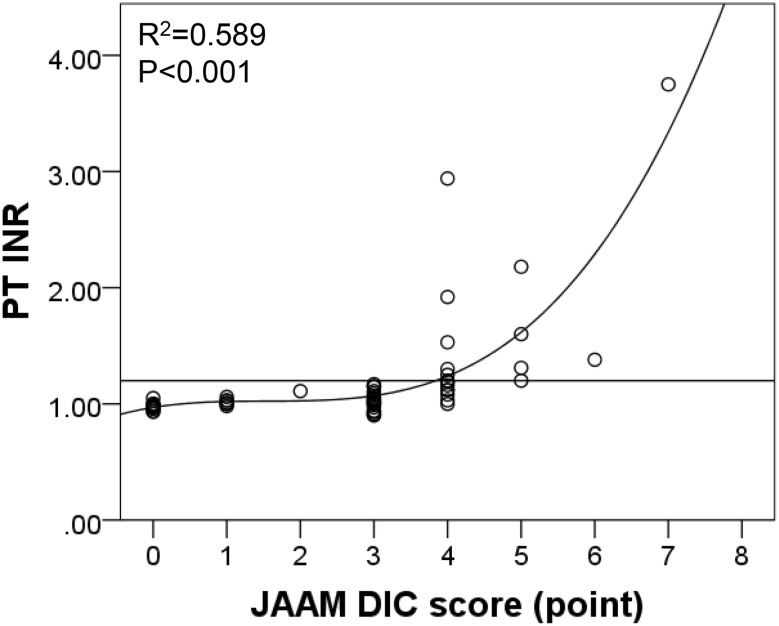
We evaluated the relationships between T-DIC and each parameter of the JAAM DIC criteria (Table 4). Almost all parameters, including SIRS ≥3 points (pts) (52.4 vs. 3.9%; p < 0.001), PLT <12 × 104/µL (23.8 vs. 0.0%; p = 0.001), PT-INR ≥1.2 (60.0 vs. 0.0%; p < 0.001), FDP ≥25 µg/mL (100.0 vs. 54.8%; p < 0.001) and FDP ≥10 µg/mL (100.0 vs. 71.4 %; p = 0.005), were significantly associated with T-DIC. Next, we calculated the diagnostic accuracy for T-DIC by utilizing every parameter of the JAAM DIC criteria (Table 4). Higher sensitivity was observed in FDP ≥25 µg/mL (100.0%) and FDP ≥10 µg/mL (100.0%). In addition, higher specificity was observed for SIRS ≥3 pts (96.1%), PLT <8 × 10⁴/µL (100.0%), PLT <12 × 104/µL (100.0 %) and PT-INR ≥1.2 (100.0%). Interestingly, PT-INR ≥1.2 showed the highest accuracy among all parameters. Moreover, Spearman correlation analysis revealed that PT-INR was statistically correlated with the JAAM DIC score more than other clinical scores (r = 0.709, p < 0.001). Curve fitting for the combination of PT-INR and the JAAM DIC score detected a significant non-linear association. The most obvious correlation was observed in the cubic curve-fitting equation (R 2 = 0.589, p < 0.001) (Fig. 3).
Table 4.
The relationships between T-DIC and each parameter of JAAM DIC criteria
| Univariate analysis | Diagnostic accuracy for T-DIC | |||||||
|---|---|---|---|---|---|---|---|---|
| T-DIC group (n = 21) | Control group (n = 51) | p values | Sensitivity | Specificity | PPV | NPV | Accuracy | |
| SIRS ≥3 pts, n (%) | 11 (52.4) | 2 (3.9) | <0.001 | 52.4 | 96.1 | 84.6 | 83.1 | 83.3 |
| PLT <8 × 10⁴/µL, n (%) | 1 (4.8) | 0 (0.0) | 0.292 | 4.8 | 100.0 | 100.0 | 71.8 | 72.2 |
| PLT <12 × 104/µL, n (%) | 5 (23.8) | 0 (0.0) | 0.001 | 23.8 | 100.0 | 100.0 | 76.1 | 77.8 |
| PT-INR ≥1.2, n (%) | 12/20 (60.0) | 0 (0.0) | <0.001 | 60.0 | 100.0 | 100.0 | 86.4 | 88.7 |
| FDP ≥25 µg/mL, n (%) | 20/20 (100.0) | 23/42 (54.8) | <0.001 | 100.0 | 45.2 | 46.5 | 100.0 | 62.9 |
| FDP ≥10 µg/mL, n (%) | 20/20 (100.0) | 30/42 (71.4) | 0.005 | 100.0 | 28.6 | 40.0 | 100.0 | 51.6 |
Fig. 3.
Spearman correlation analysis between PT-INR and JAAM DIC score
Thromboelastometric analysis of trauma induced coagulopathy based on 1.2 of PT-INR value
We divided patients into 2 groups based on their PT-INR value: PT-INR ≥1.2 group (n = 12) and PT-INR <1.2 group (n = 59), (Table 5). Univariate analyses were performed to evaluate the trauma-induced coagulopathy which was diagnosed by ROTEM in each group. In the EXTEM test, PT-INR ≥1.2 group showed statistically longer CT (99 vs. 61; p < 0.001), lower A10 (46 vs. 53; p = 0.034), lower A20 (54 vs. 60; p = 0.038) and lower ML (8 vs. 14; p = 0.006) than the PT-INR <1.2 group. Furthermore, significant differences were found in all parameters of the INTEM test as follows: CT (PT-INR ≥1.2 group: 231 vs. PT-INR <1.2 group: 197; p = 0.037), CFT (127 vs. 85; p = 0.012), α (66 vs. 73; p = 0.013), A10 (44 vs. 52; p = 0.001), A20 (52 vs. 58; p = 0.002), A30 (53 vs. 59; p = 0.010), MCF (54 vs. 59; p = 0.004), LI30 (100 vs. 100; p = 0.035) and ML (7 vs. 13; p = 0.002). In the FIBTEM test, a significantly lower MCF was confirmed in the PT-INR ≥1.2 group (8 vs. 13; p = 0.037). However, the percentage of patients with hyperfibrinolysis was not related to the PT-INR value.
Table 5.
Thromboelastometric evaluations of trauma induced coagulopathy based on 1.2 of PT-INR value
| PT-INR ≥1.2 group (n = 12) | PT-INR <1.2 group (n = 59) | p values | |
|---|---|---|---|
| EXTEM | |||
| CT, s, median (IQR) | 99 (74–128) | 61 (50–74) | <0.001 |
| CFT, s, median (IQR) | 123 (80–134) | 94 (80–110) | 0.168 |
| α, ˚, median (IQR) | 66 (65–74) | 72 (68–75) | 0.159 |
| A10, mm, median (IQR) | 46 (38–56) | 53 (49–59) | 0.034 |
| A20, mm, median (IQR) | 54 (47–62) | 60 (56–64) | 0.038 |
| A30, mm, median (IQR) | 57 (49–63) | 61 (56–64) | 0.096 |
| MCF, mm, median (IQR) | 58 (50–63) | 61 (57–65) | 0.109 |
| LI30, %, median (IQR) | 100 (100–100) | 100 (99–100) | 0.052 |
| ML, %, median (IQR) | 8 (5–14) | 14 (10–17) | 0.006 |
| INTEM | |||
| CT, s, median (IQR) | 231 (216–314) | 197 (156–273) | 0.037 |
| CFT, s, median (IQR) | 127 (97–157) | 85 (69–119) | 0.012 |
| α, ˚, median (IQR) | 66 (62–72) | 73 (67–77) | 0.013 |
| A10, mm, median (IQR) | 44 (38–49) | 52 (48–57) | 0.001 |
| A20, mm, median (IQR) | 52 (46–57) | 58 (55–62) | 0.002 |
| A30, mm, median (IQR) | 53 (48–58) | 59 (54–60) | 0.010 |
| MCF, mm, median (IQR) | 54 (48–58) | 59 (56–62) | 0.004 |
| LI30, %, median (IQR) | 100 (100–100) | 100 (99–100) | 0.035 |
| ML, %, median (IQR) | 7 (5–11) | 13 (9–17) | 0.002 |
| FIBTEM | |||
| MCF, mm, median (IQR) | 8 (6–13) | 13 (9–16) | 0.037 |
| Hyperfibrinolysis by ROTEM, n (%) | 1 (8.3) | 4 (7.4) | 0.646 |
The thromboelastometric parameters with significant differences evaluated by each parameter of the JAAM DIC criteria
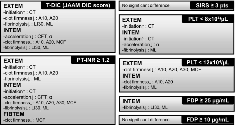
We evaluated the correlation between ROTEM data and each parameter of the JAAM DIC criteria (Fig. 4). The left two panels (T-DIC and PT-INR ≥1.2) show results of the analyses described above. In addition, the same analyses were performed and results are shown in the 5 right-hand side panels (SIRS ≥3 pts, PLT <8 × 10⁴/µL, PLT <12 × 104/µL, FDP ≥25 µg/mL, FDP ≥10 µg/mL) (detailed data not shown). The PT-INR of 1.2 was the most reliable detector for various kinds of coagulation and fibrinolytic abnormalities in trauma patients within all parameters of the JAAM DIC criteria.
Fig. 4.
The thromboelastometric abnormalities associated with either T-DIC (JAAM DIC score ≥4 pts) or each parameter of the JAAM DIC criteria including PT-INR ≥1.2, SIRS ≥3 pts, PLT <8 × 10⁴/µL, PLT <12 × 104/µL, FDP ≥25 µg/mL, FDP ≥10 µg/mL
Discussion
This study demonstrates that a PT-INR of 1.2 is the most accurate diagnostic tool for T-DIC among the JAAM DIC criteria. The strong correlation between PT-INR ≥1.2 and the JAAM DIC score suggests that PT-INR is not only a reliable predictor, but is also a severity indicator for T-DIC. Furthermore, our results also enable many physicians working at community hospitals who have limited access to the data needed for the JAAM DIC criteria, to refer to PT-INR for detecting T-DIC.
PT-INR is a standard monitoring parameter for prophylaxis and thrombolytic therapy by anticoagulation agents that has been used for decades [23, 24]. There are also several evidences of this marker in trauma cases. An initial abnormality of PT on admission to a Level I trauma center is an independent risk factor for mortality (35% risk increase) [9]. Another retrospective cohort using more than 3000 trauma cases from five international hospitals concluded that the prothrombin time ratio >1.2 is a threshold level for higher mortality and transfusion requirements [11]. Our results also indicated that PT-INR could specifically detect the changes of trauma induced coagulopathy.
More and more reports about trauma-induced coagulopathy utilizing ROTEM have been published in this decade [19, 25, 26]. A recent retrospective study in Zurich reported that Hb ≥10 g/dL and abnormal MCF of INTEM were reliable predictors for massive transfusion in severely injured patients [25]. Another study in Salzburg concluded A10 and MCF of FIBTEM upon admission to the ED had high predictive value for massive transfusion [26]. The degree of hyperfibrinolysis diagnosed by ROTEM was correlated with clinical outcome, which was published by the same group in Salzburg [27]. ROTEM is able to separate the coagulation cascade into four main categories and patients with T-DIC showed these characteristic features in most categories of the EXTEM and INTEM tests in this study. Interestingly, the same tendency was found when the patients were divided by PT-INR ≥1.2 or <1.2. However, other parameters of the JAAM DIC criteria could not detect coagulopathy correctly compared with PT-INR. Considering these results, the viscoelastic testing might be able to reveal detailed differences in the coagulation and fibrinolytic status, whereas standard coagulation tests may fail to detect these differences.
This study has some limitations which will be addressed in future work. First, this is a retrospective study and a small sample size may cause a selection bias. Second, since trauma patients tend to have multiple medical conditions with various severities, the coagulation and fibrinolytic status of trauma patients drastically changes during their clinical course [28]. Finally, we could not control for medication use as a potential confounding factor, which may affect PT-INR values. There were significantly more patients on warfarin in the T-DIC group and higher PT-INR group (data not shown) than others.
This study reveals a tendency to diagnose T-DIC with respect to each parameter of the JAAM DIC criteria. Among the parameters, higher specificity was confirmed in the subject of SIRS ≥3 pts, PLT <8 × 10⁴/µL, PLT <12 × 104/µL, and PT-INR ≥1.2 whereas higher sensitivity was observed in FDP ≥25 µg/mL and FDP ≥10 µg/mL. This means that SIRS, PLT and PT-INR are useful parameters to make the diagnosis of T-DIC, while FDP is informative to rule out the diagnosis. Most importantly, if we encounter a severe trauma patient with high PT-INR, the patient is highly likely to present DIC and immediate curative interventions should be considered.
Conclusions
Among the JAAM DIC criteria, the PT-INR ≥1.2 possesses the highest accuracy for the diagnosis of trauma-induced DIC and for an evaluation of its severity utilizing thromboelastometry.
Acknowledgements
I would like to express a great sense of gratitude to Dr. Janet Markman who has offered continuing support for English editing. This retrospective study was approved by institutional review board of Saga University Hospital (Protocol Identification Number: 2014-09-08).
Compliance with ethical standards
Conflict of interest
Hiroyuki Koami, Yuichiro Sakamoto, Kosuke Chris Yamada, Tomoya Matsuda, Junpei Nishi, Kento Nakayama, Ryota Sakurai, Miho Ohta, Hisashi Imahase, Mayuko Yahata, Mitsuru Umeka, Toru Miike, Futoshi Nagashima, Takashi Iwamura and Satoshi Inoue have no conflict of interest.
References
- 1.Krug EG, Sharma GK, Lozano R. The global burden of injuries. Am J Public Health. 2000;90(4):523–526. doi: 10.2105/AJPH.90.4.523. [DOI] [PMC free article] [PubMed] [Google Scholar]
- 2.Global Burden of Disease Study C Global, regional, and national incidence, prevalence, and years lived with disability for 301 acute and chronic diseases and injuries in 188 countries, 1990–2013: a systematic analysis for the Global Burden of Disease Study 2013. Lancet. 2015;386(9995):743–800. doi: 10.1016/S0140-6736(15)60692-4. [DOI] [PMC free article] [PubMed] [Google Scholar]
- 3.Sauaia A, Moore FA, Moore EE, Haenel JB, Read RA, Lezotte DC. Early predictors of postinjury multiple organ failure. Arch Surg. 1994;129(1):39–45. doi: 10.1001/archsurg.1994.01420250051006. [DOI] [PubMed] [Google Scholar]
- 4.Kauvar DS, Lefering R, Wade CE. Impact of hemorrhage on trauma outcome: an overview of epidemiology, clinical presentations, and therapeutic considerations. J Trauma. 2006;60(6 Suppl):S3–11. doi: 10.1097/01.ta.0000199961.02677.19. [DOI] [PubMed] [Google Scholar]
- 5.Liberman M, Mulder D, Sampalis J. Advanced or basic life support for trauma: meta-analysis and critical review of the literature. J Trauma. 2000;49(4):584–599. doi: 10.1097/00005373-200010000-00003. [DOI] [PubMed] [Google Scholar]
- 6.Billeter AT, Miller FB, Harbrecht BG, Bowen W, Stephens MJ, Postel GC, et al. Interhospital transfer of blunt multiply injured patients to a level 1 trauma center does not adversely affect outcome. Am J Surg. 2014;207(4):459–466. doi: 10.1016/j.amjsurg.2013.04.015. [DOI] [PubMed] [Google Scholar]
- 7.Werman HA, Nelson RN, Campbell JE, Fowler RL, Gandy P. Basic trauma life support. Ann Emerg Med. 1987;16(11):1240–1243. doi: 10.1016/S0196-0644(87)80231-7. [DOI] [PubMed] [Google Scholar]
- 8.Esposito TJ, Sanddal ND, Hansen JD, Reynolds S. Analysis of preventable trauma deaths and inappropriate trauma care in a rural state. J Trauma. 1995;39(5):955–962. doi: 10.1097/00005373-199511000-00022. [DOI] [PubMed] [Google Scholar]
- 9.MacLeod JB, Lynn M, McKenney MG, Cohn SM, Murtha M. Early coagulopathy predicts mortality in trauma. J Trauma. 2003;55(1):39–44. doi: 10.1097/01.TA.0000075338.21177.EF. [DOI] [PubMed] [Google Scholar]
- 10.Brohi K, Cohen MJ, Davenport RA. Acute coagulopathy of trauma: mechanism, identification and effect. Curr Opin Crit Care. 2007;13(6):680–685. doi: 10.1097/MCC.0b013e3282f1e78f. [DOI] [PubMed] [Google Scholar]
- 11.Frith D, Goslings JC, Gaarder C, Maegele M, Cohen MJ, Allard S, et al. Definition and drivers of acute traumatic coagulopathy: clinical and experimental investigations. J Thromb Haemost JTH. 2010;8(9):1919–1925. doi: 10.1111/j.1538-7836.2010.03945.x. [DOI] [PubMed] [Google Scholar]
- 12.Gando S, Nanzaki S, Kemmotsu O. Disseminated intravascular coagulation and sustained systemic inflammatory response syndrome predict organ dysfunctions after trauma: application of clinical decision analysis. Ann Surg. 1999;229(1):121–127. doi: 10.1097/00000658-199901000-00016. [DOI] [PMC free article] [PubMed] [Google Scholar]
- 13.Kobayashi T, Terao T. Preeclampsia as chronic disseminated intravascular coagulation. Study of two parameters: thrombin–antithrombin III complex and D-dimers. Gynecol Obstet Invest. 1987;24(3):170–178. doi: 10.1159/000298799. [DOI] [PubMed] [Google Scholar]
- 14.Taylor FB, Jr, Toh CH, Hoots WK, Wada H, Levi M, Scientific Subcommittee on Disseminated Intravascular Coagulation of the International Society on T et al. Towards definition, clinical and laboratory criteria, and a scoring system for disseminated intravascular coagulation. Thromb Haemost. 2001;86(5):1327–1330. [PubMed] [Google Scholar]
- 15.Wada H, Gabazza EC, Asakura H, Koike K, Okamoto K, Maruyama I, et al. Comparison of diagnostic criteria for disseminated intravascular coagulation (DIC): diagnostic criteria of the International Society of Thrombosis and Hemostasis and of the Japanese Ministry of Health and Welfare for overt DIC. Am J Hematol. 2003;74(1):17–22. doi: 10.1002/ajh.10377. [DOI] [PubMed] [Google Scholar]
- 16.Bakhtiari K, Meijers JC, de Jonge E, Levi M. Prospective validation of the International Society of Thrombosis and Haemostasis scoring system for disseminated intravascular coagulation. Crit Care Med. 2004;32(12):2416–2421. doi: 10.1097/01.CCM.0000147769.07699.E3. [DOI] [PubMed] [Google Scholar]
- 17.Gando S, Iba T, Eguchi Y, Ohtomo Y, Okamoto K, Koseki K, et al. A multicenter, prospective validation of disseminated intravascular coagulation diagnostic criteria for critically ill patients: comparing current criteria. Crit Care Med. 2006;34(3):625–631. doi: 10.1097/01.CCM.0000202209.42491.38. [DOI] [PubMed] [Google Scholar]
- 18.Gando S, Saitoh D, Ogura H, Mayumi T, Koseki K, Ikeda T, et al. Disseminated intravascular coagulation (DIC) diagnosed based on the Japanese Association for Acute Medicine criteria is a dependent continuum to overt DIC in patients with sepsis. Thromb Res. 2009;123(5):715–718. doi: 10.1016/j.thromres.2008.07.006. [DOI] [PubMed] [Google Scholar]
- 19.Davenport R, Manson J, De’Ath H, Platton S, Coates A, Allard S, et al. Functional definition and characterization of acute traumatic coagulopathy. Crit Care Med. 2011;39(12):2652–2658. doi: 10.1097/CCM.0b013e3182281af5. [DOI] [PMC free article] [PubMed] [Google Scholar]
- 20.Momeni M, Carlier C, Baele P, Watremez C, Van Dyck M, Matta A, et al. Fibrinogen concentration significantly decreases after on-pump versus off-pump coronary artery bypass surgery: a systematic point-of-care ROTEM analysis. J Cardiothorac Vasc Anesth. 2013;27(1):5–11. doi: 10.1053/j.jvca.2012.07.008. [DOI] [PubMed] [Google Scholar]
- 21.Alamo JM, Leon A, Mellado P, Bernal C, Marin LM, Cepeda C, et al. Is “Intra-operating room” thromboelastometry useful in liver transplantation? A case–control study in 303 patients. Transplant Proc. 2013;45(10):3637–3639. doi: 10.1016/j.transproceed.2013.11.008. [DOI] [PubMed] [Google Scholar]
- 22.Schochl H, Nienaber U, Hofer G, Voelckel W, Jambor C, Scharbert G, et al. Goal-directed coagulation management of major trauma patients using thromboelastometry (ROTEM)-guided administration of fibrinogen concentrate and prothrombin complex concentrate. Crit Care. 2010;14(2):R55. doi: 10.1186/cc8948. [DOI] [PMC free article] [PubMed] [Google Scholar]
- 23.Poller L. A national standard for anticoagulant therapy. The Manchester comparative reagent. Lancet. 1967;1(7488):491–493. doi: 10.1016/S0140-6736(67)91109-9. [DOI] [PubMed] [Google Scholar]
- 24.Loeliger EA, Lewis SM. Progress in laboratory control of oral anticoagulants. Lancet. 1982;2(8293):318–320. doi: 10.1016/S0140-6736(82)90283-5. [DOI] [PubMed] [Google Scholar]
- 25.Leemann H, Lustenberger T, Talving P, Kobayashi L, Bukur M, Brenni M, et al. The role of rotation thromboelastometry in early prediction of massive transfusion. J Trauma. 2010;69(6):1403–1408. doi: 10.1097/TA.0b013e3181faaa25. [DOI] [PubMed] [Google Scholar]
- 26.Schochl H, Cotton B, Inaba K, Nienaber U, Fischer H, Voelckel W, et al. FIBTEM provides early prediction of massive transfusion in trauma. Crit Care. 2011;15(6):R265. doi: 10.1186/cc10539. [DOI] [PMC free article] [PubMed] [Google Scholar]
- 27.Schochl H, Frietsch T, Pavelka M, Jambor C. Hyperfibrinolysis after major trauma: differential diagnosis of lysis patterns and prognostic value of thrombelastometry. J Trauma. 2009;67(1):125–131. doi: 10.1097/TA.0b013e31818b2483. [DOI] [PubMed] [Google Scholar]
- 28.Gando S. Disseminated intravascular coagulation in trauma patients. Semin Thromb Hemost. 2001;27(6):585–592. doi: 10.1055/s-2001-18864. [DOI] [PubMed] [Google Scholar]


