Abstract
Background
Gluteal strength plays a role in injury prevention, normal gait patterns, eliminating pain, and enhancing athletic performance. Research shows high gluteal muscle activity during a single-leg bridge compared to other gluteal strengthening exercises; however, prior studies have primarily measured muscle activity with the active lower extremity starting in 90 ° of knee flexion with an extended contralateral knee. This standard position has caused reports of hamstring cramping, which may impede optimal gluteal strengthening.
Hypothesis/Purpose
The purpose of this study was to determine which modified position for the single-leg bridge is best for preferentially activating the gluteus maximus and medius.
Study Design
Cross-Sectional
Methods
Twenty-eight healthy males and females aged 18-30 years were tested in five different, randomized single-leg bridge positions. Electromyography (EMG) electrodes were placed on subjects’ gluteus maximus, gluteus medius, rectus femoris, and biceps femoris of their bridge leg (i.e., dominant or kicking leg), as well as the rectus femoris of their contralateral leg. Subjects performed a maximal voluntary isometric contraction (MVIC) for each tested muscle prior to performing five different bridge positions in randomized order. All bridge EMG data were normalized to the corresponding muscle MVIC data.
Results
A modified bridge position with the knee of the bridge leg flexed to 135 ° versus the traditional 90 ° of knee flexion demonstrated preferential activation of the gluteus maximus and gluteus medius compared to the traditional single-leg bridge. Hamstring activation significantly decreased (p < 0.05) when the dominant knee was flexed to 135 ° (23.49% MVIC) versus the traditional 90 ° (75.34% MVIC), while gluteal activation remained similarly high (51.01% and 57.81% MVIC in the traditional position, versus 47.35% and 57.23% MVIC in the modified position for the gluteus maximus and medius, respectively).
Conclusion
Modifying the traditional single-leg bridge by flexing the active knee to 135 ° instead of 90 ° minimizes hamstring activity while maintaining high levels of gluteal activation, effectively building a bridge better suited for preferential gluteal activation.
Level of Evidence
3
Keywords: Gluteus maximus, gluteus medius, muscle recruitment, rehabilitation exercise
INTRODUCTION
Gluteal muscle strength and endurance play a significant role in injury prevention, normalizing gait patterns and posture, eliminating pain, and enhancing athletic performance.1-5 Weakness of the hip abductors and external rotators appears to be a risk factor for injury in collegiate and track athletes.5 Gluteus medius strengthening has been shown to improve functional recovery and pain reduction in patients following knee meniscus surgery.1 Gluteus medius endurance and active hip abduction tests are predictive of individuals at risk for low back pain during prolonged standing.2,3 Additionally, gluteus maximus strengthening has resulted in a decrease in low back pain and disability.4 The strength of these two gluteal muscles, which comprise about 33% of the hip musculature,6 is essential for athletic, non-athletic, and post-surgical populations.
Gluteal muscle activation during common therapeutic exercises has been the focus of numerous studies.7-11 The single-leg bridge can yield sufficient gluteus maximus and gluteus medius muscle activity for strengthening without external loading and thereby provide individuals a means to safely and conveniently increase hip joint stability. Prior electromyographic (EMG) analysis of hip muscles during the single-leg bridge demonstrated 40% activation when normalized to the maximum voluntary isometric contraction (MVIC) for the gluteus maximus, and 47% MVIC for the gluteus medius.11 The single-leg bridge produced the second-highest activation of gluteal muscles among the nine rehabilitation exercises examined.
In the same study, the single-leg bridge produced the highest level of hamstring activation (40% MVIC).11 A common clinical finding during the single-leg bridge is hamstring cramping, perhaps due to the combination of gluteal muscle weakness and high hamstring activation. In clinical practice, hamstring cramping during the single-leg bridge often prevents sufficient repetitions of the exercise to be performed, which may diminish the contribution to gluteal strengthening. In one EMG study of gluteal exercises, there were multiple subject reports of hamstring cramping during the single-leg bridge on both stable and unstable surfaces.9
To the authors’ knowledge, EMG activity of hip muscles during the single-leg bridge has primarily been studied in the traditional starting position characterized by 90 ° of knee flexion on the stance leg, with the ipsilateral foot flat and the contralateral knee fully extended. Based on clinical experience and subjective reports from patients and athletes, the authors hypothesized that alterations to the single-leg bridge exercise could result in more efficacious gluteal muscle outcomes. Therefore, the purpose of this study was to determine which modified position for the single-leg bridge is best for preferentially activating the gluteus maximus and medius.
METHODS
Twenty-eight subjects (16 females, 12 males) were recruited for this cross-sectional study from a sample of convenience. The average age was 23.43 ± 2.28 years. The average height and weight were 1.73 ± 0.11 meters and 72.57 ± 13.93 kilograms, respectively. Healthy subjects between 18 and 30 years of age able to perform one hour of low-intensity exercise completed testing in a biomechanics laboratory at the local university. Subjects completed an informed consent form and health questionnaire prior to testing and were excluded if they had any of the following: current pain or pain with exercise in their low back or lower extremity; numbness or tingling in their low back or lower extremity; back or lower extremity surgery within the past two years; pacemaker; or current pregnancy.
Subjects were familiarized with the testing procedures, including the familiarization trial, electrode placement, stationary bicycle warm-up, MVIC testing, and single-leg bridge variations. Subjects were asked to remove shoes and perform the bridging exercises in socks. Subjects underwent a familiarization trial for each of the five variations of bridges, and performed two repetitions of each variation prior to formal testing.
Surface EMG electrodes were used to record muscle activity as the subjects performed the single–leg bridge exercises. Prior to electrode placement, the skin was shaved and abraded with alcohol wipes. Surface electrodes were placed by two researchers. One used a standard tape measure to identify landmarks for electrode placement, while the other placed the electrodes. Surface EMG data were collected at 3000 Hz using a Noraxon TeleMyo 2400T GT (Noraxon, Scorrsdale, AZ). Bi-polar electrodes were placed on the quadriceps femoris, biceps femoris, gluteus maximus, and gluteus medius of the bridge leg, i.e., the dominant leg as determined by which leg would be used to kick a ball. One electrode was placed on the contralateral quadriceps femoris. This was the only electrode placed contralaterally because the only modification made to that limb (simultaneous hip flexion to a vertical position and passive knee flexion) was intended to reduce activity in this quadriceps muscle. Specific electrode placement is described in Table 1 and was based on similar studies and standard practice.11-13
Table 1.
Electrode Placements
| Muscle | Electrode Placement |
|---|---|
| Gluteus Maximus | Midway between the lateral border of the sacrum and the posterosuperior edge of the greater trochanter on the muscle belly |
| Gluteus Medius | Anterosuperior to the gluteus maximus, inferior to the lateral aspect of the iliac crest on a line towards the greater trochanter on the muscle belly |
| Biceps Femoris | Midway between the gluteal fold and the popliteal line on the posterior surface of the knee in the center of the posterior thigh |
| Rectus Femoris | Halfway between the anterior superior iliac spine and the superior part of the patella |
| Ground electrode | Anterior superior iliac spine |
Subjects pedaled a stationary bicycle at 60 rpm with a work rate of 60 W for five minutes as a warm-up. Following the warm-up, electrodes were placed in the aforementioned positions and secured with paper tape to ensure adherence to the skin. Muscle MVIC testing was then performed in the following order: ipsilateral gluteus medius, ipsilateral gluteus maximus, ipsilateral biceps femoris, ipsilateral rectus femoris, and contralateral rectus femoris. A strap attached to an immobile object was used to standardize resistance during MVIC testing. Descriptions of positions used for MVIC testing are displayed in Table 2. Subjects completed three MVIC trials for each muscle. Subjects were asked to complete each MVIC for seven seconds, with 30 seconds of rest between each trial.11
Table 2.
MVIC Testing Positions
| Muscle | Subject Position | Site of Resistance |
|---|---|---|
| Gluteus Maximus | Prone with the knee flexed to 90 ° | Distal femur |
| Gluteus Medius | Sidelying with the hip to be tested nearest to the ceiling and slightly extended | Proximal to the ankle |
| Biceps Femoris | Prone with the knee flexed to 45 ° | Proximal to the ankle |
| Dominant and Non-Dominant Rectus Femoris | Short sitting with the knee flexed to 90 ° | Proximal to the ankle |
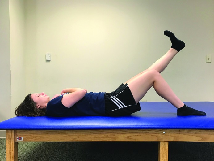
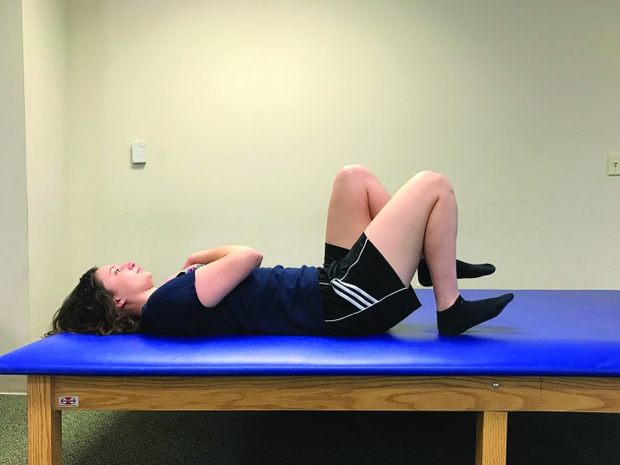
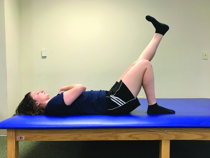
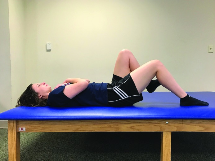
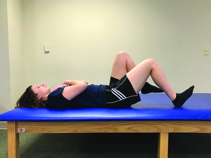
Each subject performed five variations of the single-leg bridge. The positions of these five variations (Positions A-E) are described in Table 3 and Figures 1-5. Position order was randomized, and the data collector was blind to the position being performed. Subjects were blind to the EMG activity of their hip muscles. Subjects performed eight trials of each bridge variation to the beat of a metronome set at 60 beats per minute, extending their hip to neutral during each trial. Recorded data from trials three through seven were post-processed and included in the data analysis.
Table 3.
EMG Activity of Hip Muscles in Different Single-Leg Bridge Positions (Mean ± SD of %MVIC)
| Gluteus Maximus | Gluteus Medius | Biceps Femoris | Dominant Rectus Femoris | Non-Dominant Rectus Femoris | Position Description | |
|---|---|---|---|---|---|---|
| Position A | 51.02 ± 28.12 | 57.81 ± 20.72 | 75.34 ± 24.25 | 5.83 ± 3.91 | 20.49 ± 13.04 | Dominant leg: 90 ° knee flexion with foot flat; Non-dominant leg: knee extended and hip neutral |
| Position B | 47.35 ± 27.47 | 57.23 ± 27.82 | 23.49 ± 9.30* | 8.44 ± 6.25* | 19.21 ± 10.07 | Dominant leg: 135 ° knee flexion with foot flat; Non-dominant leg: knee extended and hip neutral |
| Position C | 47.16 ± 28.07 | 55.05 ± 20.71 | 69.18 ± 18.49 | 5.11 ± 3.38* | 7.00 ± 5.64* | Dominant leg: 90 ° knee flexion with foot flat; Non-dominant leg: knee relaxed in flexion with the femur vertical |
| Position D | 49.12 ± 26.37 | 54.27 ± 20.01 | 58.71 ± 19.72* | 5.22 ± 3.62* | 7.67 ± 6.43* | Dominant leg: 90 ° knee flexion with dorsiflexed ankle; Non-dominant leg: knee relaxed in flexion with the femur vertical |
| Position E | 40.38 ± 24.55* | 41.63 ± 18.19* | 20.84 ± 12.81* | 12.05 ± 9.38* | 7.25 ± 6.51* | Dominant leg: 135 ° knee flexion with dorsiflexed ankle; Non-dominant leg: knee relaxed in flexion with the femur vertical |
indicates a statistically significant change from position A, the traditional single-leg bridge
Figure 1.
Position A. Subjects started with their dominant knee flexed to 90 ° and foot flat. The contralateral knee was extended and its hip remained in neutral. Arms were folded across the chest.
Figure 5.
Position E. Subjects started with their dominant knee flexed to 135 ° and ankle in full dorsiflexion. The contralateral knee was relaxed in flexion with the femur held vertical. Arms were folded across the chest.
Figure 2.
Position B. Subjects started with their dominant knee flexed to 135 ° and foot flat. The contralateral knee was extended and its hip remained in neutral. Arms were folded across the chest.
Figure 3.
Position C. Subjects started with their dominant knee flexed to 90 ° and foot flat. The contralateral knee was relaxed in flexion with the femur held vertical. Arms were folded across the chest.
Figure 4.
Position D. Subjects started with their dominant knee flexed to 90 ° and ankle in full dorsiflexion. The contralateral knee was relaxed in flexion with the femur held vertical. Arms were folded across the chest.
All EMG data were rectified and filtered using a 15 Hz high-pass and 500 Hz low-pass fourth-order Butterworth digital filter. The filtered EMG data were smoothed with a 50-millisecond moving average. Data plots were then inspected twice by a set of five researchers to remove movement artifacts. For each position and each participant, five bridges were recorded and five %MVICs were calculated. MVIC values for each muscle were identified as the maximum value of a 50-millisecond moving average within the three corresponding seven–second MVIC trials. The mean and standard deviation values of these %MVICs were used in final analysis. All data processing was performed using custom written code (Matlab, The Mathworks, Natick, MA). Repeated measures analyses of variance (ANOVAs) (α = 0.05) with Bonferroni corrections were performed for each muscle tested in the five bridge variations. SPSS v23.0 (SPSS Inc, Chicaco, IL) was used for data analysis.
RESULTS
Data from twenty-six subjects were included. Data from two subjects were excluded due to faulty data from the EMG leads for the gluteal muscles. Means and standard deviations of EMG activity expressed as the %MVIC of each analyzed muscle in each of the five bridge positions are presented in Table 3. Significant differences in muscle activation between the modified bridge positions and traditional bridge position (position A) are also noted in Table 3.
Hamstring activity (i.e., percent MVIC) was minimized in positions B (23.49%) and E (20.84%), in which the dominant knee was flexed to 135 ° instead of 90 °. These positions appeared to preferentially activate both gluteal muscles, as gluteus maximus and gluteus medius activation surpassed and essentially doubled biceps femoris activation. Of the two positions, position B displayed higher %MVIC for both gluteal muscles (47.35% and 57.23% versus 40.38% and 41.63% for the gluteus maximus and gluteus medius, respectively). Gluteus maximus and gluteus medius percent MVIC in position E (40.38% and 41.63%, respectively) was significantly (p < 0.05) less than position A (51.02% and 57.81%, respectively), the traditional single-leg bridge position. Gluteus maximus and gluteus medius activation in position B (47.35% and 57.23%, respectively), however, did not significantly change compared to position A.
DISCUSSION
The primary objective of this study was to examine hip muscle activity during five variations of the single-leg bridge and determine which variation preferentially activated the gluteal muscles. The positions and procedures of this study were similar to those used by Boren et al.9 and Ekstrom et al.11 Boren et al. found the traditional single-leg bridge elicited 54% MVIC of the gluteus maximus and 54% MVIC of the gluteus medius.9 Ekstrom et al. found the traditional single-leg bridge elicited 40% MVIC of the gluteus maximus and 47% MVIC of the gluteus medius.11 These data are similar to the results of the current study which showed 51.01% MVIC for the gluteus maximus and 57.81% MVIC for the gluteus medius in the traditional single-leg bridge position (position A).
A difference was seen between EMG activity of the bridge leg biceps femoris during the traditional single-leg bridge (position A) in this study (75.34% MVIC) and that found by Ekstrom et al. (40% MVIC).11 As the MVIC of the biceps femoris was achieved in a similar manner, the difference in activation may be due to the variation in upper extremity placement. Subjects in the study by Ekstrom et al. placed their upper extremities flat on the testing surface to their side.11 Subjects in the current study were instructed to fold their arms across the chest to ensure they did not aid lower extremity muscle activity during the exercise. This position difference may explain the difference in biceps femoris activity between studies. Placement of the upper extremities by the side would have permitted the subjects to use the arms to apply a downward force on the testing surface. Such a force would have been manifested as an extensor moment about the shoulder joints that would have reduced the weight force supported by the bridge leg and thereby reduced the biceps femoris muscle activity required to maintain the bridge position. Folding the arms across the chest prevented the subjects from generating an extensor moment about the shoulder joints that contributed to maintaining the bridge position.
Significant decreases in biceps femoris activity occurred in this study when the knee was flexed to 135 ° versus 90 °. Biceps femoris activity decreased from 75.34% in the traditional position (position A) to 23.49% in position B. This significant decrease in biceps femoris activity following increased knee flexion may be due to the lower leg being aligned parallel with the ground reaction force vector at the foot. In this case, there is a reduced knee extensor moment and a reduced need for the hamstrings to actively maintain the knee flexion angle during the bridge. Reduced hamstring activity would decrease the incidence of hamstring cramping, as one theory of exercise-associated muscle cramps is that they occur due to muscle overload and neuromuscular fatigue.14
Muscle activation during the bridge with a starting position of 135 ° knee flexion is unable to be compared to other studies as it appears to be a novel starting position. A study by Youdas et al. describes EMG activity of a single-leg bridge that requires a subject to achieve a 90 ° flexion angle of the knee at the height of the bridge, presumably starting the exercise with a higher degree of knee flexion.15 However, the starting knee angle of that single-leg bridge was not measured or described. Subjects in that study were also allowed to place their hands on the floor by the sides, potentially assisting the activity.
Significant changes in hip muscle activation occurred with different ankle positioning. Dorsiflexing the ankle of the dominant leg appeared to significantly decrease biceps femoris activity in the current study. Dorsiflexion of the ankle was the only difference between positions C and D, and biceps femoris activity significantly decreased from 69.18% MVIC (position C) to 58.71% MVIC (position D) when the ankle was dorsiflexed. With ankle dorsiflexion, the gastrocnemius was lengthened and thereby no longer actively insufficient to generate a knee flexion moment. Gastrocnemius activity would reduce the need for the hamstrings to be active to prevent knee extension. Additionally, in a study by Chon et al., ankle dorsiflexion was shown to enhance transversus abdominus activity during an abdominal draw-in maneuver (i.e., hook-lying position).16 Thus, ankle dorsiflexion may have caused a decrease in biceps femoris activity during the bridge by activating alternate core musculature on the anterior side of the body, in turn lessening the demand on the hamstring muscles posteriorly. In position E, a significant decrease in gluteal activity compared to position A was observed when dorsiflexion, increased knee flexion to 135 °, and contralateral hip flexion were all added to the exercise.
In the current study, exercise order was randomized and both researchers and subjects were blinded when possible, yet limitations remain. There is potential that subjects did not generate a true MVIC of each muscle tested due to lack of effort or suboptimal positioning. Muscle length during MVIC testing may also be a factor. Attempts were made to minimize these potential limitations by standardizing instructions to subjects, standardizing positioning methods, and using methods for the traditional single-leg bridge and MVIC testing similar to previous studies. Hip muscle activity during single-leg bridge positions was not studied in subjects with pathology, so generalizing results to an injured population should be performed with caution. Future research should examine the effects of strength training using the modified single-leg bridge with preferential gluteal activation (position B) and its effects on pathology and performance.
CONCLUSION
The modified single-leg bridge position with 135 ° of knee flexion (position B) displayed preferential activation of the gluteus maximus and gluteus medius over the biceps femoris. This position maintained gluteal activity while significantly decreasing biceps femoris activity compared to the traditional single-leg bridge position. This modified single-leg bridge, potentially called “the gluteal bridge,” using 135 ° of knee flexion on the dominant side may allow more optimal training of the gluteal muscles than the traditional position since the biceps femoris is less likely to be the limiting muscle of the exercise.
References
- 1.Kim EK. The effect of gluteus medius strengthening on the knee joint function score and pain in meniscal surgery patients. J Phys Ther Sci. 2016;28(10):2751-2753. [DOI] [PMC free article] [PubMed] [Google Scholar]
- 2.Nelson-Wong E Flynn T Callaghan JP. Development of active hip abduction as a screening test for identifying occupational low back pain. J Orthop Sports Phys Ther. 2009;39(9):649-57. [DOI] [PubMed] [Google Scholar]
- 3.Marshall PW Patel H Callaghan JP. Gluteus medius strength, endurance, and co-activation in the development of low back pain during prolonged standing. Hum Mov Sci. 2011;30(1):63-73. [DOI] [PubMed] [Google Scholar]
- 4.Jeong U-C Sim J-H Kim C-Y, et al. The effects of gluteus muscle strengthening exercise and lumbar stabilization exercise on lumbar muscle strength and balance in chronic low back pain patients. J Phys Ther Sci. 2015;27(12):3813-3816. [DOI] [PMC free article] [PubMed] [Google Scholar]
- 5.Leetun DT Ireland ML Willson JD, et al. Core stability measures as risk factors for lower extremity injury in athletes. Med Sci Sports Exerc. 2004;36(6):926-34. [DOI] [PubMed] [Google Scholar]
- 6.Ito J Moriyama H Inokuchi S, et al. Human lower limb muscles: an evaluation of weight and fiber size. Okajimas Folia Anat Jpn. 2003;80(2-3):47-55. [DOI] [PubMed] [Google Scholar]
- 7.Distefano LJ Blackburn JT Marshall SW, et al. Gluteal muscle activation during common therapeutic exercises. J Orthop Sports Phys Ther. 2009;39(7):532-40. [DOI] [PubMed] [Google Scholar]
- 8.Reiman MP Bolgla LA Loudon JK. A literature review of studies evaluating gluteus maximus and gluteus medius activation during rehabilitation exercises. Physiother Theory Pract. 2012;28(4):257-68. [DOI] [PubMed] [Google Scholar]
- 9.Boren K Conrey C Le Coguic J, et al. Electromyographic analysis of gluteus medius and gluteus maximus during rehabilitation exercises. Int J Sports Phys Ther. 2011;6(3):206-23. [PMC free article] [PubMed] [Google Scholar]
- 10.Selkowitz DM Beneck GJ Powers CM. Which exercises target the gluteal muscles while minimizing activation of the tensor fascia lataϿ. Electromyographic assessment using fine-wire electrodes. J Orthop Sports Phys Ther. 2013;43(2):54-64. [DOI] [PubMed] [Google Scholar]
- 11.Ekstrom RA Donatelli RA Carp KC. Electromyographic analysis of core trunk, hip, and thigh muscles during 9 rehabilitation exercises. J Orthop Sports Phys Ther. 2007;37(12):754-62. [DOI] [PubMed] [Google Scholar]
- 12.Cram JR Kasman G. Introduction to Surface Electromyography. Gaithersburg, MD: Aspen Publishers, Inc; 1997. [Google Scholar]
- 13.Finni T Cheng S. Variability in lateral positioning of surface EMG electrodes. J Appl Biomech. 2009;25:396-400. [DOI] [PubMed] [Google Scholar]
- 14.Miller KC Stone MS Huxel KC, et al. Exercise-Associated Muscle Cramps: Causes, Treatment, and Prevention. Sports Health. 2010;2(4):279-283. [DOI] [PMC free article] [PubMed] [Google Scholar]
- 15.Youdas JW Hartman JP Murphy BA, et al. Magnitudes of muscle activation of spine stabilizers, gluteals, and hamstrings during supine bridge to neutral position. Physiother Theory Pract. 2015;31(6):418-27. [DOI] [PubMed] [Google Scholar]
- 16.Chon SC Chang KY You JS. Effect of the abdominal draw-in manoeuvre in combination with ankle dorsiflexion in strengthening the transverse abdominal muscle in healthy young adults: a preliminary, randomised, controlled study. Physiotherapy. 2010;96(2):130-6. [DOI] [PubMed] [Google Scholar]


