Abstract
Background
Medial knee collapse can signal an underlying movement issue that, if uncorrected, can lead to a variety of knee injuries. Placing a band around the distal thigh may act as a proprioceptive aid to minimize medial collapse of the knee during squats; however, little is known about EMG and biomechanics in trained and untrained individuals during the squat with an elastic band added.
Hypothesis/Purpose
To investigate the effects of the TheraBand® Band Loop on kinematics and muscle activity of the lower extremity during a standard barbell back squat at different intensities in both trained and untrained individuals.
Study Design
Cross-sectional, repeated measures.
Methods
Sixteen healthy, male, university aged-participants were split into two groups of eight, consisting of a trained and untrained group. Participants performed both a 3-repetition maximum (3-RM) and a bodyweight load squat for repetitions to failure. Lower extremity kinematics and surface electromyography of four muscles were measured bilaterally over two sessions, an unaided squat and a band session (band loop placed around distal thighs). Medial knee collapse, measured as a knee width index, and maximum muscle activity were calculated.
Results
During the 3-RM, squat weight was unaffected by band loop intervention (p = 0.486) and the trained group lifted more weight than the untrained group (p<0.007). The trained group had a greater squat depth for both squat conditions, regardless of the band (p = 0.0043). Knee width index was not affected by the band during the eccentric phase of bodyweight squats in the trained (band: 0.76 ± 0.08, no band: 0.73 ± 0.08) or untrained group (band: 0.77 ± 0.70, no band: 0.75 ± 0.13) (p = 0.670). During the concentric phase, knee width index was significantly lower for 3-RM squats, regardless of group.
Conclusion
Despite minimal changes in kinematics for the untrained group, increased muscle activity with the band loop may suggest that a training aid may, over time, lead to an increase in barbell squat strength by increasing activation of agonist muscles more than traditional, un-banded squats. Greater maximal muscle activity in most muscles during band loop sessions may provide enhanced knee stability via increased activation of stabilizing muscles.
Level of Evidence
3
Keywords: Elastic resistance, electromyography, knee valgus, kinematics, squat, TheraBand
INTRODUCTION
During a barbell squat, there is a tendency for novice or untrained users to have a medial collapse of the knees under load, especially during the concentric phase of movement. Medial knee collapse, or knee valgus, occurs when there is excessive medial movement of the knee in the frontal plane.1 It can be the result of valgus force, in the frontal plane, that ultimately results in the knee joint center moving towards the midline of the body.2,3 This knee movement will generally be away from alignment with the vertical ground reaction force (GRF) vector,4,7 resulting in hip adduction and internal rotation. Medial collapse is often identified in resistance training activities such as the barbell squat, during explosive jumping,8,9 landing,4,9 and cutting8 tasks. Medial knee collapse can signal an underlying movement issue that, if uncorrected, can lead to a variety of knee injuries, including patellofemoral syndrome,11,3 anterior cruciate ligament tears12 and degeneration of the meniscus due to altered compressive forces on the femur and the patella.1
Coordinated muscle action and movement patterns are essential for optimizing barbell squat efficiency and reducing injury risk. Increasing the load on a barbell squat when medial knee collapse is already evident will likely increase medial knee loading and exacerbate the risk of injury. Previous authors have suggested that hip muscle activity, particularly gluteus maximus, is directly linked to valgus knee angles and internal hip rotation.1,13 High activity in all the gluteal muscles prior to concentric movements produces a robust (and stable) linked system through the hip and knee, resisting valgus knee collapse, and reducing injury risk.13 Quadriceps/hamstring strength ratios may also contribute to valgus knee moments, as both weak hamstrings14 and reduced muscle co-contraction15 can influence valgus knee moments and internal tibial rotation. Lastly, distal movement dysfunction in the kinetic chain, such as decreased ankle dorsiflexion can both increase valgus knee loads and decrease quadriceps activation.16 These mechanisms of valgus knee loading suggest that a detailed biomechanical analysis is required to fully determine the effect of elastic resistance on squat performance.
Training aids include a category of fitness equipment designed to coach proper movement patterns through various types of biofeedback or proprioception. These types of tools are popular in sports and rehabilitation that demand complex movement patterns for repeated success in sport and work. Strength coaches and fitness professionals use training aids to more quickly and efficiently correct dysfunctional movements in athletes/patients. Previous research has investigated the use of an elastic loop, or band, around the distal thigh during body weight squats.7 Gooyers et al.12 concluded that the band was unsuccessful at correcting medial collapse or valgus moments; however, the authors acknowledged that results may be task and/or context specific. It is worth noting that the participant population in that study12 was recreationally active students, likely familiar with, but not well trained in proper squat technique and mechanics. Barbell squats provide a similar movement as the body weight squat investigated by Gooyers et al.,12 however, much greater load bearing is required. It is possible that underlying mechanical deficiencies could be exacerbated when the lower extremity is placed under greater load. If a loaded barbell squat results in greater medial knee collapse, this scenario may be more conducive to determining if using a resistance band can be parlayed into a biomechanical benefit. If a band placed around the distal thighs can affect squat mechanics, then it is likely that muscle activity is also affected. Gooyers and colleagues did not measure lower extremity muscle activity during their investigation.
Therefore, the purpose of this study was to investigate the effects of the TheraBand® Band Loop on kinematics and muscle activity of the lower extremity during a standard barbell back squat at different intensities in both trained and untrained individuals. It was hypothesized that the trained groups’ kinematics (medial knee collapse) would be unaffected by the band, while the untrained group would experience medial knee alignment changes when using the band. Additionally, it was hypothesized that external hip rotator muscle activity would be greater for the trained participants during the band condition.
METHODS
Participants
Sixteen male, university aged-participants were split into two groups of eight: Trained (25.4 ± 4.4 years, 179.83 ± 8.81 cm, 88.36 ± 12.52 kg) and untrained (22.8 ± 1.6 years, 180.81 ± 6.04 cm, 76.91 ± 9.29 kg). The trained group was defined as regularly participating in barbell back squat training for the last year. The untrained group consisted of individuals with no barbell back squatting experience, but who were able to squat a bodyweight load. All participants completed a PAR-Q + to determine physical readiness for exercise as well as a custom exercise history questionnaire and had no previous history of musculoskeletal pain or injury in the past 12 months. Participants provided informed consent and the study was approved by the University of Ontario Institute of Technology Research Ethics Board (REB#:14-057) and in accordance with the declaration of Helsinki.
Protocol
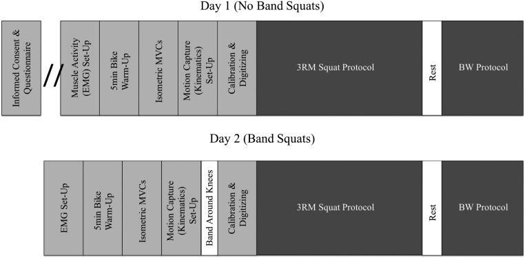
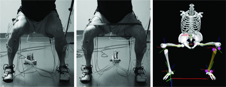
This study was designed as a mixed-model repeated measures, with both groups (trained and untrained) performing two squat conditions during two separate sessions (Figure 1). The first session included the control or no band day, where squats were performed with no instruction given to participants. After a minimum of 48 hours rest, but no more than four weeks, participants repeated the protocol with a red, medium-resistance Theraband® Band Loop (The Hygenic Corporation, OH, USA) placed around the distal thigh, just proximal to the lateral epicondyle of the femur (Figure 2). Trained and untrained participants were given the same coaching on the use of the band; an explanation that the intent is to “keep the band tight throughout the entire squat” but limited verbal cues were given during the actual protocol.
Figure 1.
Timeline of protocol. Each participant (trained or untrained) participated in two data collection sessions. The first session consisted of background information, set-up, calibration, normalization, and a 3RM and BW squat protocol. No earlier than 48 hours later the participants participated in an identical bout of data collection, using a band placed around the proximal knees.
Figure 2.
Left: EMG and rigid body placement during the no band session. Middle: Band placement during the band session. Right: Anatomical reconstruction and Visual3D model of participant performing a squat.
Aside from the Theraband® Band Loop intervention, both sessions followed an identical protocol, using the medium resistance band that requires 4.5lbs of pull to stretch a 12-inch band to 24-inches. Data collection began with muscle specific isometric maximal voluntary contractions (MVCs) for each of the eight muscles recorded. Two MVCs were collected for each muscle, with rest given between trials. Participants then performed a warm up consisting of five minutes of cycling at 75 watts. Participants first performed a 3-repetition maximum (3-RM) protocol, starting with a weight approximately half of their estimated 1-repetition maximum, and taking no less than three sets, and no more than five sets to attain a true 3RM. A ‘true’ 3RM was noted as achieved when the participant attempted a further set at the smallest weight increment possible (10 lbs), ending in failure (referred to as 3RM condition). Rest periods between sets were participant determined and regulated to two to four minute breaks. After a mandatory 10-minute rest period, participants completed a second squat condition, this time with a bodyweight load for maximum repetitions to failure (referred to as BW condition). Heavy encouragement was given to ensure maximum effort and repetitions for each participant. Termination of the BW condition was determined by failure to squat the load, requiring assistance, or a significant deviation in tempo or form with inter-repetition pauses exceeding three seconds. Foot position was the participant's natural squatting stance and was not standardized in order to avoid altering incoming mechanics and finally, to minimize injury risk when performing 3RM repetitions.
Muscle Activity
Muscle activity was recorded from four muscles bilaterally: gluteus maximus (GMa), gluteus medius (GMe), vastus lateralis (VL) and biceps femoris (BF). Ag-AgCl disposable electrodes (MediTrace 130, Kendall, Mansfield, MA, USA) were placed over each muscle belly in-line with muscle fiber orientation, according to previous work.17 Prior to electrode placement, muscle specific locations were shaved, skin was abraded using NuPrep abrasive gel (Weaver & Company Inc., CO, USA) and cleaned with an isopropyl alcohol. Electromyography (EMG) signals were differentially amplified, band pass filtered (CMRR > 115 dB at 60Hz; input impedance ∼10GΩ; 10-1000 Hz; AMT-8, Bortec Biomedical Ltd., Calgary, AB, Canada) and sampled at 2000 Hz. All MVCs included the participant exerting a 3-second maximal effort contraction. Two MVCs were performed for each muscle. Vastus lateralis MVCs were conducted with the participant seated and the leg positioned at 90˚ and secured using an ankle cuff and non-deforming steel cable for a maximal isometric knee extension effort. Biceps femoris used the same procedure but the knee flexion contraction was resisted at the ankle by the experimenter. Gluteus maximus MVCs were conducted with the participant prone on a padded exercise mat. The experimenter resisted the thigh segment while the participant maximally extended their hip, while maintaining knee flexion of 90˚. Gluteus medius MVCs were conducted with the participant lying on their side with both knees flexed to 90˚. The hip of the participants’ top leg was maximally abducted against experimenter resistance.
Kinematics and Ground Reaction Forces
3D kinematics were collected using three 3D Investigator Active Motion Capture Systems (Northern Digital Inc., Waterloo, ON, Canada). Custom rigid bodies, consisting of at least three non-collinear markers were placed on the participant's foot, shank and thigh bilaterally as well as the pelvis and thorax using double sided carpet tape (3M, London, ON, Canada) and Hypafix® (BSN Medical Inc., Hamburg, Germany). Anatomical landmarks were digitized on each participant, assuming a fixed spatial relationship with the rigid body affixed to each segment. Kinematics were sampled at 50 Hz and synchronized with EMG data. The global coordinate system was determined as X, medial-lateral, Y, anterior-posterior and Z, superior-inferior.
Data Analysis
EMG was full wave rectified and Butterworth low pass filtered (3Hz cut-off, dual pass, 2nd order). Peak activity was determined from each muscle specific MVC and muscle activity during each condition was normalized as a percentage of maximal voluntary contraction (%MVC). Kinematic data was used to determine the start, bottom (maximum knee flexion) and end of each squat repetition such that mean and maximum muscle activity could be determined for the concentric and eccentric phases (MatLab 2015b, Mathworks Inc., Natick, MA, USA). The concentric and eccentric phases were determined as frame numbers that marked peak knee flexion and peak knee extension. Additionally, knee angle end points were corroborated using absolute coordinate system data from the thigh markers. Affirming that peak knee-flexion angle occurred at the frame that corresponded to the lowest vertical marker distance. EMG was synchronized with kinematics, so the kinematic frame numbers representing each phase were corrected for sampling rate differences and used for EMG analysis.
Kinematic data were processed using Visual3D (C-Motion, Germantown, MD, USA). Local anatomical frames of reference were created for each segment and used in the kinematic and kinetic calculations. Raw kinematic data were low pass Butterworth filtered at 6Hz. Knee joint angles were calculated as the thigh relative to the shank, using an XYZ rotation sequence. Maximum and minimum knee flexion angles were used to corroborate the start of the eccentric and concentric phases for each repetition. From these angles, the distal joint coordinates (XYZ) were calculated for each segment. Knee Width Index (KWI) was calculated from the three-dimensional position data from each segment as the ratio of the distance between the right and left distal thigh, and ankle (distal shank points).4,7,18 This analysis was replicated for each repetition of the 3RM and body weight (BW) squats for both the band and no band conditions.
Statistical Analyses
An a priori power analysis was performed using G-Power software indicating a need for 16 participants, achieving an actual power of 0.97.5,6 Statistical analyses were conducted using Statistica® (Dell Software Inc., Nashua, NH, USA). A mixed model, General Linear Measures ANOVA was used to determine effect of training (Trained or Untrained), rep/load (3RM or BW), and condition (Band or No Band). All main effects of group or interaction were tested for significance using an alpha of p<0.05 determined a priori. Planned comparisons between conditions for KWI, average and maximum muscle activity, were conducted on Band-Group interaction. Mixed-model ANOVAs (group x squat type x band condition) were performed for knee angle (maximum knee flexion), KWI (bottom) and EMG for each muscle bilaterally. All data are presented as mean ± SD.
RESULTS
Squat Kinematics
There was no effect of band on weight lifted for 3RM intensity (trained, band: 132.7 ± 21.2 kg, no band: 130.3 ± 19.5 kg, p = 0.758; untrained, band: 104.5 ± 10.8 kg, no band: 104.0 ± 9.7 kg, p = 0.486). The trained group lifted significantly more weight than the untrained group for both the band (p = 0.005) and no band (p = 0.007) conditions. There were no significant differences in the number of repetitions performed by the trained participants during the BW band and no band conditions (band: 17.7 ± 9.1 repetitions; no band: 18.1 ± 10.0 repetitions, p = 0.935). There were also no significant differences in the number of repetitions performed by the untrained participants during the BW band and no band conditions (band: 14.9 ± 9.5 repetitions; no band: 16.1 ± 6.5 repetitions, p = 0.762). The trained group had a significantly greater squat depth (knee flexion angle) than the untrained group (trained: 100.94 ± 13.63 °; untrained: 89.40 ± 15.03 °; p = 0.0043) for both 3RM and BW conditions, regardless of the band. Participant and squat demographics can be found in Table 1.
Table 1.
Summary of squat outcomes
| Squat Condition | Training Status | No band | Band | ||||
|---|---|---|---|---|---|---|---|
| Weight (kg) | Reps | Squat depth (°) | Weight (kg) | Reps | Squat depth (°) | ||
| 3RM | Trained | 133.4 ± 23.4 | 3.0 | 95.2 ± 13.8 | 129 ± 19.9 | 3.0 | 104.5 ± 18.2 |
| Untrained | 102.2 ± 4.4 | 3.0 | 90.5 ± 21.7 | 104 ± 8.9 | 3.0 | 87.9 ± 12.5 | |
| BW | Trained | 91.2 ± 12.6 | 15.7 ± 6.1 | 102.3 ± 9.5 | 83.2 ± 9.0 | 15.5 ± 6.3 | 101.9 ± 12.8 |
| Untrained | 76 ± 8.9 | 16.3 ± 5.2 | 87.4 ± 12.5 | 76.0 ± 8.9 | 15.3 ± 10.4 | 91.7 ± 14.9 | |
Knee Width Index
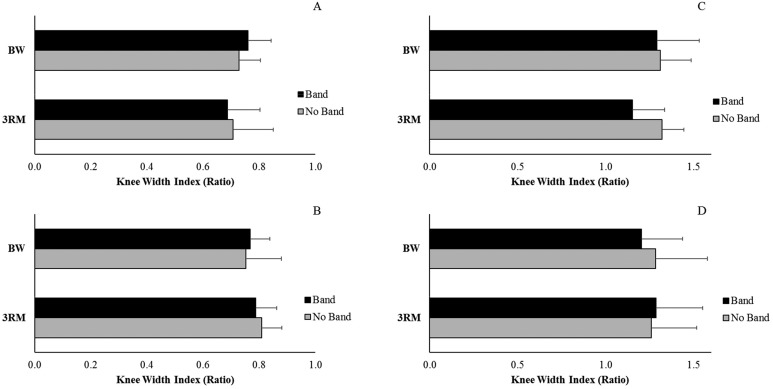
KWI was not affected by the band during the eccentric phase of 3RM squats in either the trained (band: 0.69 ± 0.12, no band: 0.71 ± 0.14) or untrained group (band: 0.79 ± 0.08, no band: 0.81 ± 0.07) (p = 0.482). Similarly, KWI was not affected by the band during the eccentric phase of BW squats in the trained (band: 0.76 ± 0.08, no band: 0.73 ± 0.08) or the untrained group (band: 0.77 ± 0.70, no band: 0.75 ± 0.13) (p = 0.670). However, during the concentric phase, there was a main effect of squat type (3RM or BW) with an overall lower KWI for 3RM squats (Figure 3) (p = 0.046).
Figure 3.
Peak Knee Width Index (mean ± SD) for the trained group during the eccentric (A) and concentric (C) phases. Peak Knee Width Index (mean ± SD) for the untrained group during the eccentric (B) and concentric (D) phases.
Muscle Activity
There was a significant main effect of the band condition, with use of the band increasing muscle activity in the majority of muscles during the eccentric (LVL, p = <0.001; RVL, p = <0.001; LBF, p = 0.001; RBF, p = 0.048; LGMe, p = 0.001; RGMe, p = <0.001; LGMa, p = <0.001; RGMa p = <0.001; Table 2) as well as the concentric phase (LVL, p = <0.001; RVL, p = <0.001; LBF, p = 0.044; RBF, p = 0.025; LGMe, p = 0.038; RGMe, p = 0.049; RGMa, p = 0.017; Table 3). For select muscles, a squat type by band interaction (LVL, p = 0.019; LBF, p = 0.035; LGMe, p = 0.020) was found for the eccentric phase of movement. Only LGMa (p = 0.035) showed a training level X band interaction for the concentric phase of movement. Bonferoni post-hoc analysis showed differential results; while most muscles had increased muscle activity with band use BW squats, LVL demonstrated that band squats elicited lower EMG activity overall and this was more pronounced in the BW condition (p = 0.026) regardless of phase (concentric vs eccentric). During BW squats, the LVL had significantly lower maximum muscle activity when performed with the band, regardless of trained (band: 115.03 ± 36.66% MVC, no band: 130.47 ± 44.54% MVC or untrained (band: 99.74 ± 53.61% MVC, no band: 118.09 ± 36.28% MVC) status.
Table 2.
Normalized maximum muscle activity (mean ± SD) for each muscle during the eccentric phase of all conditions (band, no band) and sessions (3RM, BW). Shaded bars represent trained group. *denotes a main effect of band (p<0.05) for that variable and ∧∧denotes a significant (p<0.05) interaction effect.
| Eccentric Phase | ||||
|---|---|---|---|---|
| Band 3RM | No Band 3RM | Band BW | No Band BW | |
| LVL* | 156.63 ± 76.68∧ | 168.69 ± 41.25∧ | 115.03 ± 36.66∧ | 130.47 ± 44.54∧ |
| 103.20 ± 66.99∧ | 142.18 ± 42.95∧ | 99.74 ± 53.61∧ | 118.09 ± 36.28∧ | |
| RVL* | 169.51 ± 84.75 | 159.76 ± 41.10 | 135.17 ± 64.87 | 114.85 ± 45.43 |
| 163.99 ± 94.35 | 136.93 ± 41.57 | 126.04 ± 68.25 | 118.16 ± 32.82 | |
| LBF* | 49.57 ± 39.54∧ | 47.48 ± 29.27 | 44.53 ± 52.01∧ | 26.65 ± 13.06∧ |
| 18.48 ± 11.29 | 18.23 ± 10.73 | 16.62 ± 9.98 | 14.44 ± 8.42∧ | |
| RBF* | 53.73 ± 61.20 | 46.28 ± 34.20 | 54.31 ± 91.23 | 25.36 ± 13.55 |
| 22.87 ± 8.93 | 17.91 ± 10.25 | 17.00 ± 7.28 | 11.07 ± 5.50 | |
| LGMe* | 59.92 ± 68.34 | 80.04 ± 50.53 | 29.40 ± 16.57∧ | 46.37 ± 34.20 |
| 55.35 ± 27.56 | 48.72 ± 17.41 | 43.13 ± 20.59∧ | 29.57 ± 16.45 | |
| RGMe* | 64.20 ± 35.08 | 69.88 ± 40.63 | 46.93 ± 33.70 | 45.00 ± 22.94 |
| 45.92 ± 22.35 | 54.66 ± 44.21 | 33.18 ± 16.39 | 39.96 ± 37.14 | |
| LGMa* | 126.09 ± 98.87 | 106.71 ± 48.15 | 56.97 ± 41.12 | 76.92 ± 45.39 |
| 128.89 ± 116.97 | 101.91 ± 31.92 | 103.47 ± 88.36 | 78.62 ± 32.10 | |
| RGMa* | 114.02 ± 73.10 | 119.02 ± 51.10 | 80.27 ± 66.60 | 79.91 ± 37.71 |
| 111.85 ± 81.80 | 81.91 ± 43.87 | 85.64 ± 54.78 | 57.50 ± 29.76 | |
Note: L, left; R, right. VL, vastus lateralis; BF, biceps femoris; GMe, gluteus medius; GMa, gluteus maximus.
Table 3.
Normalized maximum muscle activity (mean ± SD) for each muscle during the concentric phase of all conditions (band, no band) and sessions (3RM, BW). Shaded bars represent trained group. *denotes a main effect of band (p<0.05) for that variable and ∧∧denotes a significant (p<0.05) interaction effect.
| Concentric Phase | ||||
|---|---|---|---|---|
| Band 3RM | No Band 3RM | Band BW | No Band BW | |
| LVL* | 139.88 ± 77.77 | 123.18 ± 45.20 | 101.42 ± 45.65 | 107.05 ± 52.18 |
| 98.05 ± 66.96 | 140.51 ± 57.12 | 96.33 ± 57.07 | 116.86 ± 51.09 | |
| RVL* | 155.97 ± 69.08 | 123.72 ± 40.67 | 119.83 ± 53.83 | 88.30 ± 42.25 |
| 160.04 ± 109.73 | 131.37 ± 34.83 | 132.97 ± 105.97 | 113.89 ± 28.46 | |
| LBF* | 26.98 ± 13.46 | 19.24 ± 4.03 | 17.92 ± 7.23 | 13.17 ± 2.61 |
| 34.17 ± 57.58 | 28.29 ± 26.74 | 27.21 ± 39.68 | 20.49 ± 17.04 | |
| RBF* | 37.09 ± 31.71 | 17.8 ± 6.55 | 25.42 ± 21.95 | 12.79 ± 2.67 |
| 29.88 ± 21.49 | 20.65 ± 19.70 | 25.84 ± 16.57 | 15.44 ± 11.40 | |
| LGMe* | 35.36 ± 24.60 | 33.68 ± 19.84 | 23.57 ± 14.42 | 26.45 ± 17.33 |
| 50.38 ± 39.06 | 38.99 ± 17.91 | 38.96 ± 28.02 | 36.31 ± 29.80 | |
| RGMe | 42.02 ± 25.19 | 36.71 ± 22.27 | 28.94 ± 18.16 | 23.09 ± 10.22 |
| 39.64 ± 29.52 | 63.40 ± 91.75 | 30.55 ± 23.75 | 57.02 ± 69.87 | |
| LGMa* | 89.03 ± 56.50 | 66.74 ± 67.06 | 46.47 ± 40.07 | 59.42 ± 67.65 |
| 92.29 ± 60.41 | 86.50 ± 47.54 | 77.00 ± 47.70 | 97.41 ± 87.53 | |
| RGMa* | 76.22 ± 43.13 | 94.14 ± 140.17 | 55.04 ± 46.79 | 66.79 ± 105.72 |
| 97.49 ± 91.35 | 76.28 ± 59.15 | 70.80 ± 56.31 | 80.81 ± 73.08 | |
Note: L, left; R, right. VL, vastus lateralis; BF, biceps femoris; GMe, gluteus medius; GMa, gluteus maximus.
DISCUSSION
This study investigated the effects of a resistance loop band, placed around the distal thigh, on medial knee collapse and muscle activity during the barbell back squat. More specifically, the band was evaluated in regards to training status (trained or untrained) and load (3RM or BW). Interestingly, there was a significant effect of load intensity (3RM or BW) on KWI, but no effect of band or training level conditions. Somewhat conversely, for the majority of muscles monitored, there was significantly greater muscle activity during the band conditions than no band conditions and this was not specific to training status or load.
KWI, the primary measure of medial knee collapse for this investigation, showed no significant difference with respect to a band intervention. While many strength coaches indicate that the band helps promote a more neutral knee alignment and prevent medial knee collapse, the results of this study showed no significant effect of the band intervention, regardless of training status. This is in agreement with Gooyers et al.,12 who found similar conclusions during a bodyweight squat exercise. As suggested previously by Gooyers et al.,12 a longer-term intervention (with an untrained group) using a band may result in plastic changes to squat mechanics and performance, however a single set, regardless of load (3RM or BW), showed no change with the band in the current study. During the concentric phase of the squat, there was a main effect of squat type (3RM or BW) with an overall lower KWI for 3RM squats. This could be interpreted in two ways. One, the increased mechanical demand on the lower extremity due to greater squat load likely contributed to the lower KWI (more medial collapse) and, two, demands associated with the 3RM may have resulted in muscle fatigue, resulting in a lower KWI. Even though neither group showed improvements in KWI during the band loop conditions compared to no band, there is likely still room for improvement in KWI, possibly through the use of a longer training exposure using the band.
Despite few changes in KWI during the 3RM or BW squat, the placement of a resistance band around the distal thighs did increase lower extremity muscle activity in both trained and untrained participants when compared to using no band. The band affected peak muscle activity for muscles in both phases of movement (eccentric: LVL, RVL, LBF, RBF, LGMe, RGMe, LGMa, RGMa; concentric phase: LVL, RVL, LBF, RBF, LGMe, RGMe, RGMa). The most consistent change across training level was for VL, which demonstrated consistently greater muscle activity with the band, across both the 3RM and BW conditions. For example, for the trained group, LVL had activity of 156.6, 168.7, 115.0 and 130.5 %MVC compared to 103.2, 142.2, 99.7, 118.1 %MVC for the untrained group during the 3RM band, 3RM no band, BW band and BW no band conditions, respectively. While Gooyers et al.12 did not measure muscle activity during their body weight squat exercise, the current findings support their hypothesis that the band may differentially change muscle activity patterns even though no reduction in medial collapse was quantified when using the band. Other studies have found no difference in hip abductor muscle activity when a resistance band is applied to a hip eccentric exercise.19 This is contradictory to the findings of this investigation, which demonstrate that the band increased activity across groups and conditions (effect of p<0.001 for eccentric: LVL, RVL, RGMe, LGMa, RGMa; concentric: LVL, RVL). This difference could be the results of a strategic difference when performing a heavy barbell squat as opposed to bodyweight hip centric exercises where smaller stabilizers can assume the roll of band resistance while the agonist muscles do not alter activity. Spracklin et al. 20, also demonstrated that the use of a looped resistance band increases hip muscle activity during a barbell back squat. The authors also concluded that squat performance (measured as number of repetitions completed) were not affected by the band. We also demonstrate no change in the number of repetitions completed, but also show few differences in lower extremity kinematics with respect to a band intervention.
The band increased gluteal muscle activity (GMe and GMa) but only with consistency in the untrained participants. Trained participants demonstrated no difference in peak gluteal muscle activity (with the exception of LGMa in the 3RM squat) when using the band. It was hypothesized that only untrained participants would benefit from the resistance band preferentially, based on the premise that the trained participants have already achieved the muscle activation patterns required to promote neutral knee alignment and to resist medial collapse. The GMa and GMe are the major hip abductors acting to counter the band pull across the knees, but also the major initiator of hip extension, so it may be plausible that trained participants, when told to ‘keep the band tight’ are actually hindering their practiced and efficient gluteal activation patterns.19 The use of resistance band training aids is usually employed by coaches to benefit novice squatters, and these results confirm that trained groups would probably see little benefit.
There are a few limitations that should be discussed. The trained group had a mean 3RM weight that was 125% greater than the untrained group. The authors did not explicitly aim for weight categories in this work, only focusing on training frequency and a minimum load of bodyweight. It is likely that there may not have been a large enough disparity between our trained and untrained groups. Because of the study design, participants in the untrained group had to squat their bodyweight. This means fairly active and strong males were recruited, and a group of lesser-trained participants might have benefited more from the band intervention. Both our groups presented with KWI ratios close to 1, having little room for improvement with the band intervention. Gooyers et al.,12 demonstrated that at maximum squat depth participants achieved a KWI of almost 1.0. This was for a body weight squat and demonstrates that the increased load during a barbell squat could negatively effect KWI. Additionally, it was deemed unacceptable to allow the untrained group to perform a 3RM with the band, prior to a regular unaided squat. Therefore, experimental days were not randomized and the trained group followed the same timeline for parity. The only cue given during the band intervention was ‘keep the band tight’. Perhaps with additional instruction, further differences would have been found in KWI and hip abductor activity with participants knowing when to activate abductors and extensors. Additionally, only one level of resistance band was used during this study. It is possible that using a gradient of resistances could have demonstrated a directional effect and it is possible that using heavier resistance would have elicited a greater response in peak muscle activation. Finally, it may be of interest to measure medial collapse under load in a general population. Even being able to barbell back squat one's own weight (the untrained group) requires a higher level of training than the greater population.
CONCLUSIONS
This study reports the first data on the neuromechanics of the lower limb during a barbell back squat with and without a band loop in both trained and untrained individuals. Squatting with a band increases lower limb muscle activation but does not change knee width index, and these observations were not training status or load dependent. Specifically, this work suggests that there is no evidence that a one-time exposure to a resistance band training aid changes the biomechanics of a barbell back squat as determined by knee width index, kinematics and muscle activity. Furthermore, this null result with respect to resistance band efficacy in reducing medial knee collapse was consistent across training experience. Despite little change in KWI for the untrained group, increased lower extremity muscle activity may suggest a more effective training paradigm for this population.
Knee valgus, if left uncorrected, could lead to a variety of knee injuries, which has been shown to occur in a variety of sports that require squatting, explosive force and quick directional changes. Thus, it is critical to find ways to correct knee valgus and avoid these injuries. Future studies should incorporate different loop band resistances and incorporate both male and female participants. In addition, calculating knee collapse via changes in frontal plane kinematics and knee moments could provide further interpretation. This knowledge could, in turn, be used to develop training programs for athletes and rehabilitation patients who require a reduction in knee valgus loading in order to avoid potential injury or re-injury.
REFERENCES
- 1.Bell DR Vesci BJ DiStefano LJ Guskiewicz KM Hirth CJ Padua DA. Muscle Activity and Flexibility in Individuals With Medial Knee Displacement During the Overhead Squat. Athl Train Sport Health Care. 2012;4(3):117–125. [Google Scholar]
- 2.Claiborne TL Armstrong CW Gandhi V Pincivero DM. Relationship between hip and knee strength and knee valgus during a single leg squat. J Appl Biomech. 2006;22(1):41–50. [DOI] [PubMed] [Google Scholar]
- 3.Holden S Boreham C Doherty C Delahunt E. Two-dimensional knee valgus displacement as a predictor of patellofemoral pain in adolescent females. Scand J Med Sci Sport. (2015). 10.1111/sms.12633. [DOI] [PubMed] [Google Scholar]
- 4.Ford KR Myer GD Hewett TE. Valgus knee motion during landing in high school female and male basketball players. Med Sci Sports Exerc. 2003;35(10):1745–1750. [DOI] [PubMed] [Google Scholar]
- 5.Faul F Erdfelder E Lang A-G Buchner A. G*Power 3: a flexible statistical power analysis program for the social, behavioral, and biomedical Sciences. Behav Res Methods. 2007;39:175–91. [DOI] [PubMed] [Google Scholar]
- 6.Faul F Erdfelder E Buchner A Lang A-G. Statistical power analyses using G*Power 3.1: tests for correlation and regression analyses. Behav Res Methods. 2009;41:1149–60. [DOI] [PubMed] [Google Scholar]
- 7.Gooyers CE Beach TA Frost DM Callaghan JP. The influence of resistance bands on frontal plane knee mechanics during body-weight squat and vertical jump movements. Sport Biomech. 2012;11(3):438–439. [DOI] [PubMed] [Google Scholar]
- 8.Myer GD Ford KR Mclean SG Hewett TE. The Effects of Plyometric Versus Dynamic Stabilization and Balance Training on Lower Extremety Biomechanics. Am J Sport Med. 2006;34(3):445–455. [DOI] [PubMed] [Google Scholar]
- 9.Geiser CF O’Connor KM Earl JE. Effects of isolated hip abductor fatigue on frontal plane knee mechanics. Med Sci Sports Exerc. 2010;42(3):535–545. [DOI] [PubMed] [Google Scholar]
- 10.Alenezi F Herrington L Jones P Jones R. The reliability of biomechanical variables collected during single leg squat and landing tasks. J Electromyogr Kinesiol. 2014;24(5):718–721 [DOI] [PubMed] [Google Scholar]
- 11.Kritz M Cronin J Hume P. The Bodyweight Squat: A Movement Screen for the Squat Pattern. Strength Cond J. 2009;31(1):76–85. [Google Scholar]
- 12.Hewett TE Myer GD Ford KR Heidt RS Colosimo AJ McLean SG van den Bogert AJ Paterno MV Succop P. Biomechanical Measures of Neuromuscular Control and Valgus Loading of the Knee Predict Anterior Cruciate Ligament Injury Risk in Female Athletes: A Prospective Study. Am J Sports Med. 2005;33(4):492–501. [DOI] [PubMed] [Google Scholar]
- 13.Hollman JH Galardi CM Lin IH Voth BC Whitmarsh CL. Frontal and transverse plane hip kinematics and gluteus maximus recruitment correlate with frontal plane knee kinematics during single-leg squat tests in women. Clin Biomech. 2014;29(4):468–474. [DOI] [PubMed] [Google Scholar]
- 14.More RC Karras BT Neiman R Fritschy D Woo SL Daniel DM. Hamstrings--an anterior cruciate ligament protagonist. An in vitro study. Am J Sports Med. 1993;21(2):231–237. [DOI] [PubMed] [Google Scholar]
- 15.Lloyd DG Buchanan TS. Strategies of muscular support of varus and valgus isometric loads at the human knee. J Biomech. 2011;34(10):1257–1267. [DOI] [PubMed] [Google Scholar]
- 16.Macrum E Bell DR Boling M Lewek M Padua D. Effect of limiting ankle-dorsiflexion range of motion on lower extremity kinematics and muscle-activation patterns during a squat. J Sport Rehabil. 2012;21(2):144–150. [DOI] [PubMed] [Google Scholar]
- 17.Hermens HJ Freriks B Disselhorst-Klug C Rau G. Development of recommendations for SEMG sensors and sensor placement procedures. J Electromyography Kinesiolology. 2000;10:361–384. [DOI] [PubMed] [Google Scholar]
- 18.Noyes FR Barber-westin SD Fleckenstein C Walsh C West J. The Drop-Jump Screening Test Difference in Lower Limb Control By Gender and Effect of Neuromuscular Training in Female Athletes. Am J Sort Med. 2005;33(2):197-207. [DOI] [PubMed] [Google Scholar]
- 19.Frost DM Beach T Fenwick C Callaghan J Mcgill S. Is there a low-back cost to hip-centric exerciseϿ. Quantifying the lumbar spine joint compression and shear forces during movements used to overload the hips. J Sports Sci. 2012;30(9):859-870. [DOI] [PubMed] [Google Scholar]
- 20.Spracklin KF Button DC Halprin I. Looped band placed around thighs increases EMG of gluteal muscles without hindering performance during squatting. J Perf Health Res. 2017;1(1):60-71. [Google Scholar]


