

Abstract
Syphilis such as the other Sexually transmitted diseases are a cultural background of physician. The authors have presented this case of nodular secondary syphilis for three main reasons. The first one is that, in the last years, syphilis has re-emerged as the problem of public health. The second one is to underline how secondary syphilis, also known as the great imitator, may present itself with numerous manifestations, mimicking different dermatological diseases. Finally, because we want to remember how syphilis and the other sexual transmitted diseases must to be in the cultural background of a dermatologist, and have to be considered in the dermatological differential diagnosis.
Keywords: Syphilis, Sexually transmitted diseases, diagnosis, physician, dermatologist
An otherwise healthy, 38-years old man showed up to our clinic for nodular lesions in the genital area (Fig.1-2), which was present by eight months. At first, they were localised on his gland and, afterwards, also on the scrotum. History for drug assumption or contact with local irritants was negative. He didn’t report any contact with local irritants. The patient did not refer any episodes of high-risk sex and any relevant pathology. He showed no familiarity for lymphoma, lupus and another skin disease.
Figure 1.

Man, 38 years old, with asymptomatic nodular lesions in the genital area
Figure 2.
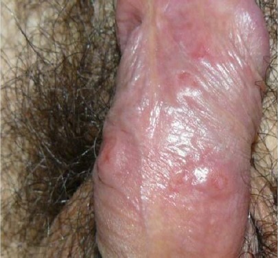
Nodular lesions on the penis
Because of the chronic disease, the patient had been previously visited by several doctors, not dermatologist ones, without obtaining clinical results. At first, the patient had been visited by his general doctor, who prescribed to him a topical therapy with fusidic acid. Due to the lack of clinical results, the man had a visit with a urologist. The doctor, after examining the lesions, prescribed to the patient a swab test which resulted in being negative. Because of the suspect of an infectious disease, the urologist prescribed to the man a systemic therapy with ciprofloxacin twice a day for a week. For the second time, the patient did not observe any beneficial effects and decided to consult an andrologist. The doctor, after having evaluated the lesions, performed an ultrasound exam, without significant results. Finally, he prescribed to the man a systemic therapy with levofloxacin 500 once a daily for ten days. No clinical effects had been observed. Frustrated by the disease, the patient decided to consult a dermatologist and referred to us.
At genitals examination, we observed numerous subcutaneous hard nodules, with a diameter ranging from a few mm to 2 cm. The skin over the lesions was reddish to brown in colour. No local signs of inflammation or infection were detected. Lesions were asymptomatic. During the clinical evaluations, except for a diffuse lymph-adenopathy, we did not observe significant skin lesions in the other body areas. The patient did not report any disturbance of other kinds. As a result of the clinical and anamnestic evaluation, we advised the patient to perform routine blood tests and specific tests for syphilis.
The latter were positive, confirming our suspect of syphilis (RWt +++, RWl ++, VDRL ++, TPHA ++ 1:5120, FTA-ABS-IgG ++, FTA-ABS-IgM +). Only at this moment, the patient admitted having unprotected casual sex. We prescribed to the patient diaminocillina therapy (2400000 U.I./week) for four weeks. Finally, we advised him to abstain from sexual activity and to suggest serological tests to his partners. The patient has been monitored for the duration of treatment. At the end of antibiotic therapy, the patient returned to our observation for a checkup. The lesions were completely healed (Fig.3-4), and serological tests confirmed the improvement of the disease.
Figure 3.

The patient after the treatment
Figure 4.
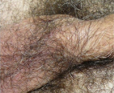
Complete resolution of the lesions of the penis, after the treatment
The authors have presented this case of nodular secondary syphilis for three main reasons. The first one is that, in the last years, syphilis has re-emerged as the problem of public health [1-2]. The second one is to underline how secondary syphilis, also known as the great imitator, may present itself with numerous manifestations, mimicking different dermatological diseases [3-6].
Finally, because we want to remember how syphilis and the other sexual transmitted diseases must to be in the cultural background of a dermatologist, and have to be considered in the dermatological differential diagnosis.
Footnotes
Funding: This research did not receive any financial support.
Competing Interests: The authors have declared that no competing interests exist.
References
- 1.Repiso B, Frieyro M, Rivas-Ruiz F, De Troya M. Condom use and number of sexual partners among male syphilis patients who report having sex with men. Actas Dermosifiliogr. 2010;101(10):847–52. https://doi.org/10.1016/j.ad.2010.06.014 PMid:21159260. [PubMed] [Google Scholar]
- 2.Jebbari H, Simms I, Conti S, Marongiu A, Hughes G, Ward H, Powers C, Thomas DR, Evans B. Variations in the epidemiology of primary, secondary and early latent syphilis, England and Wales 1999 to 2008. doi: 10.1136/sti.2009.040139. [DOI] [PubMed] [Google Scholar]
- 3.Schnirring-Judge M, Gustaferro C, Terol C. Vesiculobullous syphilis:a case involving an unusual cutaneous manifestation of secondary syphilis. J Foot Ankle Surg. 2011;50(1):96–101. doi: 10.1053/j.jfas.2010.08.015. https://doi.org/10.1053/j.jfas.2010.08.015 PMid:21106408. [DOI] [PubMed] [Google Scholar]
- 4.Breznik V, Potočnik M, Miljković J. Papulonodular secondary syphilis in a 52-year-old non-HIV heterosexual patient. Acta Dermatovenerol Alp Panonica Adriat. 2010;19(4):27–30. [PubMed] [Google Scholar]
- 5.Furlan FC, Oliveira AP, Yoshioka MC, Enokihara MM, Michalany NS, Porro AM. Leukocytoclastic vasculitis:another condition that mimics syphilis. An Bras Dermatol. 2010;85(5):676–9. doi: 10.1590/s0365-05962010000500011. https://doi.org/10.1590/S0365-05962010000500011 PMid:21152792. [DOI] [PubMed] [Google Scholar]
- 6.Mullooly C, Higgins SP. Secondary syphilis:the classical triad of skin rash, mucosal ulceration and lymphadenopathy. Int J STD AIDS. 2010;21(8):537–45. doi: 10.1258/ijsa.2010.010243. https://doi.org/10.1258/ijsa.2010.010243 PMid:20975084. [DOI] [PubMed] [Google Scholar]