Fig. 5.
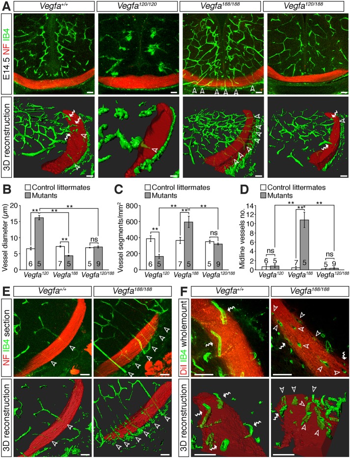
Vascular patterning defects in the diencephalon of Vegfa isoform mutants. (A) Confocal z-stacks (top panels) and 3D reconstructions of z-stacks (bottom panels) of coronal sections through the diencephalon of E14.5 control embryos and mutants expressing specific Vegfa isoforms. Blood vessels were labelled with IB4 (green) and nerves with antibodies against neurofilaments (NF; red). (B-D) Vessel diameter (B), vessel density (C) and number of midline vessel sprouts (D) in mutants expressing specific Vegfa isoforms and control littermates (mean±s.e.m.). **P<0.01; ns, not significant (one-way ANOVA with post-hoc Tukey). Numbers on bars indicate the number of embryos analysed for each genotype. (E,F) Confocal z-stacks (top images) and 3D reconstructions of z-stacks (bottom images) of optic tracts from E14.5 control embryos and Vegfa188/188 mutants, shown in coronal sections (E) or as whole-mounts (F). Samples were labelled with antibodies against neurofilaments (E, red) or DiI (F, red) to label the nerves and IB4 (green) to label blood vessels. The red channel has been made semi-transparent in the 3D reconstructions in A,E,F. Unfilled arrowheads indicate vessel sprouts extending through the RGC axon bundles, white wavy arrows vessels growing along the surface of the RGC axon bundles. Scale bars: 50 µm.

