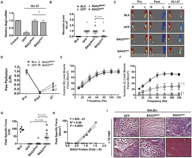
Figure 6. Systemic BAG3Ile81 delivery rescues BALB/c limb muscle blood flow and function after ischemia.
BL6 (black bars, N=5) and BALB/c (gray bars) mice injected IV with AAVs encoding GFP (N=5), BAG3Met81 (N=10), or BAG3Ile81 (N=9) were subjected to modified HLI with collateral vessels left intact. A. Quantification of skeletal muscle BAG3 mRNA expression by qRT-PCR, corrected for GAPDH and normalized to contralateral control. B. Limb necrosis score data plotted per mouse, with means ± SEM. *P<0.05 vs. BL6. #P=NS (0.07). †P<0.05 vs. GFP. C. Representative LDPI images of paw blood flow. D. Quantification of paw perfusion by LDPI. E–F. Force-frequency analysis of isolated extensor digitorum longus (EDL) muscles from the contralateral control (E) and ischemic (F) limbs at HLI d7. G. Peak specific EDL muscle force (expressed as a % of the contralateral EDL). BL6 peak specific force values presented only as a frame of reference. #P=NS (0.068), Ile v GFP. #P=NS (0.057), Ile v Met. H. Correlational analysis between BAG3 protein expression and muscle peak specific force. Line fit by least square regression. I. Representative H&E stains for TA muscle morphological analysis (scale bar=100μm). P<0.05 vs. BL6; ‡ P<0.05 vs. GFP or BAG3Met81. All other data are means ± SEM.

